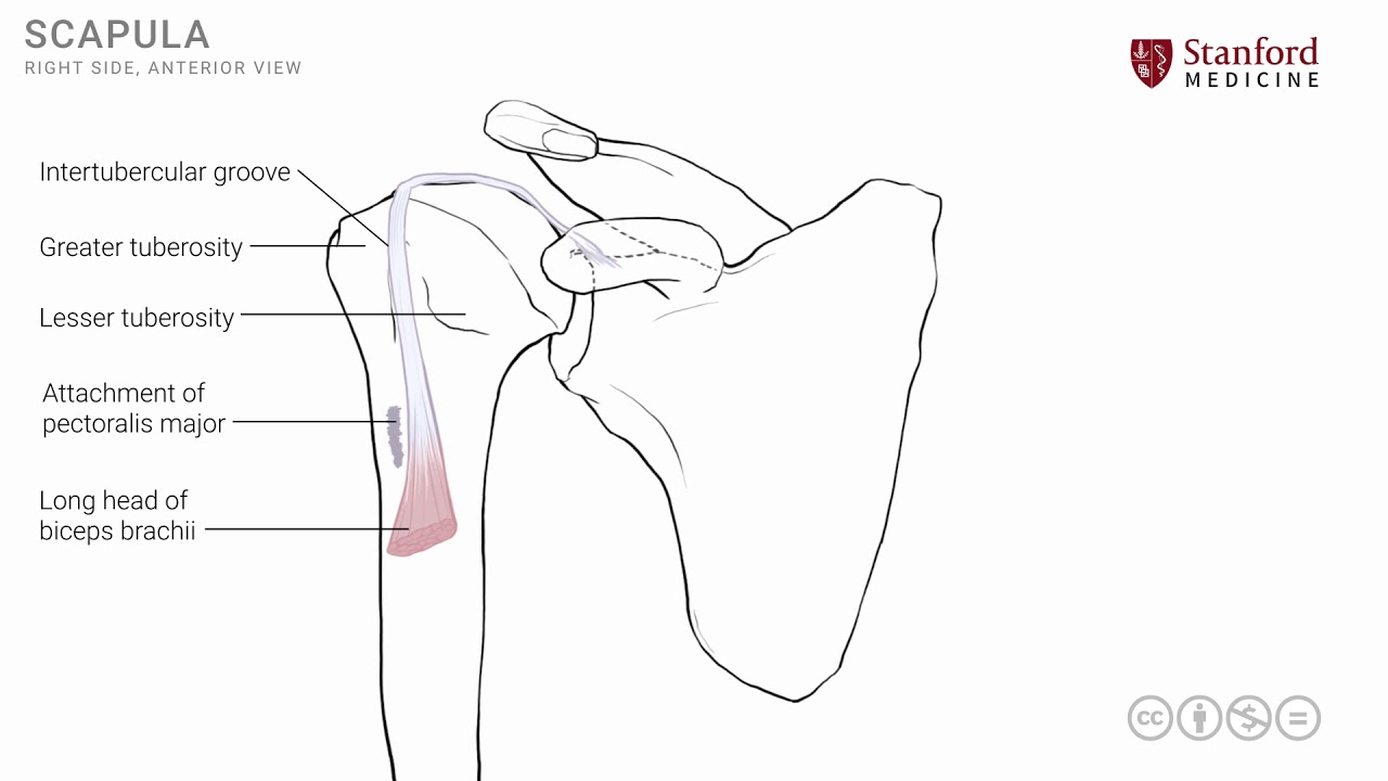
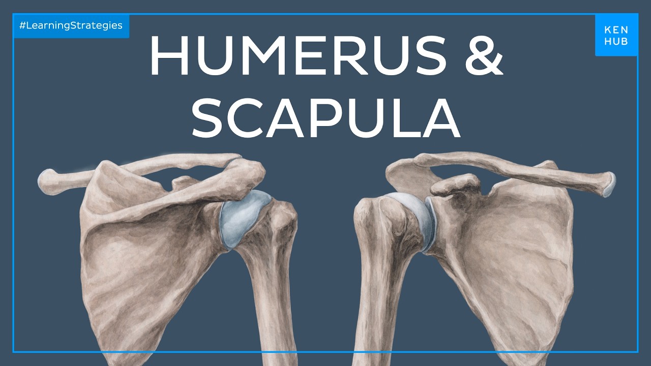
[Music] in this lesson we will look at the posterior view of the scapular shoulder area and consider the various muscles that are located in this region so let's start by looking at a simple line drawing and look at the right shoulder area from an anterior vantage point in order to orient ourselves I'm going to isolate the scapular bone and we will now rotate it around so that we have a posterior view note that we still have the right side of scapula and so the midline structures the vertebral column and the scapula will be in the
relationship as shown here I will now convert this into a very simple schematic line diagram the dashed vertical line is representing the vertebral column and the scapula is as shown here note again this is the posterior view of the right scapula and we can put some structures in order to orient ourselves on to the scapula the first one of those is the glenoid which is the area of articulation that is on the lateral side of the scapula for the shoulder joint the other important structure we see is this shelf like structure known as the spine
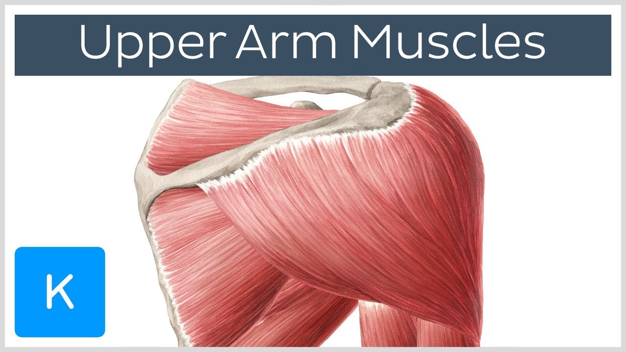
of scapula the spine is a structure that emanates from the posterior side of the scapula and it ends in the formation of the acromion process the acromion process is in fact the terminal part of the spine of the scapula and from the acromion we have the clavicle that articulates with it to complete this particular bony Anatomy now the first muscle that is in this area is a muscle that is known as the trapezius it's a large muscle it's flat muscle and there is a right side and a left side each muscle is triangular in shape
and the two muscles together become trapezoidal in shape to give the name trapezius and the trapezius muscle had a very extensive midline attachment all the way from the base of the skull to the thoracic 12th vertebra and the fibers go laterally and attach onto the area of the spine of the scapula the acromion and the lateral end of the clavicle as shown in the diagram note that the fibers that are of the trapezius have varying angles of inclination the fibers which are more superiorly they go downwards and laterally whereas the fibers that are in the
inferior part of the trapezius go upwards and laterally and hence the fibers of the trapezius have slightly different directions of pull when they contract this muscle is innervated by the spinal accessory nerve and a nicer view of this is seen in the little photograph diagram that we have here where we can see the anterior oblique view to show that for pzs as it is attaching onto the chromium and the lateral end of the clavicle I briefly mentioned about the pole of the fibers and if you observe and conceptualize how these fibers might contract at the
same time simultaneously we up the fibers which are in the upper part of the muscle and those in the lower part of the muscle they will end with a movement that is rotation of the scapula which is also an integral part of shoulder movements it is difficult if you note in your ownselves it is difficult to abduct the shoulder to its full 180 degrees without scapular movement and hence this is an important part of shoulder movements there's another important muscle in this area of the superficial back and in order to understand that muscle we need
to put the humerus in place the second muscle is known as the latissimus dorsi muscle and it also has a very extensive attachment on to the midline thoracotomy ria and on to the pelvic hip bone and from that extensive attachment the fibers go upwards or superiorly and laterally they have some attachments onto the ribs and the inferior angle of the scapula and ultimately attach on to the anterior side of the proximal humerus we will see the exact location of its attachment in a subsequent lesson the latissimus dorsi muscle is innervated by a nerve from the
brachial plexus known as the thorak or dorsal nerve we will see exactly where this nerve comes from the brachial plexus in a subsequent lesson let's let's now take a look at a dissection photograph that shows the two muscles trapezius and latissimus dorsi this here is the midline just to get ourselves oriented this is the midline we're mostly seeing the right side of the back and the neck area and the first muscle that we can see here is this triangular muscle on the right side the trapezius and combining it with the left side muscle which we
can see a little portion of it in the left side of our photograph it gives us the trapezoidal shape note that is fibers go laterally and attach onto the spine of the scapula and its associated structures the second muscle is the latissimus dorsi muscle which is seen here you can see the extensive attachment more approximately and inferiorly and then the fibers go laterally and superiorly and disappear out of the view of this photograph we will see exactly where it hatches onto the humerus in a subsequent lesson let's move on and now consider some of the
muscles that lie deep to the trapezius and are also attached to the scapula for that we will remove these two muscles the trapezius and latissimus and we're back to our very simple line drawing in order to understand the deeper muscles the first one of these three muscles is the levator scapulae muscle and this muscle is attached to the superior medial angle of the scapula and it connects it to the cervical spine the more proximal cervical spine c1 c2 c3 and c4 and it has a pole which is in an upward and medial direction it is
often a muscle that becomes painful and goes into spasm if the posture is not correct and if neck the neck position is not ideal with poor ergonomics for example the second muscle in this region is known as the rhomboid and there are two of them the first one is known as the rhomboid minor the second one is the rhomboid major the minor is smaller as the name will suggest while the major is the larger of the two these two muscles often appear as one muscle rather than two distinct muscles and the separation is sometimes rather
artificial these two muscles are attached more approximately on the midline for the rhomboid minor at about the c7 or t1 level and for the rhomboid major between the t2 to t5 level that's roughly the level at which it attaches on to the midline and then it from there the fibers go laterally and inferiorly to attach on to the medial border of the scapula the rhomboid minor typically attaches at the site of what is called as the root of the spine of the scapula where the root where the spine of the scapula ends on the medial
border whereas the rhomboid major attaches a little bit more inferior to that note from a reference standpoint that the inferior angle of the scapula is at the t7 vertebral level this is this is an important landmark to keep in mind especially as you are approaching a patient for physical examination or you are involved in a surgery related to this region it's a useful landmark to keep in mind let's now look at a photograph that has a dissection of this region and here again for reference we're looking at the right side of the photograph this is
the midline so we're mostly seeing the right side the trapezius has been removed this is the area of the trapezius that has been removed and cut away in order to expose those three muscles that we just described the first one of those three muscles is the levator scapulae which is seen here and it's extending from that superior medial angle of the scapula all the way up to the upper cervical region the second muscle is the rhomboid minor which is seen here and it is extending from the midline going in an inferior and lateral direction to
the medial border of the scapula much larger than the rhomboid minor is the rhomboid major which is seen here lying inferior to the minor also extending from the midline and going to the medial border of the scapula thus we see these muscles that participate in the shoulder joint area and shoulder joint movements levator scapulae in the rhomboids primarily have a stabilizing action though they can contract and result in movement of the scapula towards the midline a movement that is called retraction of the scapula [Music] you