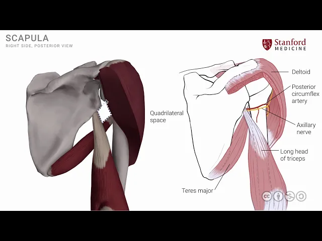
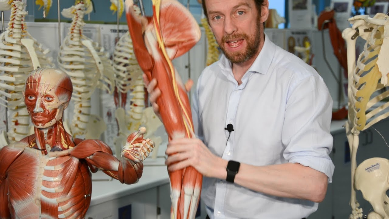
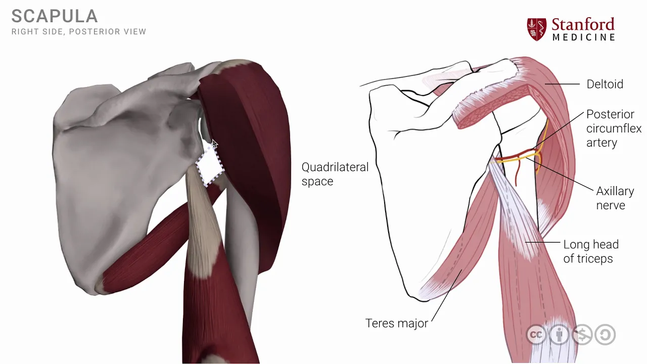
[Music] in this lesson we will look at the shoulder muscles from a post year viewpoint specifically those muscles that are attached around the shoulder joint more laterally and in order to understand these muscles let's look at a simple line drawing as shown here we see the right scapula posterior view along with the right humerus and the right clavicle in place and the first muscle that we will put on is known as the deltoid muscle this is a fairly large well-developed muscle that gives the rounded contour of the shoulder region there are post ear fibers there
are anterior fibers and there are central fibers one can see the posterior fibers taking its attachment onto the spine of the scapula the anterior fibers are seen here taking their attachment on the lateral end of the clavicle and the central fibers are seen here attaching onto the acromion process all three sets of fibers then converge and go inferiorly and attach on to the lateral surface of the shaft of the humerus in a place that is often called as deltoid tuberosity I'm going to remove a small portion of this deltoid muscle especially the posterior fibers in
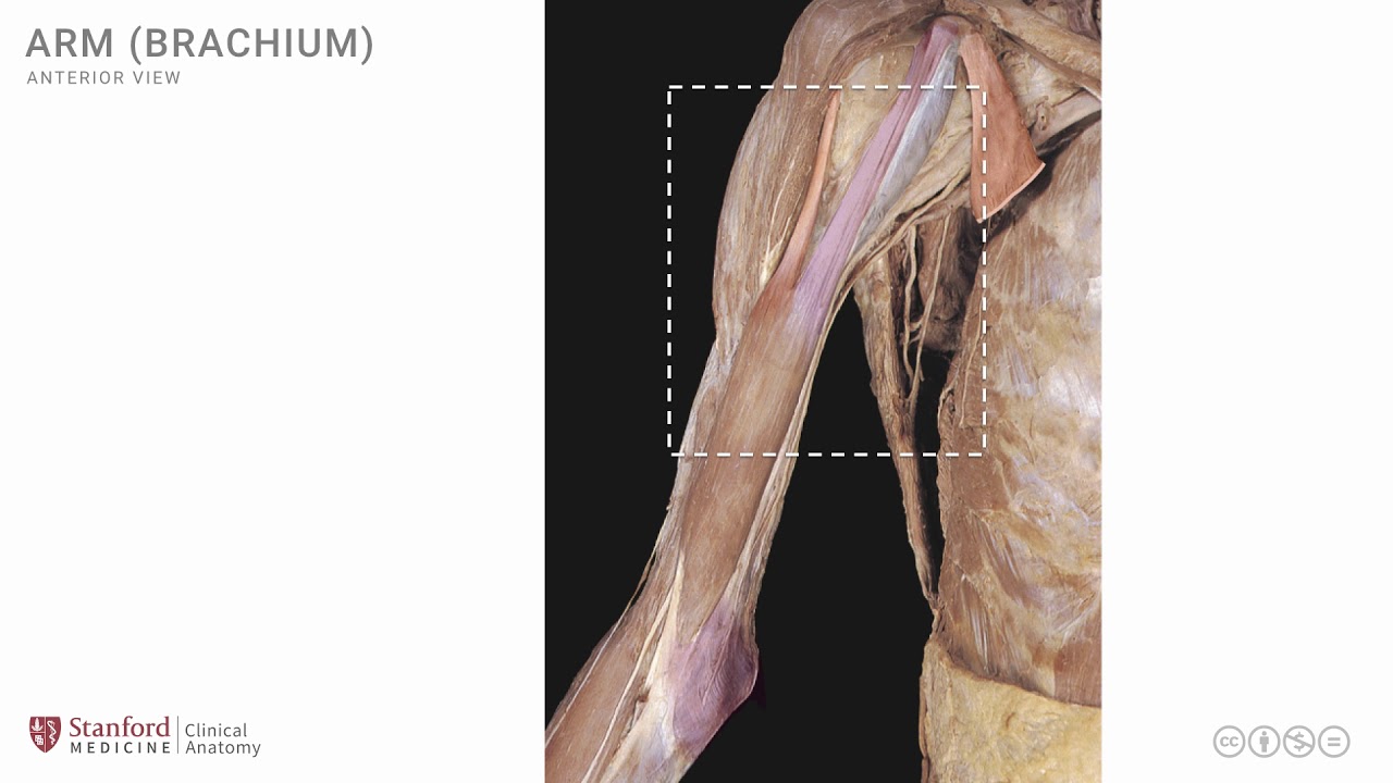
order to make the deeper structures more visible so I have now cut out the posterior fibers here and here in order to make the deeper structures visible that we're interested in the first one of these structures is known as the long head of the triceps muscle tri meaning three and sepsis head triceps is the muscle that has three heads and this is one of those three heads known as the long head of the triceps note that it has an attachment on to the scapula just below the glenoid cavity this particular location is called as infra
glenoid tubercle I'm going to put a second muscle on here known as the teres major muscle that we have looked at in a previous lesson this muscle has its attachment on to the inferior part of the poster scapula and the fibers then go laterally and superiorly to attach on to the humerus note the very important relationship of the fibers of teres major and the long head of the triceps the teres major fibers are anterior to the long head of the triceps this is an important relationship to keep in mind from this small area that you
see between the teres major and the long head of the triceps there are important structures that are coming out the first one of these is known as the axillary nerve the axillary nerve is a branch from the brachial plexus and we will see exactly where it comes out from the brachial plexus in a future lesson and it wraps around the shaft of the humerus as seen here and it supplies the deltoid muscle it innervates the deltoid muscle the axillary nerve is accompanied by an artery known as the posterior circumflex artery sometimes also called a posterior
circumflex humeral artery and this artery accompanies the axillary nerve also to supply this region with blood these two structures the posterior circumflex artery and the axillary nerve travel around or wrap around the shaft of the humerus in an area that is called the surgical neck of the humerus it's an area that has great clinical significance it's often fractured and in fractures of this area the artery and nerve can be damaged the location from where these nerves and arteries exit to gain access to the posterior part of the shoulder joint has an important relationship and an
aim in order to understand this let me show you a simple animation where we rotate this region to get a very Clearview and you will note now there's there's a space which is roughly rectangular or quadrilateral in shape and in fact is called the quadrilateral space the boundaries of this space are formed by and you can be you should be able to name these now as you can see here this is the long head of the triceps this is the teres major muscle and here we have the inferior aspect of the Deaver humeral joint and
here we have the surgical neck of the humerus which is a little bit hidden by the cut fibers of the deltoid muscle here we also have additionally two other spaces they are triangular in shape and hence they're called triangular spaces there's the medial one and there is a lateral one the medial one is adjacent to the scapula whereas the lateral triangular space is adjacent to the shaft of the humerus these spaces the triangular spaces also have structures neurovascular structures that emanate out of them and we will look at the exact structures that are coming out
from the triangular spaces in a future lesson let's now look at a simple photograph of a dissection to highlight these structures and here you'll note the quadrangular space which is seen here the quadrilateral space is the site for exit of the axillary nerve and is clearly seen here there is a muscle that you can see on this medial part of that quadrangle space and we can give it a name as you've seen in the previous slide this is in fact the long head of the triceps it's quite a substantial muscle and it forms one of
those boundaries of the quadrangular space there's the other muscle which is very closely related to this that we have looked at in the earlier image the teres major muscle and the fibers of this muscle go laterally an anterior to the long head of the triceps as very clearly seen in this dissection photograph and gain attachment on to the ante of the humerus we have seen the exact location of the teres major muscle attachment onto the humerus in a previous lesson so as that you don't get confused let me make one other point very clear here
this muscle that we see over here is a muscle known as the teres minor muscle it lies also towards the lateral side of the scapula and also attaches onto the humerus however note that the fibers of teres minor are posterior to the long head of the triceps and the teres minor is one of the four muscles of the rotator cuff and we will see the details of the rotator cuff in a subsequent lesson [Music]