[Music] if we look at the distal end of the humerus there are some shiny surfaces here and very smooth that are articular surfaces for the elbow joint the one that is on the medial side looks like a pulley and is known as the trochlea the one more laterally seen here is somewhat hemispherical and is known as the capitulum or capitellum and it is looks like a head and knobbly head if I rotate this around to look at the posterior part of the distal humerus we find a very large fossa in this area which is known
as the electron fossa and it accommodates the electron process of the ulna in full extension of this joint on the anterior surface we have two other very shallow fossa this one here is known as the coronoid fossa which accommodates the coronoid process of the ulna and the one on the radial side is known as the radial fossa which accommodates the radial head here we have the right ulna and we're looking at the proximal end of the ulna here and it tapers down through the shaft into the distal end of the ulna which is over here
contrast this with the radius which is seen here which is the second of the forearm bones and this is the right radius note that it's proximal end is much smaller and then it continues down the shaft in its more distal end becomes expanded and much wider so in that sense the radius and ulna have a reciprocal architecture the radius and ulna articulate together in the forearm like this so in the anatomical position they are situated like this and the gap between the two bones are covered by an interosseous membrane this is a thick fibrous membrane
which runs between the two bones and if you note they're adjacent surfaces this is the lateral surface of the ulna and you can see that sharp bony ridge where that interosseous membrane would be attached and similarly on the medial side of the radius you have this sharp Ridge where the interosseous membrane is attached and so that membrane runs between these two forearm bones if we focus for a moment on the proximal ulna we see a very prominent projection here which is best seen in a lateral view of the ulna and this is known as the
olecranon process the one on the anterior side seen here is known as the coronoid process and this is the area that is normally covered with articular cartilage and it articulates with the distal humerus to form the elbow joint so let me bring the humerus in and show you how these two bones articulate this is the distal end of the right humerus and this is the proximal end of the right ulna and they articulate in this fashion here and let me show you from a lateral view this is how they would articulate and this is how
the flexion would be caused and it extends and an extension note that the olecranon process enters into the olecranon fossa and in full flexion over here if we see this the coronoid process will enter into the coronoid fossa so that's the movement of flexion extension at the elbow joint or more strictly sometimes we can think of this as the articulation between the ulna and the humerus and so it's often described as they all know humeral Junction or articulation similarly we have the radius which would articulate the proximal end of the radius would articulate with that
distal humerus in what's known as the capitulum in this fashion as seen here and in full flexion that radial head would also come more anteriorly and be accommodated in that radial fossa the radial head is much smaller and so the radial fossa on the humerus is relatively shallow for the movement of pronation and supination note that this happens at the proximal and distal radioulnar joints and they move in tandem in such a way the radius rotates across the ulna in this fashion to become from a supinated to a pronated position and then the reverse of
that would take it back from the pronation to the supination position the ulna really doesn't move at all in supination pronation it is really only the radius that moves the radius will move at both the articulations there is an articulation between the proximal radius and ulna known as the proximal radioulnar joint and similarly there is an articulation between the distal radius and ulna known as the distal radioulnar joint and these two joints will move in tandem to create the movement of pronation and supination so this is now the distal end of the ulna and we're
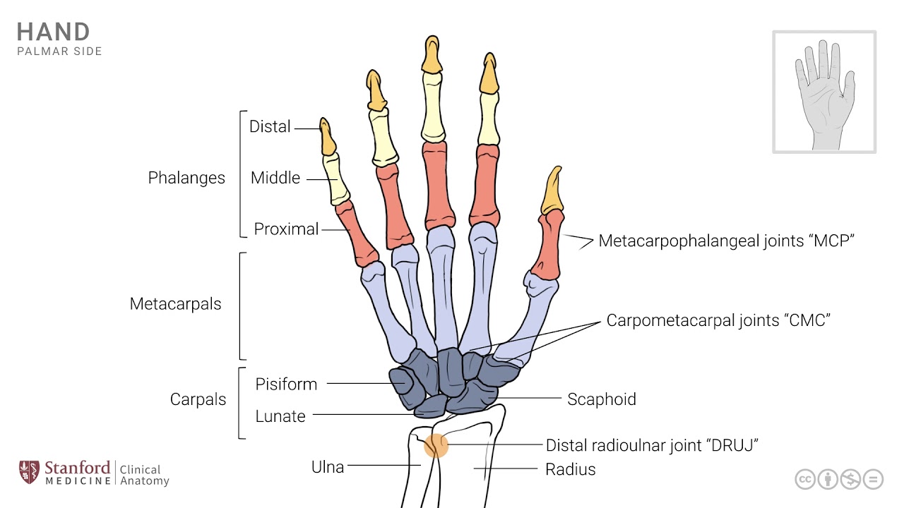
looking at it so that this is more distal and this is proximal just to get ourselves oriented and similarly we have the distal end of the radius so this is the distal articular surface and this is the proximal end just so that we don't get disoriented and this Alma here has a bony projection known as the ulnar styloid process it's a site for attachment of some ligaments and it is easily palpable on clinical examination at the wrist joint here the distal end of the radius also has a styloid process known as the radial styloid process
seen here this is also easily palpable on a clinical examination of the wrist joint there's another little bony projection here that is known as the Lister's tubercle and this is an important landmark for some of the tendons that are crossing the wrist joint the extensor tendons on their way from the forearm to the dorsum of the hand [Music] you