[Music] in this lesson we will look at the elbow joint and the structures that participate in the formation of this hinge joint let's start by looking at a simple line drawing that shows the right elbow joint from an anterior vantage point there are three bones that participate in the formation of the elbow joint the first one is the humerus and we see the distal end of this single arm bone that participates in the formation of the elbow joint more distally we see the ulna over here this is one of the two forearm bones and we
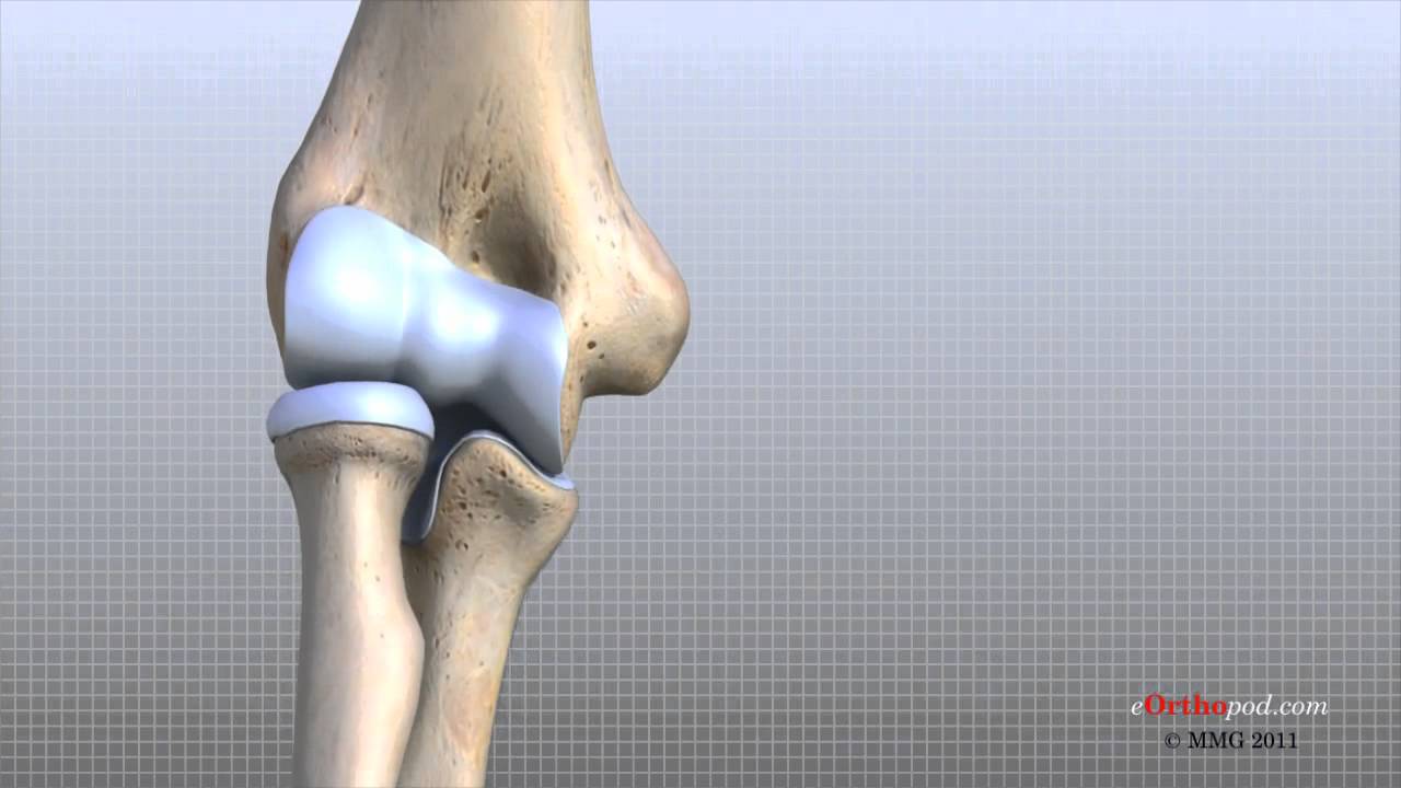
see the proximal end of the ulna and the second bone in the forearm is the radius seen here which also participates in the elbow joint if we focus on the distal end of the humerus we see certain notable bony prominences the first one is a prominent prominence on the medial side of the distal humerus known as the medial epicondyle this is an easily palpable bony landmark in a patient the similar structure on the lateral side is known as the lateral epicondyle which is far less prominent and is seen here towards the center of the distal
end are two other prominences that are covered by articular cartilage and participate in the formation of the elbow joint the first one of these is known as the trochlea and is seen here on the more medial side of the condyles the name trochlea means pulley like and it describes the shape of this structure the other condyle on the lateral side is known as the capitulum and is this structure seen here which is raw round and nobly and look resembles at head and hence the name capitulum we have similar bony prominences on the distal side as
well the first one is known as the coronoid process which is this structure seen here on the proximal end of the ulna and this articulates with the trochlear similarly on the lateral side the more proximal end of the radius is known as the radial head and is seen here which articulates with the capitulum these two structures move in the movement of flexion and require an area on the more proximal side of the distal humerus in order to be accommodated and there is indeed fossa a fairly shallow fossa known as the coronoid fossa which is seen
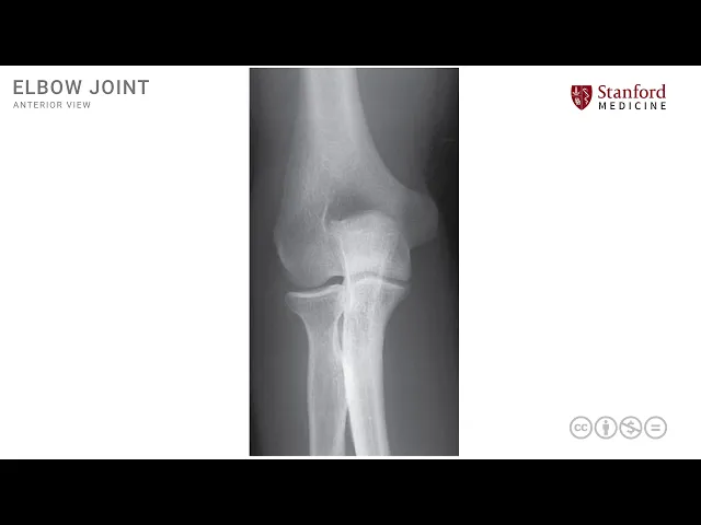
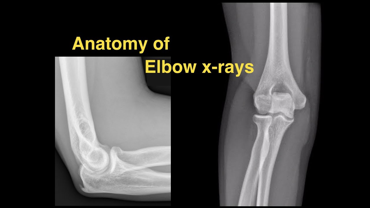
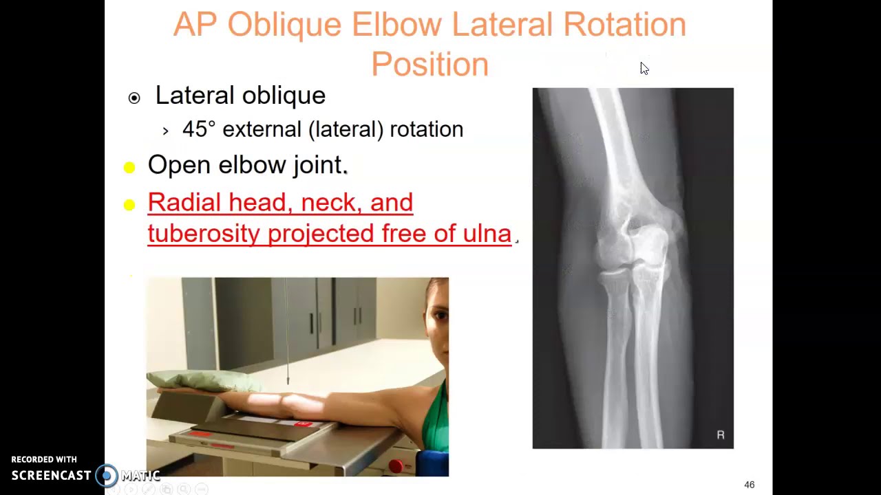
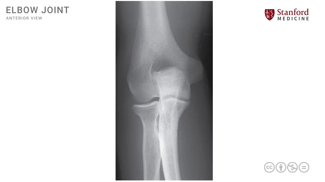
here which accommodates the coronoid process in full flexion similarly we have a radial fossa which is seen over here to accommodate the radial head in full flexion these are some of the key structures in the arab elbow region let's now look at a simple x-ray this is a plane ap x-ray in anteroposterior x-ray of the right elbow joint and we can see some of the same structures again this is the humerus which is the distal end of this bone we can also see the ulna and the radius more distally over here this is the radius
on this side and this is the ulna on this side and these three bones participate in the formation of the elbow joint which is a hinge joint the coronoid process can be seen over here and is outlined here in yellow orange color similarly the radial head is seen over here and is outlined in reddish orangish color over here these are two key structures that articulate with the distal end of the humerus to form the elbow joint or as a as known as a hinge joint the lateral epicondyle and the medial epicondyle are also seen over
here on the distal end of the humerus and over here on the lateral side of the distal end of the humerus the olecranon is seen over here which is a very interesting structure seen on the posterior side and requires a posterior vantage point view we can see it in an x-ray because the radiation goes through and through and it is overlapping with the condylar area of the humerus we will see this more clearly in a lateral view of the elbow joint in the olecranon fossa is the area on the distal humerus that accommodates this electron
here we have an x-ray of the elbow joint and this is seen from a lateral view here and we see the three bones again that participate in the formation of the elbow joint this is the distal end of the humerus seen here and we also have the proximal end of the radius seen here and the proximal end of the ulna seen here these are the three bones that participate in the formation of the hinge joint which is what an elbow joint is let's now also look at those bony prominences that we looked at earlier this
is the coronoid process which is part of the proximal end of the ulna which is right here and see how it articulates with the distal end of the humerus we can also see the radial head seen here which is the proximal end of the radius and also articulating with that distal humerus note that the coronoid process and the radial head in this view this lateral view overlap to some extent and much of the radial head seems to be hidden by the coronoid process we also can see another part of the proximal elma ulna which is
known as the olecranon process which is a large bony extension of the proximal ulna seen here this is the electron process that had been outlined so nicely here and note that the olecranon process extends posteriorly it goes behind or posterior to the Condillac condylar processes of the distal humerus any of these three processes the olecranon process or the coronoid process or the radial head can be fractured and in injuries around the elbow joints these fractures are commonly identified [Music] you