in this video we are going to be talking about the cervical spine the first part of this conversation should really center around the cervical nerves understanding the anatomical layouts and relationships between the cervical nerves and their vertebral bodies and the intervertebral discs is really a high yield concept so the first place we need to begin is talking about where these nerve roots go in relationship to the vertebrae generally speaking the rule you want to remember is that cervical nerves will exit above their corresponding cervical vertebrae and this is true of every cervical nerve the only
exception is that the c8 nerve root will exit below the c7 vertebrae and this should make a little bit of sense because there is no c8 vertebral body there's no c8 vertebrae there's only a c8 nerve root and therefore if you were going to follow the rule completely including c8 there'd be no c8 nerve root to exit above a corresponding c8 vertebral body so therefore the exception to the rule is that c8 since it has to exit somewhere it must exit below the c7 vertebral body and if you're having difficulty conceptualizing this let's let's just
illustrate it on the slide so in this example i'm showing you three vertebral bodies and their intervertebral discs which are shown in blue you see c5 c6 and c7 let's represent the cervical nerve roots in red i'm going to label these for you in just a second but see if you can test yourself and figure out which nerve roots these little red lines correspond to you can pause the video if you want a second to think about it but i'm going to go ahead and label this diagram now remember cervical nerve roots exit above above
their corresponding cervical vertebrae so you can see that the the top most or the superior cervical nerve roots shown on this diagram must correspond to c6 because they're exiting above the corresponding vertebrae so it has to be above c6 and remember the exception to the rule is that the c8 nerve root actually exits below c7 because again there's no c8 vertebral body and therefore it can't exit above a vertebral body that just doesn't exist so it has to go somewhere and therefore it exits below c7 now this is what you want to remember and the
mnemonic to remember this is seven up eight down this mnemonic will remind you that for the first seven cervical nerve roots think up or exiting above the corresponding cervical vertebrae and for the exception for c8 nerve root remember down it will exit down or below c7 now why is this so important to understand on comlex on in-class exams on whatever test you're taking where omm may show up it's pretty typical for test writers to give you a question where they describe some type of cervical spine pathology resulting in some type of cervical nerve root damage
usually this will come in the form of an intervertebral disc herniation so for example look at the slide if you have a leftward herniation of the intervertebral disc between c5 and c6 you can see that when that disc slides over it will impinge upon the c6 nerve root and then the question becomes what type of sensory problem or motor problem or loss of reflex do you expect based on the fact that the c6 nerve root is having issues so you need to know the clinical sequelae of a problem with any of these cervical nerve roots
so what does that mean that you need to know you need to know the sensory distribution of these nerves the motor distribution of these nerves or the motor innervation and the reflexes if applicable that these cervical nerve roots are responsible for so let's start with sensory dermatomes this is a map of sensory dermatomes basically it shows you the areas of the body where these cervical nerve roots provide sensory innervation what i want you to do if this seems overwhelming is to pay special attention to the hands because if you look at the hands depending on
which finger you're talking about which digit there are three different cervical nerve roots responsible for that so these are the ones you want to remember if you're feeling cramped for brain space and you think maybe you'll only be able to memorize a few but for completeness sake let's fill out this chart so we're looking at cervical nerve roots going from c3 down to c8 and we want to be able to understand the sensory dermatome of all of these cervical nerve roots so let's fill these in the place to begin i'm actually going to combine c3
c4 and c5 the reason i'm combining them is because these three cervical nerve roots if you go back to this picture of the dermatome you can see that c3 c4 and c5 all provide sensory innervation to somewhere around the neck upper shoulder area maybe slightly posterior aspect of the head but it's mostly neck and shoulder area so you can see that neck shoulder and superior chest and the interesting thing about c3 c4 and c5 is that these nerve roots collectively comprise the phrenic nerve which will refer gallbladder and diaphragm pain to the right shoulder so
if you're having true pathology in the gallbladder or the diaphragm that pain via these nerve roots will be referred to the right shoulder and again look at the dermatomal distribution of c3c4 and c5 you can see that they provide sensory innervation to not only that posterior head neck and chest and and chest area but it does go to the right shoulder as well so that's the reason that via the phrenic nerve you get that classic referred pain pattern now as far as c6 c7 and c8 you want to remember the hand i told you if
you're only going to remember one thing from this discussion remember the dermatomal distribution in the hand so c6 provides sensory innervation to the thumb c7 provides it to the index and middle finger also known as the second and third digit and c8 will provide it to the ring and pinky finger also known as the fourth and fifth digit so if you have some type of nerve root impingement usually in the form of intervertebral disc herniation and that disc buds up against one of the cervical nerve roots and sort of knocks out or impinges upon it
you want to know how how do i name that injury and again that will totally depend on which cervical nerve root is involved but generally speaking what i need you to remember and what i need you to take away from this discussion because it's really high yield is that when you have nerve root injury causing sensory loss or sensory dysfunction we call that radiculitis we refer to this as radiculitis so let's do an example to make sure we're all on the same page a 23 year old baseball player suffers a throwing injury he undergoes an
mri which reveals a moderately sized leftward herniation of the c6 to c7 intervertebral disc which nerve root will be impinged by this herniation all right so let's go back to our diagram here is our vertebrae with intervertebral discs and cervical nerve roots i'm telling you that there's leftward herniation of the c6 to c7 intervertebral disc so this is what we're going to expect that disc will slide to the left and therefore it's going to impinge upon the c7 nerve root because remember that c7 nerve root exits above corresponding c7 vertebral body so that's the nerve
root that's there to be impinged so we know it's going to be c7 now part two if the patient were to experience paresthesias due to this injury in what sensory dermatome would the discomfort be felt all right so i'm telling you that he's having paresthesia so he's having abnormal sensation maybe a little tingling maybe a little bit of pain where do you expect that to be felt well let's go back to our table we know it's the c7 cervical nerve root and according to this table and what i told you is particularly high yield it's
going to be the index and the middle finger the second and third digit because that's the sensory dermatome that maps out c7 left index finger and left middle finger because again it's the left herniation so it's just going to be on the left side part three how would you correctly name this pathology so this is going to be referred to as a left-sided c7 radiculitis left-sided because it's a left herniation c7 because that's the nerve root that's going to be impinged and radiculitis because that's the term that refers to abnormal sensory loss due to nerve
root impingement all right so that's this is the table that you want to remember but the truth is guys and girls that i didn't really give you the complete picture here because in addition to just having to know sensory dermatomes you also need to know one more thing about all of these cervical nerve roots you need to know if applicable what reflex these nerve roots control so the good news is that for c3 and c4 you actually don't need to know anything there's no high yield reflex reflex that you need to know for c5 and
c6 you want to know that these nerve roots help control the biceps and brachioradialis reflex for c7 and c8 these will control the triceps reflex so if you think about kind of your classic primary care physician using a reflex hammer to elicit a motor response to elicit that reflex and they're tapping on the biceps they're tapping on the brachioradialis these are the cervical nerve roots that are responsible for making that happen and they're somewhat easy to remember because they go in groupings right c5 and c6 both do the same reflex c7 and c8 both do
the same reflex so just remember this now we we mentioned that if there's loss of sensory right some problem with sensory we call that radiculitis but if there's loss of either muscular control or reflexes that's a different term that's actually called radiculopathy so the itis is sensory and the apathy is muscle and reflex so let's do another example it's going to be similar to the example that we already did but i'm just going to change up a few things a 23 year old baseball player suffers a throwing injury he undergoes an mri which reveals a
moderately sized leftward herniation of the c6 to c7 intervertebral disc which nerve root will be impinged by this herniation so this is literally the same question as before we've got leftward herniation of the intervertebral disc between c6 and c7 impinging upon the c7 nerve root so just as it was before you're you're knocking on the c7 nerve root if the patient were to experience muscle fatigue and loss of a reflex due to this injury what reflex might be hypoactive well we know it's c7 so let's go back to our chart the reflex that may be
hypoactive is going to be the triceps reflex all right lastly how would you appropriately name this type of injury this is a left-sided c7 radiculopathy left-sided because that's the direction of the herniation c7 because that's the cervical nerve root that gets pinched in this injury and radiculopathy because in this example we're not talking about a sensory problem we're talking about muscle fatigue and loss of a reflex or a hypoactive reflex so again if it's a muscle problem or a reflex problem it's radiculopathy if it's a sensory problem so like tingly pain it's radiculitis so the
itis means sensory the apathy means muscle and reflex memorize that it's very high yield let's continue this conversation by talking about regional anatomy so as far as the anatomy that you need to know in the cervical spine there's really just a few things first the scalene muscles there's three of them anterior middle and posterior and the highest yield thing that you need to know is just where they attach this will be more relevant in the conversation we have about ribs but briefly anterior and middle scalenes they both attach the first rib and posterior scalene that
muscle attaches to the second rib just try to memorize this it'll be very important in a future lesson the sternocleidomastoid muscle the the most important thing you need to know is the action of the muscle so forget about the attachments just remember the action the action of the scm is side bend towards rotate away also known as straw stra side bend towards rotate away this is what happens when the sternocleidomastoid muscle contracts but it's also what happens if you have a hypertonic scm that's constantly contracting or over contracting and the reason this is really important
is because you might have a picture where they show you torticollis right someone's sternocleidomastoid is just way too hypertonic and what it'll look like is what you see in this image and based on the image they give you you'll have to figure out is it the right sternocleidomastoid that's hypertonic or is it the left sternocleidomastoid that's hypertonic and it's somewhat tricky because since it has that rotation away component to it their head will be turned to the contralateral side and if you look at an image and you see the head turned away to the contralateral
side relative to the sternocleidomastoid that is actually the problem it can throw you off and incorrectly make you think that it's actually the other sternocleidomastoid that's hypertonic so just remember you're going to see what this picture looks like side bending towards shown with that blue arrow rotational way shown with that green arrow and it's going to be in reference to the sternocleidomastoid that's actually dysfunctional so if the chin is turned to the right it's the left sternocleidomastoid that's going to be the problem and it's because of that rotation away component so very important to understand
that now let's change gears and talk about cervical vertebrae motions at each of the three parts of the cervical spine there are three different primary motions so as you might imagine because there are three relevant portions of the cervical spine and each of those three portions has a different primary motion this is particularly high yield because test writers will just love to ask you this at oa the primary motion is flexion and extension at a a the primary motion is rotation in fact a a contributes at least 50 percent if not more of all of
the rotation in the cervical spine c2 to c7's primary motion is side bending now how do you remember this my mnemonic here is that the cervical spine is the first real segment or the first real section if you prefer that but f r s for flexion rotation side bending in order for your studying pleasure now what about fryet's laws if you recall from our discussion in the fundamentals section i told you that fryat's laws only apply to the thoracic and lumbar spine the reason for this is beyond the scope of comlex and frankly beyond the
scope of what you even need to know for your in-class exams but just for completeness sake this has to do with the orientation of the facets and the way that osteopathic physicians can motion test these segments so basically when fryet was formulating his laws he determined that they could only consistently apply to the thoracic and lumbar spines now despite that even though the textbook answer is that fryet's laws do not apply to the cervical spine the way that we diagnose and name somatic dysfunction in the cervical spine still follows a pseudo version of fryet's laws
so instead of saying that this segment is type one meaning it follows law number one fry it's law number one or that this is type two meaning that it follows fry at law number two we have to use a little bit different terminology and that terminology is shown here on the slide oa is considered to be type one like which means that oa follows fryet law number one not actually but you'll follow that when you try to name somatic dysfunction at oa in the cervical spine and likewise from c2 to c7 that is said to
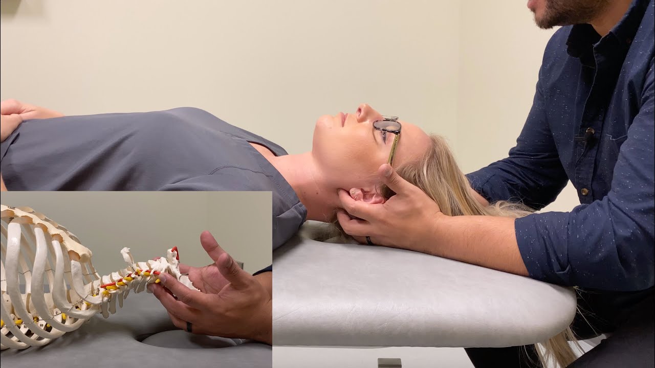
be type 2 like which means you'll use fryet's law number 2 although again the textbook answer is that this doesn't follow freya's laws but you'll still apply the law to determine the direction of side bending and rotation when you're diagnosing somatic dysfunction let's talk about something that will aid you in doing just that in order to get these questions right you have to understand the concept of translation translation is a motion that the cervical vertebrae will kind of glide in one direction or the other and usually this happens when you're doing your osteopathic structural exam
so you'll have the patient literally lying on the table and on the slide what you see is a depiction of the cervical spine while your patient is lying on the table so imagine for the purposes of this discussion that your patient is lying on the table they're lying supine their head is closest to you on the exam table and their feet are far away from you at the end of the table and you're sitting at the head of the table and you're palpating their cervical spine as you perform your structural examination and you do motion
testing if one of their vertebrae are said to translate more easily to the right what that means is that this vertebrae here shown in the middle on this slide slides more easily to the right and the reason that the concept of translation is so important to understand is because when you have translation one way it creates side bending in the opposite way as you can see shown in this red arc so as you translate a vertebrae more to the right if it prefers to translate right that segment is side bent left because it prefers side
bending to the left and you can see that evident from the red arc so this is really high yield because if you think about it on complex if they give you a question and they tell you which direction that segment prefers to translate you can figure out side bending and then knowing whether you're talking about oa and it being type 1 like or c2 to c7 and that being type 2 like you would then apply fryet's law number 1 or fryet's law number two knowing what side bending is to figure out which direction rotation goes
and using that problem-solving formula you can correctly name somatic dysfunction in the cervical spine so very very high yield make sure you understand translation type one like and type two like let's finish up this lesson by just pointing out a few clinical pearls these are things that have shown up on exams in the past and therefore i do think it would be very prudent to know these bits of information one always treat thoracics or ribs before the cervical spine oftentimes somatic dysfunction or musculoskeletal pathology just in general can originate in the thoracic or rib area
and cause radiation up into the cervical spine so you want to treat the thoracics first two the alar or transverse ligament both of those ligaments are weak in patients with down syndrome and rheumatoid arthritis so there are certain techniques that are actually contraindicated in patients with down syndrome or rheumatoid arthritis because if you accidentally tear one of those ligaments which stabilize the vertebrae you can cause really severe irreversible damage in fact it can be life-threatening so those techniques will be contraindicated more on that in a different lesson three you need to take certain cautions when
you perform any degree of cervical extension in the cervical spine due to vertebral artery insufficiency so because of the way and because of the anatomy by which the vertebral arteries exit around the vertebrae anytime you extend those segments you're kind of putting a little bit of pinch on the vertebral artery so if you're taking complex or if you're taking an in-class exam and they give you a question where somebody is treated in some way with cervical extension and then all of a sudden they get certain types of symptoms you want to think about vertebral artery
insufficiency lastly let's talk about whiplash injuries whiplash injuries to the cervical spine are particularly severe but there are two things that you want to keep in mind that are poor prognostic indicators one is loss of the normal cervical lordosis so normally the cervical spine has that nice curvy lordosis to it but if somebody has a whiplash injury and then their c-spine on radiograph becomes more straight and they lose that lordosis that is a poor prognostic indicator and likewise loss of consciousness at the scene of an accident that's also a poor prognostic indicator so if somebody
is say the restrained passenger in a motor vehicle accident and the fact that they're wearing their seatbelt causes that really severe whiplash injury and then they lose consciousness that loss of consciousness in and of itself is a poor prognostic indicator so keep both of those things in mind especially the loss of the lordosis in the cervical spine and now lastly finally i know i keep saying we're going to end but this is actually the last topic we're just going to touch on the orientation of facets and the anatomical uniqueness of cervical vertebrae so when you
palpate a cervical vertebrae you're not actually palpating the transverse process if you're down in the thoracic or the lumbar spine you're certainly palpating the transverse processes but actually up in the cervical spine what you're feeling are where those orange arrows are pointing and those are actually articular processes not transverse processes and while we're on this topic one of the really high yield parts of comlex is knowing the orientation of the superior facets and you need to know the orientation in the cervical thoracic and lumbar spines for whatever reason the directions and orientations of these superior
facets are very high yield there's an easy way to remember this and the demonic is bumble bim bumble bim b-u-m-b-u-l-b-m going down in order starting at cervical the superior facets are backwards upwards and medial in the cervical spine backwards upwards and lateral in the thoracic spine and just backwards and medial in the lumbar spine i'm not going to include the facet orientations when we talk about the thoracic spine and the low back so it's going to be here in this lecture but just recall bumble bim very very high yield