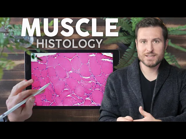
[In this video, I’ll teach you the differences between smooth, cardiac, and skeletal muscle so hopefully you can see a slide under a microscope and know exactly which kind of muscle you’re looking at. If you’re new to the channel, welcome, my name is Patrick and this channel is all about anatomy and how we learn about it. As always, I have the accompanying notes for this video linked in the description if you want to check those out.
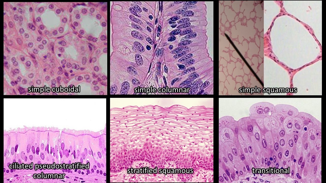
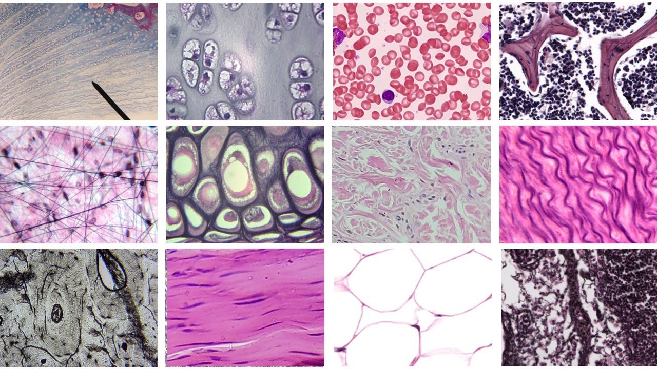
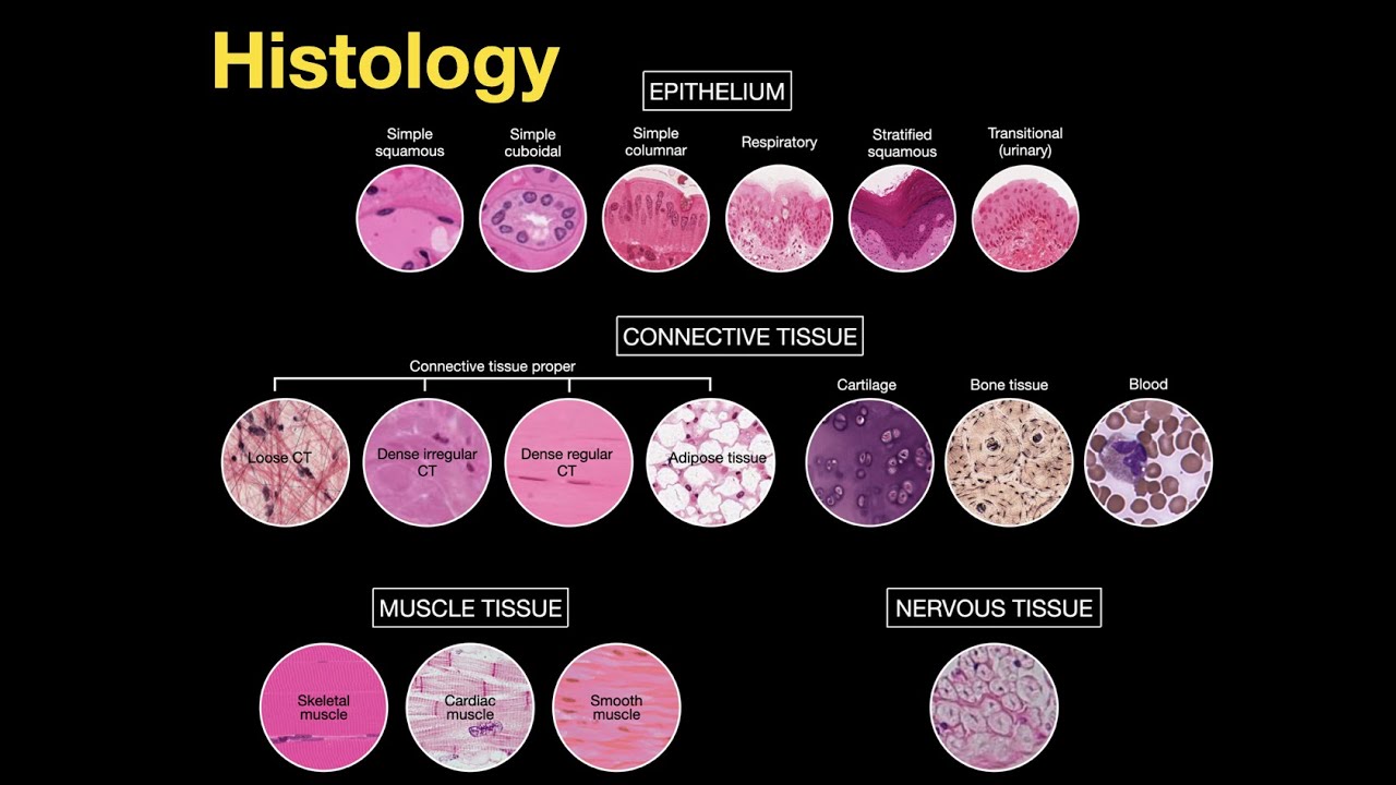
Otherwise, let’s get started. Muscle is one of the four types of tissues in the human body, and it’s the simplest one to start learning. The others, nervous, epithelial, and connective tissue all have a ton of different subcategorizations, but there are only three types of muscle tissue and they all look and behave differently.
But to make sense of the microscopic anatomy, or what we deal with in the world of histology, we need to take a step back and look at big picture anatomy where things are more familiar. Like you already know that the whole purpose of muscle is to produce force. When muscle fibers contract, no matter which type of muscle fiber it is, it will produce force.
But all three types work in slightly different ways. Skeletal muscle contracts forcefully along a set path, so it has big bundles of parallel fibers. Meanwhile smooth muscle just needs to squeeze, so it’s organized into broad sheets.
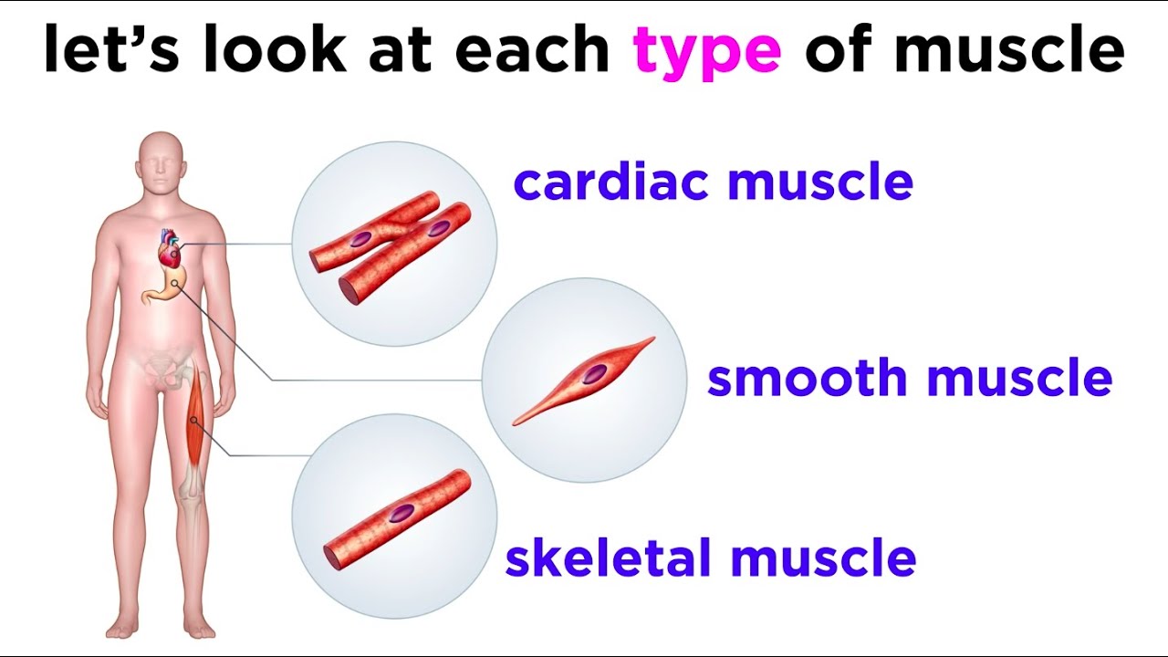
So as we go over these three muscle types, remember that form influences function. Once we know that we’re looking at /some/ kind of muscle, it’s our job to tell what kind of muscle it is, and there are only three types. You have cardiac muscle only in your heart.
. . hopefully.
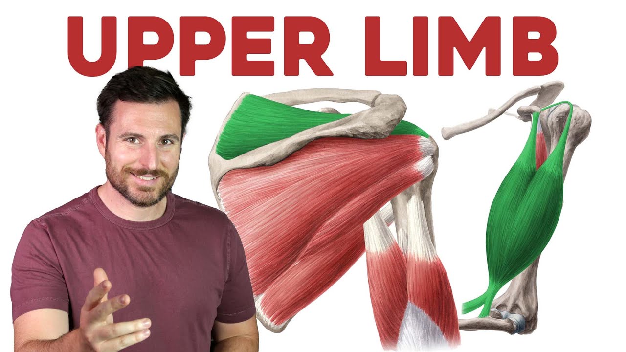
There’s skeletal muscle in all of the muscles you’re used to seeing, biceps, quads, etc. Finally you have Smooth Muscle which surrounds different organs that need to constrict and expand like your blood vessels, sphincters, or uterus. We’ll start with skeletal muscle.
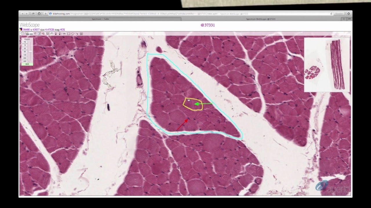
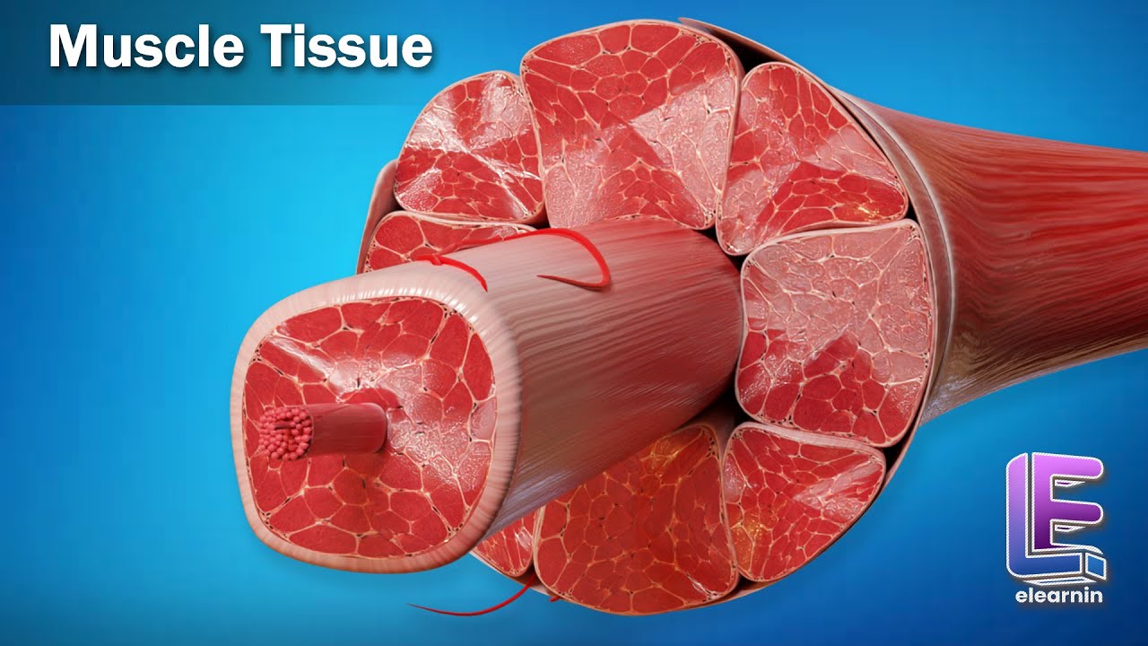
This stuff is optimized for quick and strong contractions, it’s gotta be able to move your skeleton against gravity and heavy weights. When you actually see it on a slide, you’ll typically see one of two perspectives. The first is the classic transverse cross section — it’s as if you took a chicken breast, cut it in half and looked at all the muscle fibers bundled together, which usually looks like a bunch of circles with white connective tissue wrapping them together.
The other perspective is from the side, or a longitudinal section. Here, you’ll see all the parallel fibers, and if you zoom in way further, you can see the functional unit of the muscle called the sarcomere. Most of the time when professors present the cross section bundle view, you’ll just need to identify the muscle fiber and the connective tissue around it.
But because a muscle is effectively bundles of bundles of muscle cells, your task is to figure out which layer of connective tissue and organization you’re looking at. The whole muscle is wrapped in dense connective tissue called the epimysium — epi- for upon, -mysium for muscle. But within the big bulky muscle, we see clearly segregated clusters of muscle tissue called fascicles which literally translates to bundles.
And any time you bundle something, you need something to hold it together. Each fascicle is held together by perimysium, peri- for around. Within each of these fascicles are individual muscle fibers, coated with a type of loose connective tissue called the endomysium, literally meaning within the muscle.
Each muscle fiber has a cell membrane called the sarcolemma. And this is a big stumbling block, but sarcolemma and endomysium are /not the same thing/. The sarcolemma is the cell’s membrane, it‘s part of the muscle fiber itself while the endomysium is connective tissue, it’s a different type of cell entirely.
At this point, each of these strands is a living muscle cell with nuclei and mitochondria and other organelles. And each fiber has a bunch of tiny myofibrils, which are proteins, not living things. If you have a cross sectional view of skeletal muscle, one thing I find helpful is to take a step back and think about what you’re looking at.
Is it visible with the naked eye? That’s a muscle. Is it magnified at a mid level and you can see individual nuclei?
That’s probably a bunch of muscle fibers surrounded by epimysium. Every now and then, you’ll use this view to tell the difference between Type 1 muscle, or slow twitch muscle, and Type 2 Muscle, fast twitch. The slow twitch fibers rely on aerobic metabolism, so they’ve got a /bunch/ of mitochondria and myoglobin.
This gives it a distinct darker red color compared to the pale white meat, which has less mitochondria and myoglobin. Important caveat, but you wouldn’t be able to tell the difference between the human skeletal muscle types with the naked eye. In reality, each of your skeletal muscles has a mix of muscle types and we can only see the difference with specific dyes under magnification.
Shifting perspectives, if we want to look at muscle from the longitudinal section, we’ll see all these stripes, or striations, as well as all these dots which are the multiple nuclei of each cell. Pro tip, striations refer to the stripes along the muscle cell itself. When I was a beginner, I thought the stripes being referred to were the long parallel muscle fibers, but that’s not the case.
Those striations are the visible sarcomeres , the functional unit of muscle, we have to zoom in a bit more. Each of these things is /really/ tiny, only about 2 to 3 /thousandths/ of a millimeter long, and photos of them are usually super blurry, so your professor will usually use this diagram instead. For this video, I’ll overlay the diagram on the microscopy so you can see what we’re working with.
From this view, we can see the Z line, the thick boundary of each sarcomere that anchors the thin filament, the actin. You can see the I band, which is this section in the middle where there’s only actin, and the H zone where there’s only myosin, the thick filament. Then there’s the A band where you see them overlap.
If you want more mnemonics and some tips about how this thing actually produces muscle contraction, I’ve got an entire video about that which you can find here and in the description. So for skeletal muscle, if you remember to keep in mind which view your slide is taking, and how magnified you are, you’ve already done most of the work. Next up is cardiac muscle, the muscle of the heart.
And we’re gonna do the same thing here — big picture first, then we’ll see how that plays out microscopically. The heart is just hanging out in between the lungs, pumping blood, and its connective tissue serves to separate the inside from the outside and keep it anchored in place. If you were to dissect a cadaver, the first layer you’d see is the fibrous pericardium, a layer of dense connective tissue around the whole package.
Cut that away and you’ll find a layer of serous fluid then the epicardium. Sometimes the outer layer is referred to as the parietal pericardium and this is the visceral pericardium, but it’s just a different name for the epicardium. The myocardium is the muscle tissue that we usually see histological slides of.
Finally, the innermost layer of tissue is endocardium, a combination of epithelial cells and connective tissue that lines the chambers of the heart. Usually that double layer pericardium mode is analogous to putting your first into a balloon filled with liquid. Your hand is the heart muscle while the rubber balloon is the parietal pericardium on the outside and the visceral pericardium, or epicardium on the inside — the layer touching your hand heart.
It’s one continuous sac of connective tissue, but they’re clearly separated. And of these structures, we usually focus on the myocardium because /that’s/ the tissue that contracts — it’s muscle. Quick terminology note.
Remember the old biological levels of organization? Organ systems are made of organs which are made of tissues which are made of cells? In the case of the heart organ, when we say /myocardium/ we’re talking about the tissue level, but when we talk about cardiomyocytes, we’re talking about cells.
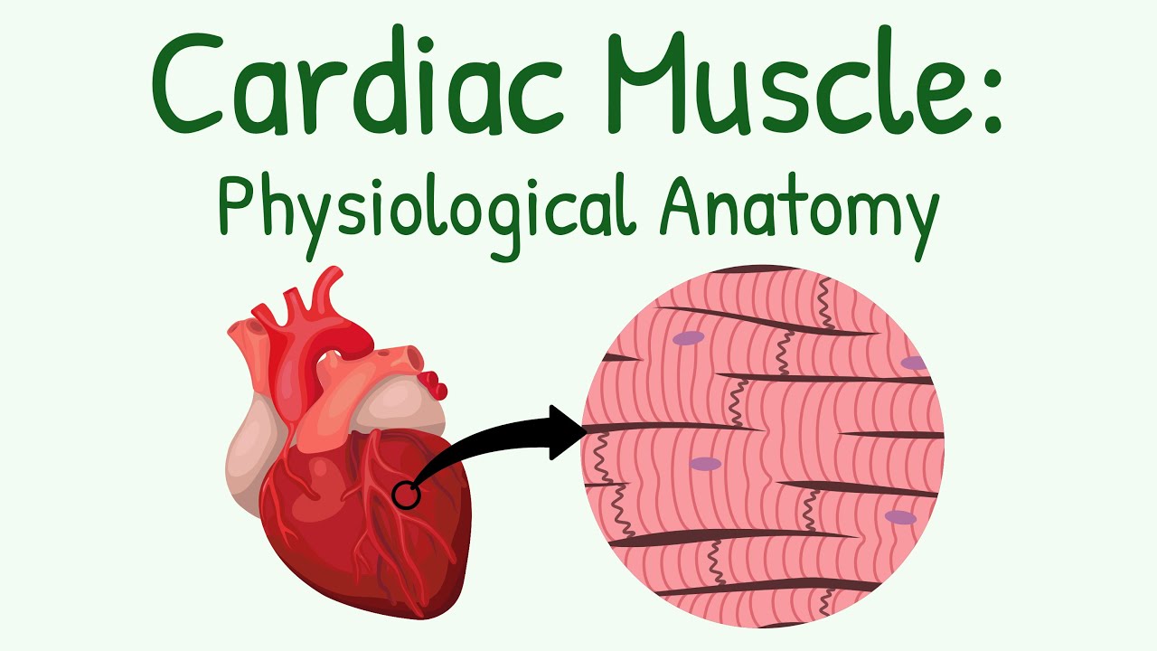
Literally heart muscle cells. And those cells have some unique characteristics compared to skeletal muscles. To me, they look like rectangles instead of cylinders.
And each of them usually has a single nucleus per cell and a boatload of mitochondria. Of course, the heart isn’t just muscle, so you’ll see some capillaries scattered around. Since each heart chamber’s muscle cells need to contract in unison, the heart needs to make sure there is like, zero lag in electrical impulses from cell to cell.
So these junctions, what are called intercalated discs, allow cardiac muscle to send transmission signals super quickly between cells. Our final type of muscle cell is smooth muscle. They’re called /smooth/ because they don’t have the striations of cardiac and skeletal muscle.
Each smooth muscle cell is spindle shaped, with narrow tips and wider cell body, and a single nucleus in each cell. So instead of neatly organized muscle cells, you get a broad mosaic of cells organized into sheets. Just like with cardiac muscle, smooth muscle uses gap junctions to contract all the cells within a muscle at the same time.
That lets a smooth muscle contract at the same time, and like cardiac muscle, smooth muscle is controlled involuntarily by the autonomic nervous system. You’ll find smooth muscle around things that expand and contract. Anatomy like your blood vessels which dilate and constrict, or the uterus, bladder, and gastrointestinal tract, all of which have to contract and expand at times.
All in all, when you’re looking at muscle histology, my number one tip is to take a step back and consider how the form of each muscle cell influences function. If you’re studying this for class and want a little extra help, I’ve got links for my notes and example problems linked in the description. You can also check out this playlist here for more histology help or this playlist for more musculoskeletal videos.
Have fun, be good, thanks for watching.