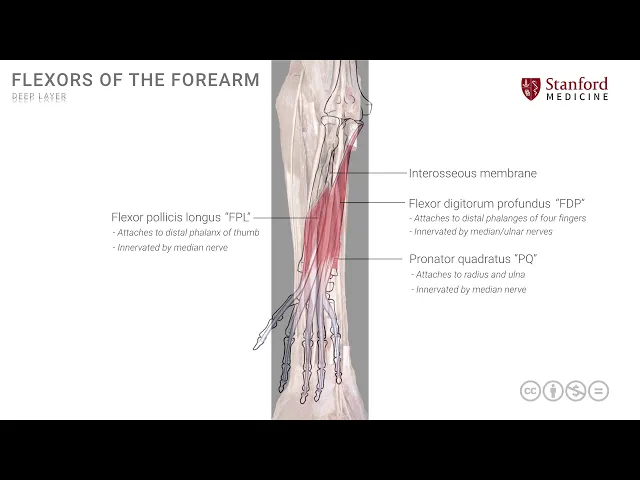
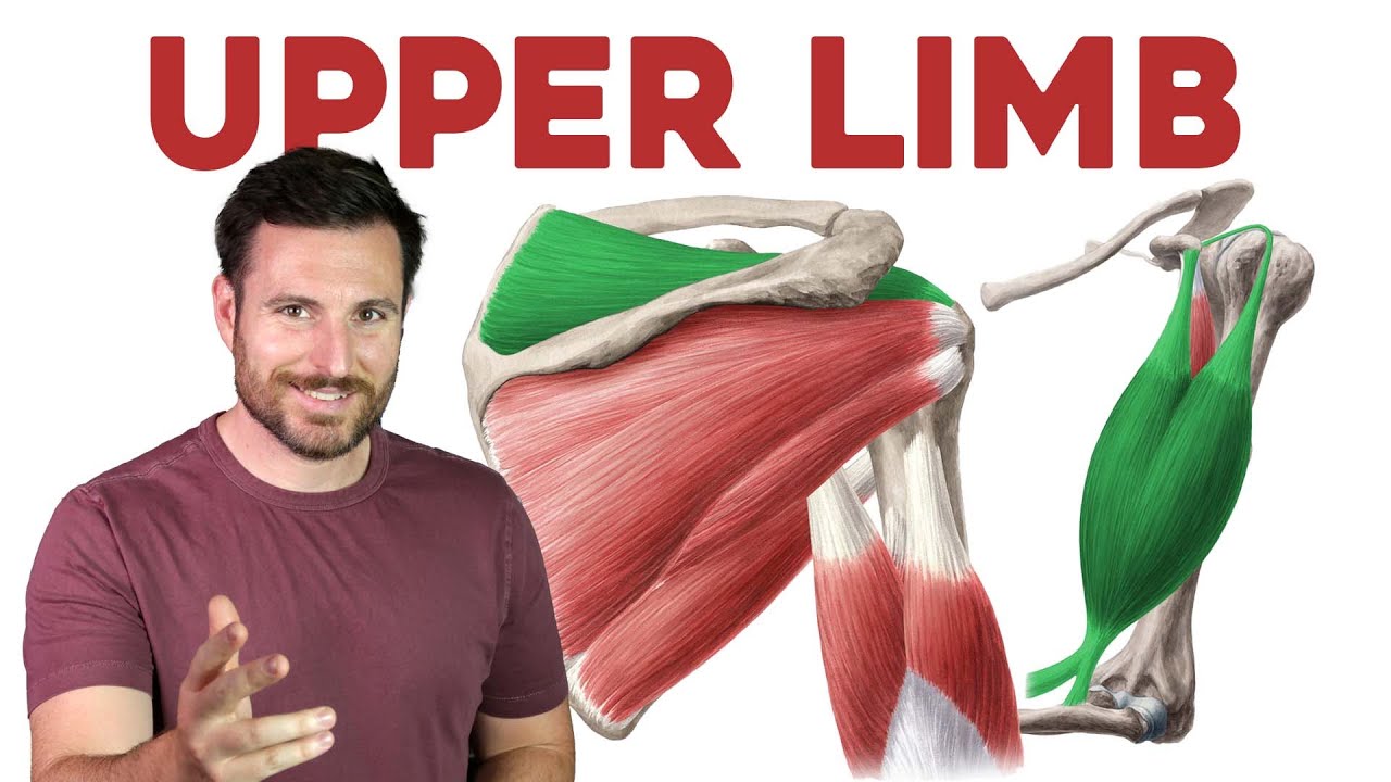
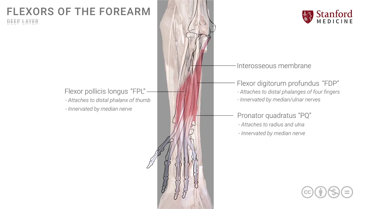
[Music] in this lesson we will review the muscles of the forearm that make the deep flexors and in order to understand that we would have to remove the superficial group and start really from the depths so we have a very simple line drawing of the forearm and in the depths of the depths there is an a membrane known as the interosseous membrane be meeting between bones which is seen here it runs between those two forearm bones the radius and the ulna and it is an interesting structure because it provides some additional support for these bones
in terms of their movements of pronation supination but also more importantly it provides a site for attachment of some of these deep muscles in order to understand these deep muscles we will go from a deep towards a superficial arrangement because even if within the deep layer there is the deepest of the deep which is the first muscle that I'm I will put on which is known as the pronator quadratus muscle or sometimes called P Q for short which is over here it's a small muscle it's roughly quadrangular in shape hence the name and it is
prone it pronates and therefore the name pronation is part of its the muscle nomenclature and it is attached on to the distal radius it goes across and attaches onto the distal ulna and by its action will assist in pronation but this muscle is covered by the other two muscles that also form the deep layer the first one of those is the muscle that we can see on the radial side known as flexor pollicis longus or FPL it attaches onto the radius and the interosseous membrane and has a tendon which goes into the thumb and is
seen here so this is the FPL flexor pollicis longus and it goes and attaches all the way up to the distal phalanx of the thumb and like any other muscle tendon it will flex or it will have an action on each joint that it crosses so it has a little bit of movement on the wrist a little movement on the small joints of the thumb as well the other muscle the third and final muscle of this deep layer is the flexor digitorum profundus or FDP this is a muscle that is more on the ulnar side
and it has attachment on to the ulna and the atrocious membrane and like its other muscle which is more superficial to it the FDS the flexor digitorum superficialis it also divides into four segments and attaches onto the phalanges the small bones of the four fingers in this case in in the case of FTP the tendons go all the way to the distal phalanges of the four fingers in terms of innervation of these muscles the FPL is innervated by the median nerve which is also the case with the pronator quadratus the flexor digitorum profundus has a
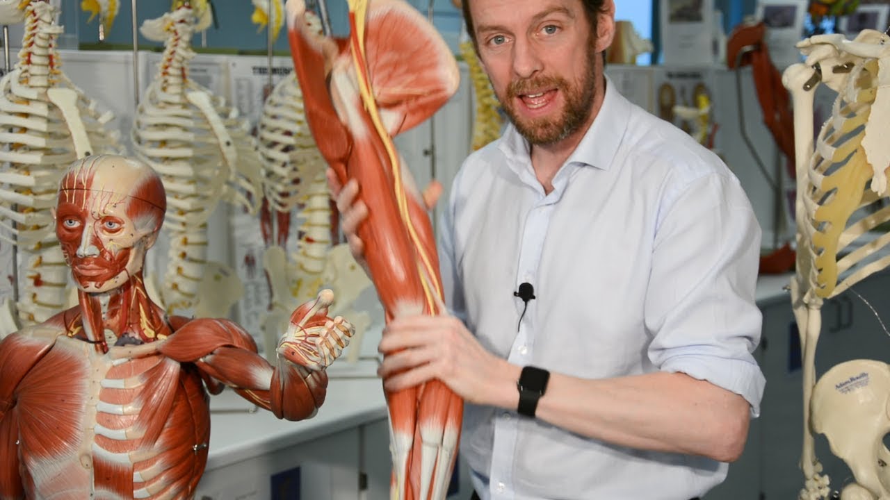
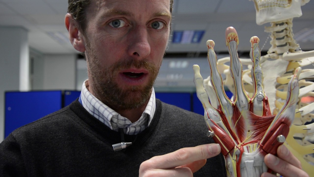
very interesting innervation supply nerve supply it is innervated by the median nerve and the ulnar nerve the median nerve supplies that segment of the muscle that ultimately provides tendons for the index and the middle fingers whereas the ulnar nerve supplies the other half of this muscle or the medial half of this muscle that provides tendons to the little finger and the ring finger let's now look at a deeper dissection a photograph of the deeper dissection of this area of the forearm the anterior forearm and just to orient we are still looking at the right forearm
and therefore this is the more lateral side this is more medial sometimes this is also known as the radial side and this is known as the ulnar side and so and the deep and superficial muscles have been removed so we can see some of these superficial cut muscles over here this is these are all the superficial cut muscles that have been cut away and we can also see their tendons more distally at the wrist and so with these official muscles removed we can now focus on the deep muscles and the first muscle that we see
from the deep group here is the flexor pollicis longus which is this muscle over here this is the flexor pollicis longus it's on the radial side of the forearm and it's tendon will make its way to the distal phalanx of the thumb the other muscle which is seen on the medial or ulnar side is the flexor digitorum profundus and we see that here it's marked in red so this is the flexor digitorum profundus muscle and it will divide into its individual tendons as well that could make their way into the four digits we see some
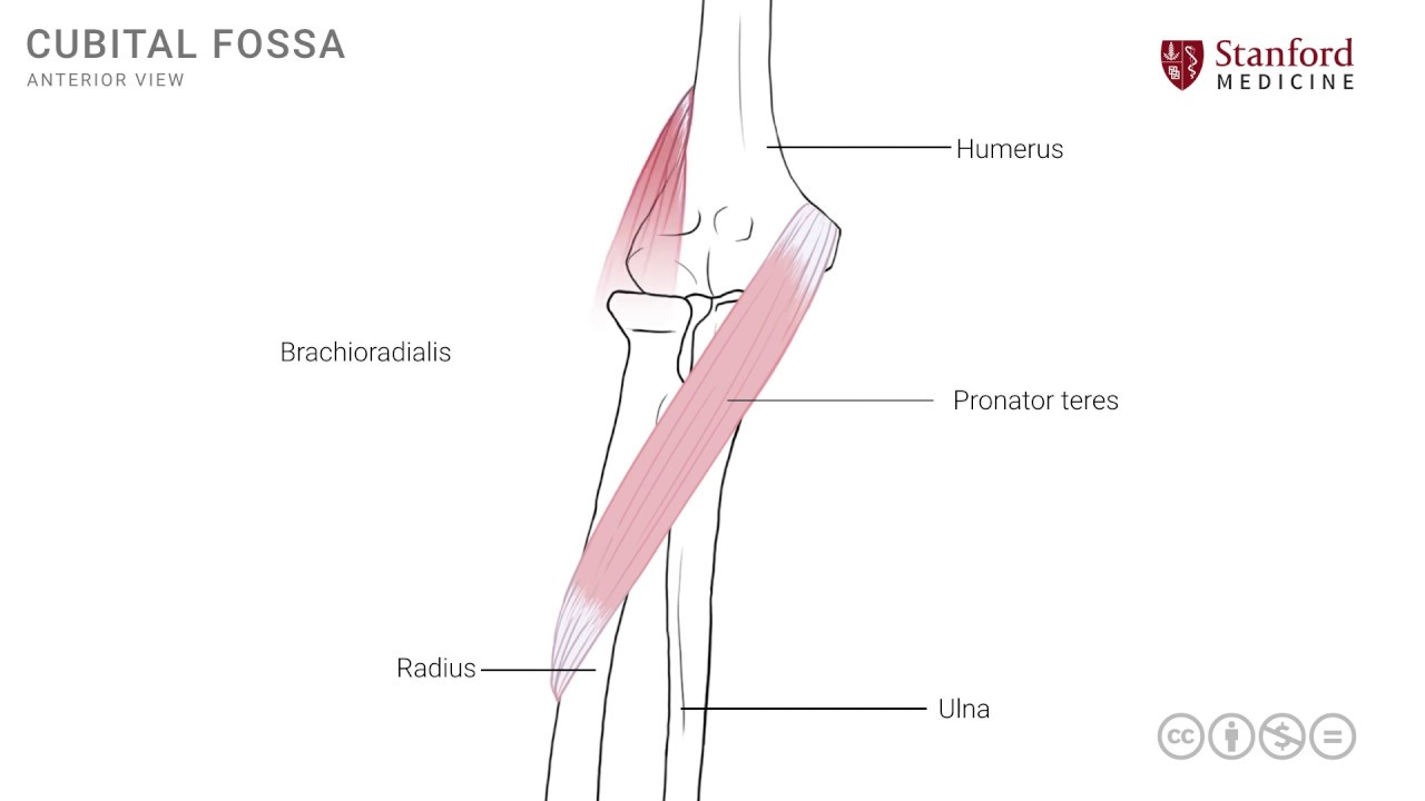
other very important structures here in terms of neurovascular structures so let's review that the first one is the brachial artery that we have seen earlier in the army and the cubital fossa and it now makes its way into the forearm and as it makes its way into the forearm it divides into its branches the radial artery is this artery that is seen here on the radial side of the forearm and it goes down all the way up to the wrist and if you focus on the ulnar or medial side you see the other branch which
is the ulnar artery and it also goes down all the way into the wrist these are the major arterial supply for the forearm and they also cross the wrist and enter the hand there's one other structure that you see here which is the median nerve and it's running in the arm next to the brachial artery just medial - it enters the cubital fossa and then interested forearm and makes its way all the way down here and enters into the wrist and we'll look at its course at the wrist and its subsequent lesson and there's one
final structure here which is the most medial of the nerves it is known as the ulnar nerve and it's seen right here it has its course from the arm into the forearm going behind or posterior to the medial epicondyle and then it goes down the medial or the inner side of the arm and is accompanied by the ulnar artery the ulnar artery and the Eleanore come close to each other somewhere in its course in the mid forearm and then they cross into the wrists across the wrist joint into the hand very close to each other
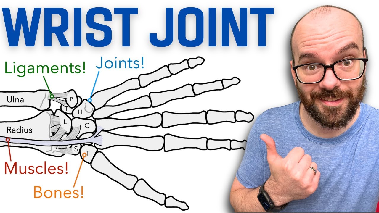
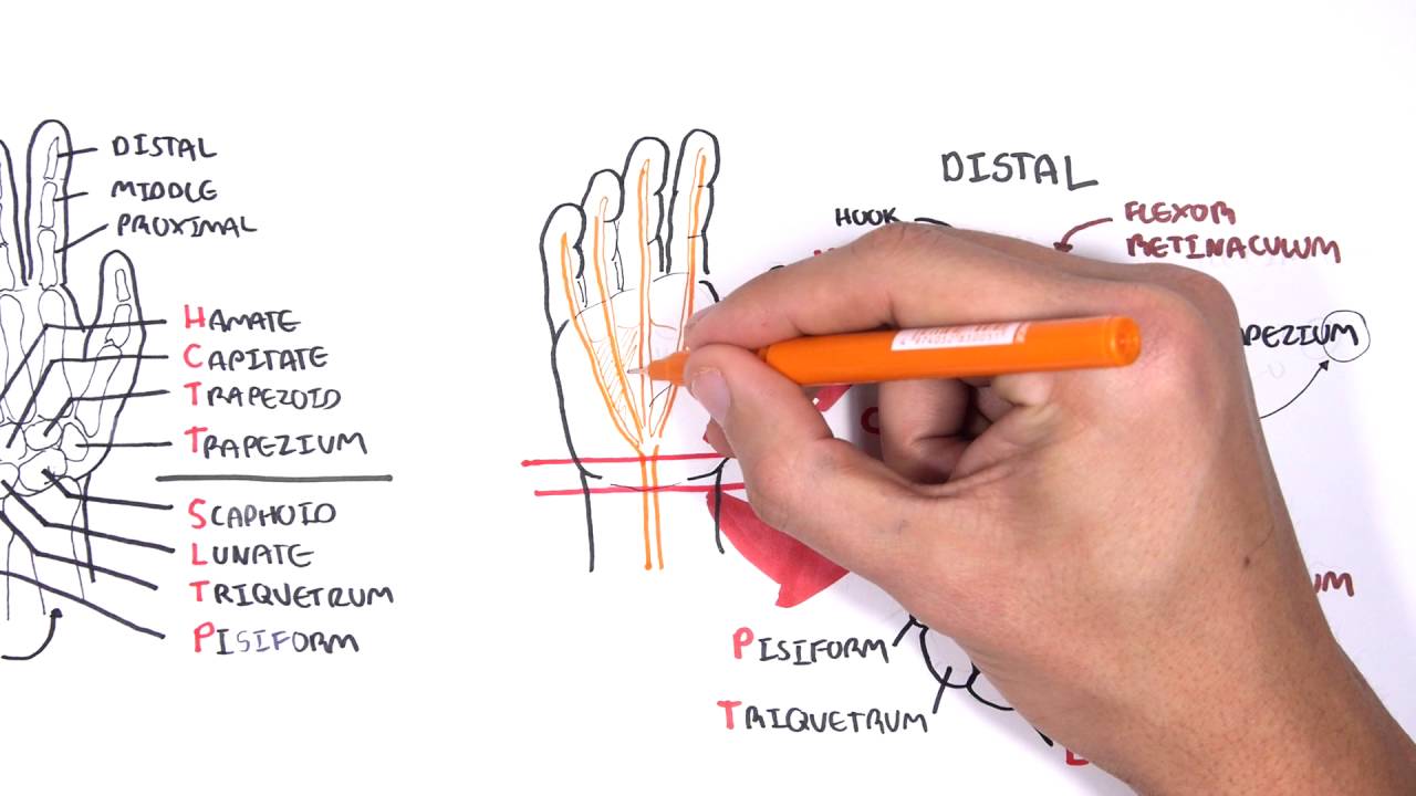
and if you look at the wrist there is a structure here which is an important structure known as the flexor retinaculum and this structure keeps most of these tendons and nerves and vessels in their place in a very specific arrangement and we'll look at that when we look at the detail anatomy of the carpal tunnel if we remove some of these structures in this area and remove the superficial structures here we can then see the deepest of the deep muscles which is this muscle here known as the pronator quadratus note its shape its quadrilateral or
rectangular in shape and it extends between the distal ulna and the distal radius this muscle is supplied also by the median nerve [Music]