hello and welcome to the review of chapter 39 of Gaytan in the halls medical physiology textbook which covers the pulmonary circulation and then how pulmonary oedema and pleural fluid that develops if you're feeling generous and you enjoy the video please don't forget to give the video a like and subscribe to the channel and as it helps us out greatly if you're in need of the textbook there is an affiliate link in the description so it starts off by talking about how the lung has two primary circulatory systems one which is a high pressure low flow
circulation which is the blood supply that actually supplies the trachea and the bronchial tree and then also the adventitia of the lung itself so this is the the blood supply that's actually supplying the lung organ itself so it has to be oxygenated to provide oxygen to the tissues so it actually comes directly off the order itself now it's a pretty low flow circulation because there's it doesn't require that much blood so but it is a high pressure because it's coming directly from the aorta from the left side of the heart whereas you also have another
circulatory system the low pressure high flow circulation and this is all the blood coming from the right ventricle from the pulmonary artery and this is all the blood that's come from the rest of the tissues around the body which have all used all the oxygen and that blood is now deoxygenated and there's also full of carbon dioxide and waste products so it comes to the lungs to be oxygenated and then remove the co2 as well so these are the pulmonary vessels so the pulmonary vessels have quite a large compliance they have a larger diameter they're
quite short and is smaller as well and that's because they have to accommodate all of the blood supply that would normally come from the left side as well because the cardiac output of both the left and the right is the same so it has to be able to accommodate the stroke volume output of the right ventricle even though there's less tissue that actually getting supplied you know it's just the lungs coming from the right side whereas the left side of the heart supplying blood to the entire body so in order to accommodate all that extra
blood then there is a large compliance the pulmonary vessels now the bronchial vessels these are the blood vessels coming directly off the aorta to supply oxygen to the lungs and the lung tissues they come off the aorta and they return back to the left atrium instead of returning back to the right atrium where normal deoxygenated blood goes to go to the pulmonary vessels to be oxygenated so that means that there's a small portion of deoxygenated blood that enters into the left atrium resulting in a slightly greater output from the left ventricle because it's taking in
that slightly additional blood volume and also the oxygen concentration within the leaf intrical is slightly lower than 100% because we have a little mix of the oxygenated blood there but that's such a small volume that is hard to tell then the other anatomy that we have is the lymphatics just like you vary where else on the body the lymphatic function to remove away in the interstitial fluid in any particulate matter as well now when we get into the pressures within the right side since the right ventricle only has to eat all the blood to a
pretty short distance you know just up to the lungs right next to the heart the pressure that's generated is much lower in an e left ventricle you can see that where the right ventricle is the black line diastolic pressures are low because it has to go as low as the right atrium to allow the opening of the atrioventricular valves and then the red line is the pulmonary artery which obviously there's a closure of the pulmonic valve so blood doesn't continue to leak into the right ventricle and diastolic pressures are higher but you can see here
that this pulmonary artery pressure is much lower than this a otic pressure because the aorta is supplying blood to the entire body so the lift heart has to generate more pressure which means that the lift muscle was going to be thicker as we know it's about three times as thick as the right heart now once it leaves the right ventricle and goes into the pulmonary artery we have a dampening effect as we go through our pulmonary vessels getting all the way to our pulmonary capillaries I showed you in Figure 39 - and you can see
that this is the systolic and diastolic in an hour Dean runs down the middle here until we eventually reach the pulmonary capillaries and then the pressure slightly reduces to our left atrium as it travels through the pulmonary veins now we can measure all of these values quite easily up until the pulmonary capillaries and that's done by placing a catheter into the femoral vein passing it all the way through the right atrium through the tricuspid valve into the right ventricle and then out into the pulmonary artery and so you can go all the way out through
the level of the capillaries and get what's called a pulmonary Ridge pressure which measures the rough pressure of the blood within the pulmonary capillary so we can measure the sliver we can't however measure the left atrial levels because it's pretty hard to get a candidate into the left atrium because it's obviously taking blood from the lungs and you can't catheterize the pulmonary veins and then if you try to catheterize the femoral artery and then go through the a order it's quite difficult - almost impossible to get up through that mitral valve and get the accurate
reading on the left atrium so the left atrial pressures are an estimation because we can measure our pulmonary wedge pressure so we can see what the pressure is here and and directly understand what the knit atrial pressures are because of left atrial pressures rise there will be a corresponding rise in our pulmonary capillary pressure now one other factor that we have to understand is that that like this the two servers like a blood reservoir because they take on about 9% of the total blood volume but they can take on more and that can occur in
the physiological setting say when you breathe out very heavy and you create a large positive pressure within the lungs resulting in a push of blood out of your pulmonary circulation and into your systemic circulation but they can also happen pathologically to where your pulmonary vessels can take on a lot more fluid if say you have mitral valve regurgitation and increase blood flow backwards and you can never increase pulmonary capillary pressure and the corresponding increase in your pulmonary blood flow so there is can be the shift in blood from one system to the other quite easily
now the fact is that control cardiac output as we've talked about also control pulmonary blood flow - so you know how functioning is a pump and then also our peripheral vascular resistance as well now one important factor to note here is that there is a difference here between the peripheral vasculature and the lungs compared to in the systemic body and the main reason here is that in the peripheral tissues whenever you have a lack of oxygen you end up with vasodilation to increase blood flow to that tissue to supply more oxygen however in the lungs
if you have a lack of oxygen that means that there is an alveoli that is not receiving much air flow so you don't want to see in blood to that region because you're not going to oxygenate your blood so there's actually the opposite effect where the blood vessels constrict so if you have a lack of oxygen in the pulmonary vasculature then these blood vessels will constrict and blood flow will cease so there is a increase in base or constriction substances such as in the filling and the reduction in vasodilator substances such as nitric oxide and
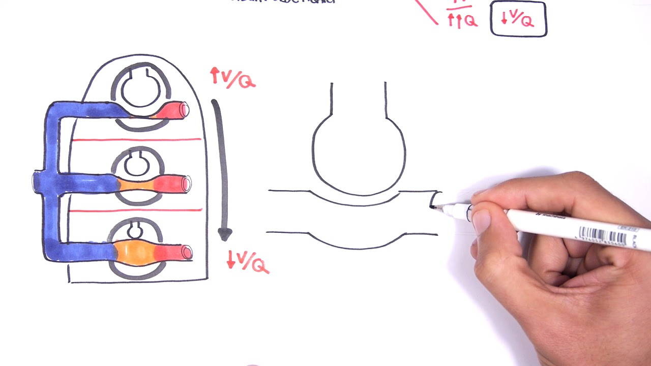
then that will help to distribute blood to other areas of the lung which are well oxygenated and send it to the alveoli which have a lot of oxygen concentration to oxygenate our blood now there is also three regions within our lungs or related to just the standard hydrostatic pressure from sitting upright so the normal gravitational force on our blood is obviously greater at the lowest level versus at the highest level so it autumn of your lungs versus the top of your lungs so blood flow is naturally going to go down with gravity and there's going
to be more blood flow in the bottom of your lungs as shown here versus the middle and in the top of your lungs which has to work against gravity is very minimal blood flow in that region and then with exercise we have an increase in cardiac output which is able to overcome that effect of gravity so then we're able to have normal blood flow in the top part of our lungs and then even greater in that bottom part of our lungs now that kind of goes on into these three zones that we have because zone
1 zone 2 and zone 3 shown here now zone 1 is showing a alveoli which has an alveolar pressure that is higher in the Hillary pressure so that means that the alveoli is coming pressing on to this capillary stopping all blood flow so zone one there is no blood flow because alveolar pressure is higher now this isn't seen in the normal lung this is only occurring in pathological states where there is either an increase in alveolar pressure or a reduction in our capillary pressure which would happen say with hemorrhage or a reduction in cardiac output
zone two is what we see in the top of our lungs where we obviously have the effect of gravity pulling away our pulmonary artery pressure and what happens in zone 2 is that the alveolar pressure is overcome during systole for the pulmonary artery pressure so during systole the pulmonary artery pressure is greater than the alveolar pressure so during systole there is blood flow however and diastole the alveolar pressure overcomes the blood pressure of the capillary so then there is a cessation of blood flow since the kiplyn theory gets collapsed so in zone 2 there is
only blood flow during systole because the systolic pressures overcome the alveolar pressure and that's what we see at the top parts of our lungs and zone 3 the pulmonary artery pressure or the capillary pressure is always greater than the alveolar pressure so there's flow during systole and diastole now during heavy exercise we've already talked about how cardiac output increases our pulmonary artery pressure or pulmonary artery blood flow now what that notes here is that is able to increase that blood flow through the lungs by three main mechanisms one an increase in the number of open
capillaries so there's more blood flow through multiple more capillaries by the standing all the capillaries which reduces our resistance the blood flow increasing the rate of flow then our third one is by increasing pulmonary arterial pressure now however it is important to note that the first two changes so that increase in the number of capillary and also distending of the capillaries which reduces our resistance both of these factors reduce our pulmonary vascular resistance which means that pulmonary arterial pressure only raises a small amount and that's shown over here with cardiac output increases our pulmonary arterial
pressure only just slightly increases because we have the opening of more capillaries more parallel circuits and in all distending officials reducing our resistance so that shows how our lungs are able to accommodate an increased blood flow without an increase in arterial pressure because if there's an increase in arterial pressure we're going to develop pulmonary edema which we'll get to very shortly and we've already briefly talked about how left atrial pressures correspond to our pulmonary arterial pressures or capillary pressures and have left atrial pressures rise over seven to eight millimeters of mercury they were actually caused
a corresponding increase in pulmonary arterial pressure which then increases our afterload of our right heart so if we have left heart failure resulting in the increased left atrial pressure we actually eventually end up with a pulmonary arterial hypertension and also right ventricular failure or a ventricular hypertrophy in response now before that may happen we this increased capillary pressure will then cause a increased hydrostatic pressure within our capillaries which promotes the movement of fluid out of the capillaries and into the interstitial space and into the alveoli resulting in pulmonary edema pulmonary edema is just fluid accumulation
within the lungs now there are some important differences between how the capillaries work to exchange fluid within the lungs versus and the tissues qualitatively they're the same but quantitatively they're different so for example there is a considerably higher functional capillary pressure in the peripheral tissues so the capillary pressure with around the lungs is lower and the interstitial fluid the pressure in the lungs is slightly more negative so there's more of a suction effect than the interstitial fluid in the lungs the colloid osmotic pressure within the pulmonary and tested is greater than in the peripheral tissues
so there is a greater tendency to hold on to water within the capillaries and not exceed eight and then the alveolar walls are extremely thin including a weak alveolar epithelium so they can quite easily rupture by any positive pressure in the interstitial space resulting and dumping of fluid within the alveoli now this diagram here is showing our capillaries and then album FedEx and then our vo lie and the different forces including the hydrostatic and automatic pressure interacting the overall there is an infiltration of fluid from the capillaries into the interstitial space and out through the
lymphatic pump resulting in a negative interstitial fluid and that helps to keep the alveoli dry because any fluid that accumulates within into the interstitial space gets sucked away right away by the lymphatics now if the distal space accumulates with fluid they're making develop pulmonary edema and that can occur due to left side failure because of an increase in pulmonary venous pressures or increased hydrostatic pressure within the capillary but then also due to damage to the blood capillary membranes as well do to say infection so increasing the permeability resulting in greater leak now there is a
safety factor to our pulmonary edema so when we have over raised left atrial pressure resulting in an increased hydrostatic pressure with the novel polarities we do have a point where it can increase without the development of pulmonary edema and then once we reach roughly twenty twenty five millimeters of mercury left atrial pressure any further increase will increase our pulmonary edema linearly and the safety factor can increase and chronic conditions as well up and to around 40 to 45 millimeters of mercury and that occurs due to an expansion of a lymphatic vessels so the safety factor
just means that a left atrial pressure can rise initially without an increase in pulmonary edema but once pulmonary edema occurs and that's quite risky because death can occur pretty shortly after that now the pleural fluid is the small tiny amount of fluid within the pleural space that helps lubricate B lungs as they move within the chest cavity but the entire space is more of a potential space because it's so narrow and isn't really a space and it has to be kept as a negative pressure because that's what keeps the lungs up against the chest wall
if that becomes a positive pressure at any point you know all collapse the lungs so if you open up the ribs or you have a leak in your pleural space and you will create a positive pressure environment within the pleural space and your lung collapse now that negative pressure is developed by the lymphatics and we can get a development of plural fusion for the exact reason that we get a beamer anywhere else and just to remind us that can occur due to blockage of the lymphatics or cardiac failure resulting in a high capillary pressures resulting
in too much excitation and to the pleural cavity we can get a greatly reduced color those Maalik pressures that do it so encouraging every movement of fluid out of the capillaries or an infection or inflammation which increases the permeability to the membranes promoting more pleural fluid accumulation and then that really summarizes our chapter I hope you enjoyed it feel free to drop a comment otherwise we'll see you in the next chapter