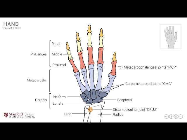
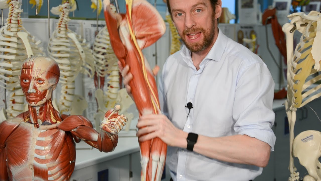
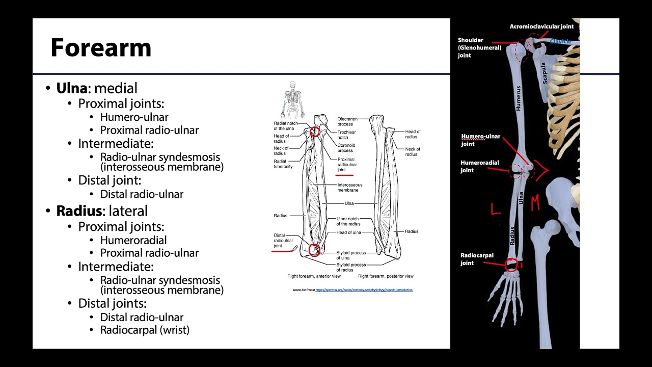
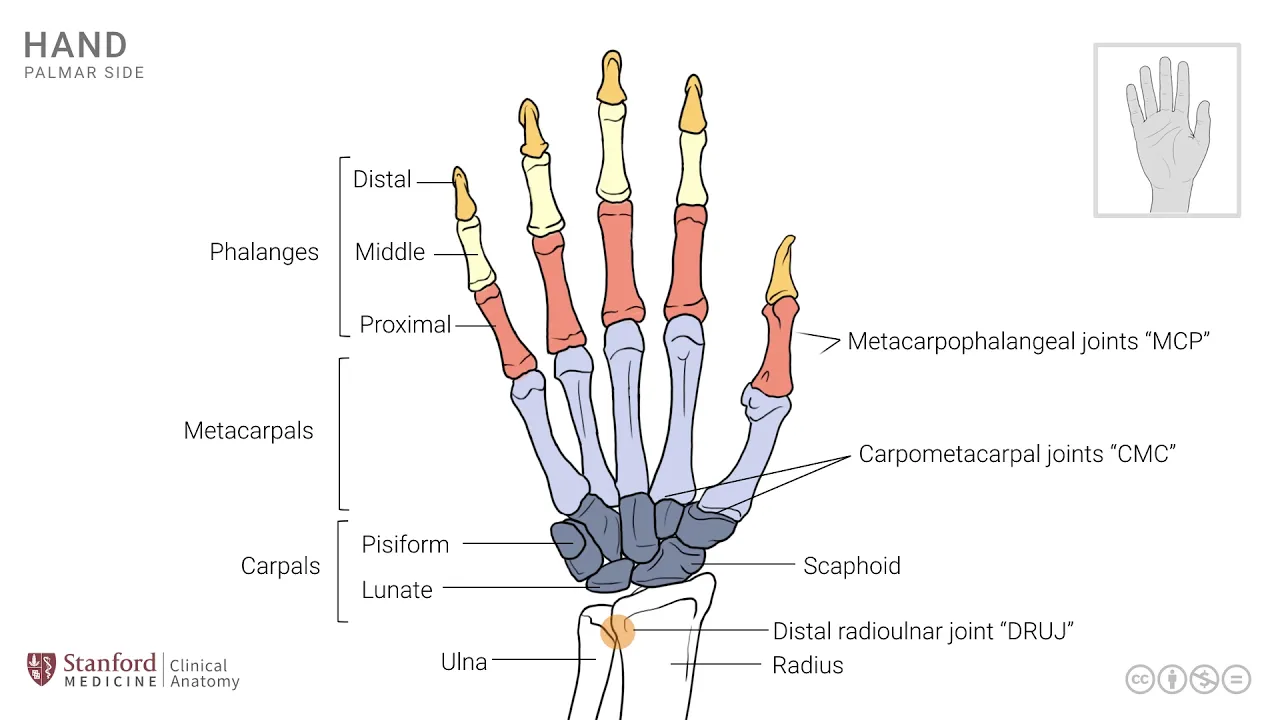
[Music] in this lesson we will review the osteology of the hand consider the various bones and joints that participate in the formation of the wrist joint as well as the remainder of the hand let's start by looking at a line drawing of the right hand and review the skeleton pieces and to begin this we will add the distal ends of the two forearm bones the radius and the ulna so this is the end of the ulna the distal end and this is the distal end of the radius and the distal ends of these two bones
the ulna and radius form a joint that is known as the distal radioulnar joint which is seen right here this joint along with its proximal counterpart the proximal radioulnar joint participate in the movement of pronation and supination the distal ends of these two bones the radius and ulna articulate with the small bones of that wrist known as the carpal bones sometimes collectively called the carpus each of these carpal bones has a unique structure it has a name we're not going to review all of these but only a few that have high level of clinical significance
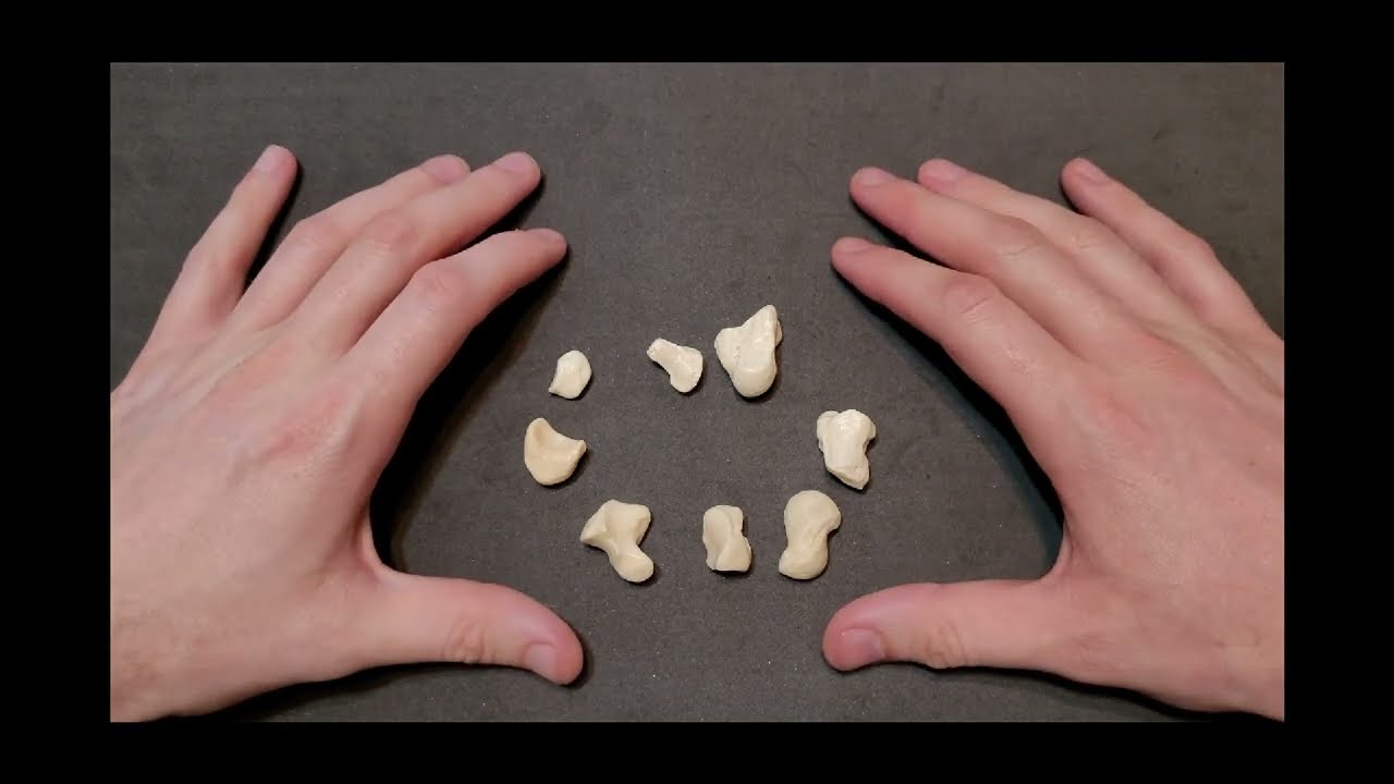
the first one of these is a bone that is seen on this side here on the medial side of the wrist and is known as the pc form bone and it's named after its shape which is a P like and it is the site for attachment of one of the forearm muscles known as flexor carpi ulnaris there's another important carpal bone known as the lunate and it is named because of its shape being a moon shaped bone as specifically as it is seen in its lateral profile this bone the lunate is often dislocated from its
anatomical position as a result of injury and it's and has some consequences with chronic wrist pain the most important arguably the most important carpal bone is this one on this side which is seen on the radial side here the scaphoid it's one of the larger of the carpal bones and is often injured and in fact results in a fracture through the scaphoid bone which is sometimes the notorious and because it doesn't heal very well and can also be a cause for chronic wrist pain and disability so these are some of the important carpal bones that
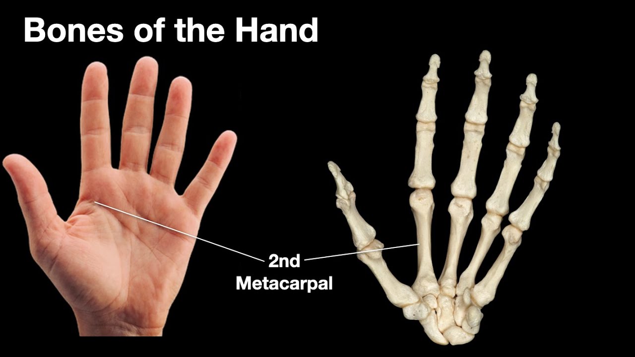
are often seen in clinical cases and therefore good to keep in mind the next set of bones are known as the metacarpals and they're seen here five in number one for the thumb and then the remaining digits the fingers and these articulate at their proximal end with the carpal bones and their distal ends these carpal these metacarpal bones articulate with the phalanges the phalanges are many they are divided or sub classified into proximal phalanges as seen here the middle phalanges as seen here and then the distal phalanges as seen here note that the thumb has
only two phalanges and are named as the proximal and distal phalanges it does not have a middle fell italics there are a number of joints I alluded to the joints between the base of the metacarpal and the carpal bones and these are collectively known as the carpal metacarpal joints there's one at the base of each of these digits the next set of joints are known as the metacarpal phalangeal joints or the mCP joints and these are at the distal end of the metacarpals where they articulate with the bases of the proximal phalanx the other remaining
joints are collectively known as interphalangeal joints in each of the fingers we have two of these joints they're known as proximal and distal interphalangeal joints p i-- p di p the thumb has only one interphalangeal joint and it simply called the interphalangeal joint it doesn't have a qualifier in terms of being proximal or distal since there's only one joint now each of these digits has a name and often in clinical practice one might be tempted to use numerical labeling of these digits and they're often described as first second third and so on it's often a
recipe for some confusion because for for many reasons one of those reasons is that the number of digits may not always be five so if you look at the little diagram in the right upper hand corner it shows five digits at the moment but it could easily be a patient with six digits and so the confusion is where do you start numbering you start numbering from the thumb aside and then towards the little finger or in the reverse direction and so to avoid these confusions in terms of number and the sequence it's often good practice
to use the name that describes the digit and so you can call the index finger as seen here the middle finger the ring finger and then the little finger by their names and then the thumb by its name as the thumb this avoids any opportunity for causing any confusion in documentation as well as in communication let's now look at a simple x-ray of the hand and the wrist joint these are primarily focused on the wrist but it has a little bit of the hand as well it doesn't have the full hand as you will note
what is apparent in this first x-ray which is an AP or an ant or posterior view are the distal ends of the radius and ulna labeled as r and u here we can also see the base of the metacarpal the carpal bones are seen as well and the one that is the label here is the scaphoid with an S let me also show you the lunate bone which is adjacent to the scaphoid and you'll wreck 9 step from the line diagram in the previous slide we can also look at an oblique view as seen here
this is an oblique view so it's not quite an AP but not quite a lateral view and in this case as well you can see most of the same bones the key element here is the scaphoid and this view is often done to actually assess the scaphoid when one is suspecting fracture the scaphoid bone obviously the other bones are also invisible such as the distal end of the radius and ulna and then the metacarpals or distally note that in a lateral view as seen here on the right side of your screen you can see a
very different architecture the key element here in order to get you oriented is a bone that you see almost sticking out it's almost out of sync with the other carpal bones and it's labeled here with AP this is the pisiform bone and you don't see this very clearly in the oblique view or in the AP view and this underscores the idea of doing and creating different views of x-rays in order to focus in on different anatomical structures and the wrist is a fairly complex structure and different views are very helpful in depicting different structures within
this complex anatomy and then being able to make a clinical diagnosis [Music] you