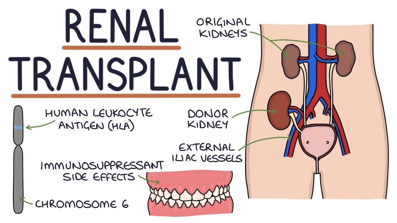
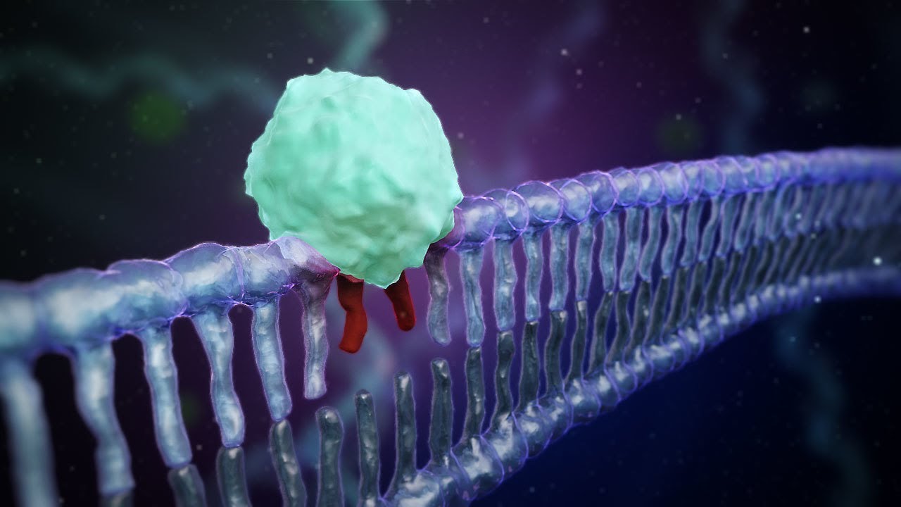
welcome back to the operating room in module one we have discussed important aspects before the transplant including patient selection and immunologic barriers once a patient has crossed these barriers what does a kidney transplantation actually look like in the following video you will see a unique visual representation of a regular and uneventful kidney transplantation into the right iliac fossa because of the animated nature small details may differ from life surgery but after the video you will be able to recognize the important steps of the kidney transplantation surgical procedure complicated cases are subject of a separate lecture
but this visualization will help you in understanding the procedure itself and its complications a regular primary kidney transplantation into the right iliac fossa show some technical details are provided that may require basic surgical and anatomical knowledge Cesar starts with opening the skin in the lower abdomen with a surgical knife the oka stick or oblique incisions are most common the length of incision depends on the habitus of the patient and ends two centimeters above the pubic bone which allows access to the retroperitoneal space the iliac vessels and the bladder subcutaneous fat is incised using electric artery
of the external oblique muscle is incised diagonally the combined fashion of the internal oblique and transverse muscles is incised in a more vertical fashion tonio space is exposed and the inferior epigastric vessels as well as the rotund ligament in females or spermatic cord in males are identified and secured with a vessel loop lowest part of the posterior rectus sheath is in size after this the retroperitoneal space is raised by gently pushing away peritoneum this contents are held by retractor the common external and internal iliac artery and vein are identified in the second from fatty tissue
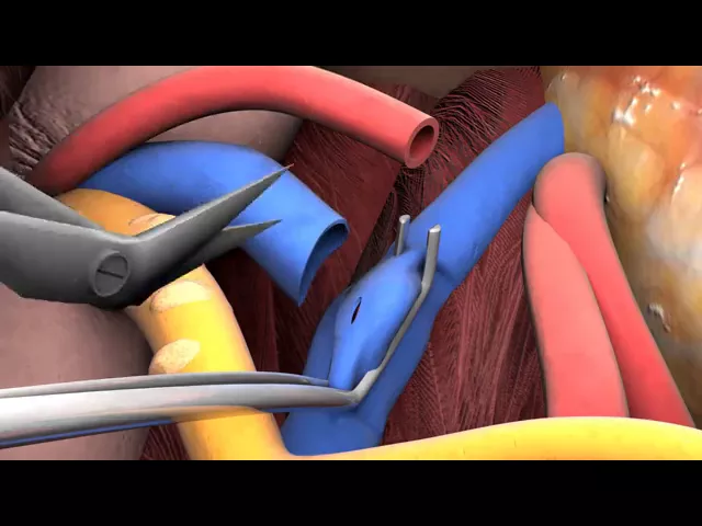
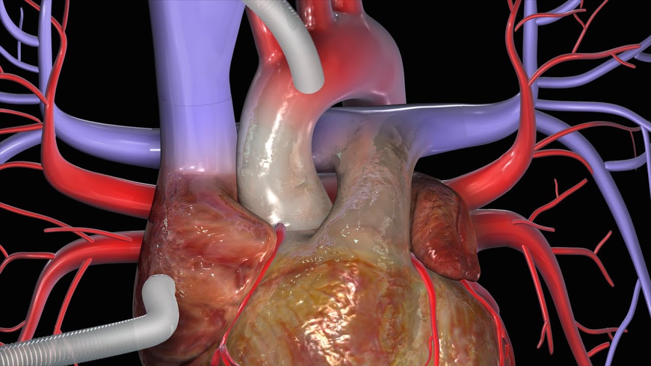
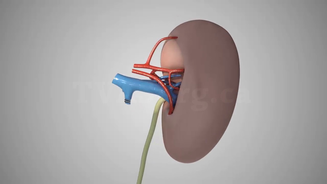
and lymphatic tissue to make clamping of the vessels possible the iliac arteries are investigated by palpation for atherosclerosis the kidney is now retrieved from cold storage in this animation it has already been prepared for transplantation and pre-transplant biopsy has been taken corner stitches are applied to the renal vein edges the kidney is placed into the iliac fossa in the best possible position and the length of the renal vein and artery may need adjustment first the vein to vein and samosas is done if needed the iliac arteries retracted very gently to improve access eally advance Clanton
cut and the incision is extended a supporting pullout stitch is placed on the medial side to keep the lumen open Lucian is used to rinse and clean the lumen from blood and small thrombotic material renal vein corner stitches are connected to the corners of the opening of the iliac vein and from one corner the venous anastomosis is performed in general by using the 500 absorbable monofilament running suture the supporting stitch is removed and both remaining sutures are knotted next the iliac arteries are clamped the artery preferably the common iliac is incised and the opening is
enlarged using a four millimeter a or tech punch is cleaned with heparin solution the arterial anastomosis is now performed using a parachutist 6o unobservable running suture before a reperfusion of the kidney the renal artery is clamped close to the anastomosis this is done to prevent blood clots to go into the kidney first the arterial clamps are removed and the anastomosis is check if there is no major bleeding the other vascular clamps are taken off and the kidneys repr fused with blood I'm the kidney color should change to a more pink aspect the kidney is warmed
up using warm saline solution and the kidney and the vessels are checked for bleeding he seems to be appropriate the anastomosis between ureter and bladder is performed through the urinary catheter the bladder is filled with saline the ureter is cut to the right length and the you blumen is special ated by a longitudinal incision direct under the epigastric vessels and spermatic or to the bladder production of urine may be observed at this stage the bladder is now opened first a muscular layer and second to sera mucosal layer corner stitches are placed between the urinary bladder
and the ureter the ureter extent is inserted the ureter bladder anastomosis is now performed using semi absorbable 500 running shoes the anastomosis is retracted into the bladder and the muscular layer is closed over the ureter anastomosis to create a valve effect that minimizes urinary reflux when he must asus and kidney perfusion are adequate the peritoneum and its contents are placed over the kidney the posterior rectus sheath is closed first then the internal transverse fecha is closed and the external fashion is closed all of them using semi absorbable suture the subcutaneous layer and the skin are
closed using absorbable sutures you so we have now seen that kidney transplantation consists of different complex steps involved major blood vessels and requires advanced technical skills and expertise important steps include clear exposition of the iliac fossa and its vasculature meticulous anastomosis techniques of vein artery and ureter and early recognition of possible problems as you can imagine the result of the procedure is depending on patient and donor kidney conditions as well as the performance of the surgical team the challenges and possible complications of this procedure will be discussed in another lecture you