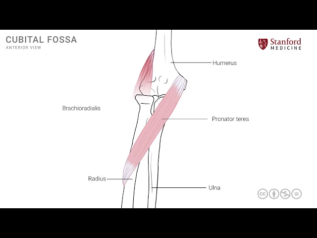
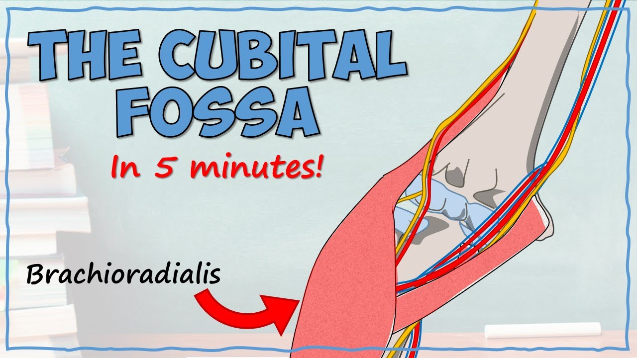
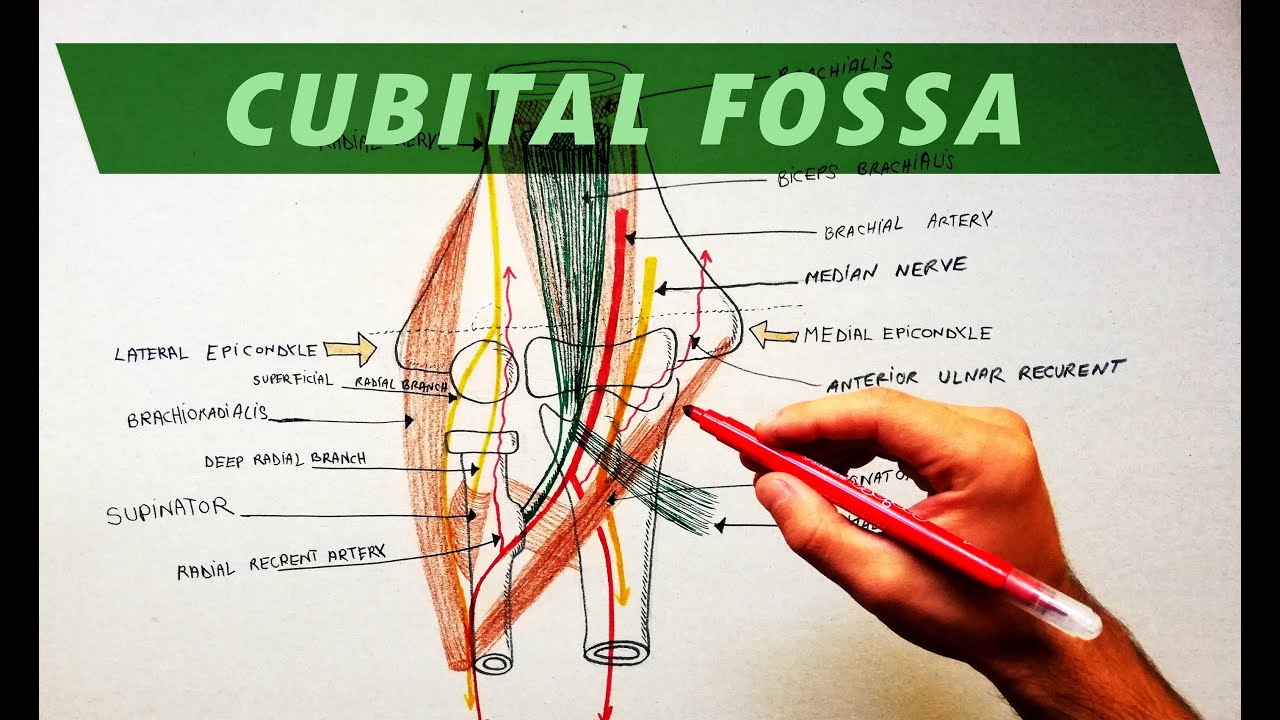
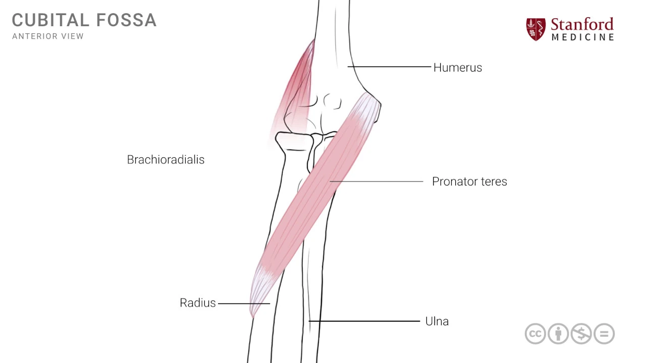
[Music] in this lesson we will review the boundaries and the structures that participate in the formation of the cubital fossa the cubital fossa is a triangular area on the front or the anterior side of the elbow joint and we can start by looking at an anterior vantage point of the right elbow joint the humerus the ulna and the radius participate in the formation of the elbow joint and the first key structure of the cubital fossa is a muscle known as the pronator teres muscle this muscle has this attachment onto the medial epicondyle of the humerus
more approximately and then the fibers of this muscle extend laterally and inferiorly to reach the mid shaft of the radius and the attached onto the middle third of the radius there's a second muscle known as the brachioradialis muscle and this muscle extends from the lateral epicondyle and the region above it known as the supracondylar Ridge of the humerus and then the fibers extend distally and attach on to the distal radius the area of attachment is beyond what the diagram depicts this muscle is known as the brachial radialis as the name suggests break ium is the
arm and radialis refers to the radius so this muscle extends from the humerus the distal humerus in this case to the distal radius printer Therese just to give you the description of the colander Therese is a muscle that pronates the forearm antares refers to the shape of this muscle specifically in a transverse section it has a round shape these two muscles form the boundaries of this triangular space that we call the cubital fossa as shown here one boundary the lateral boundary of this cubital fossa is formed by the medial border of the brachioradialis muscle the
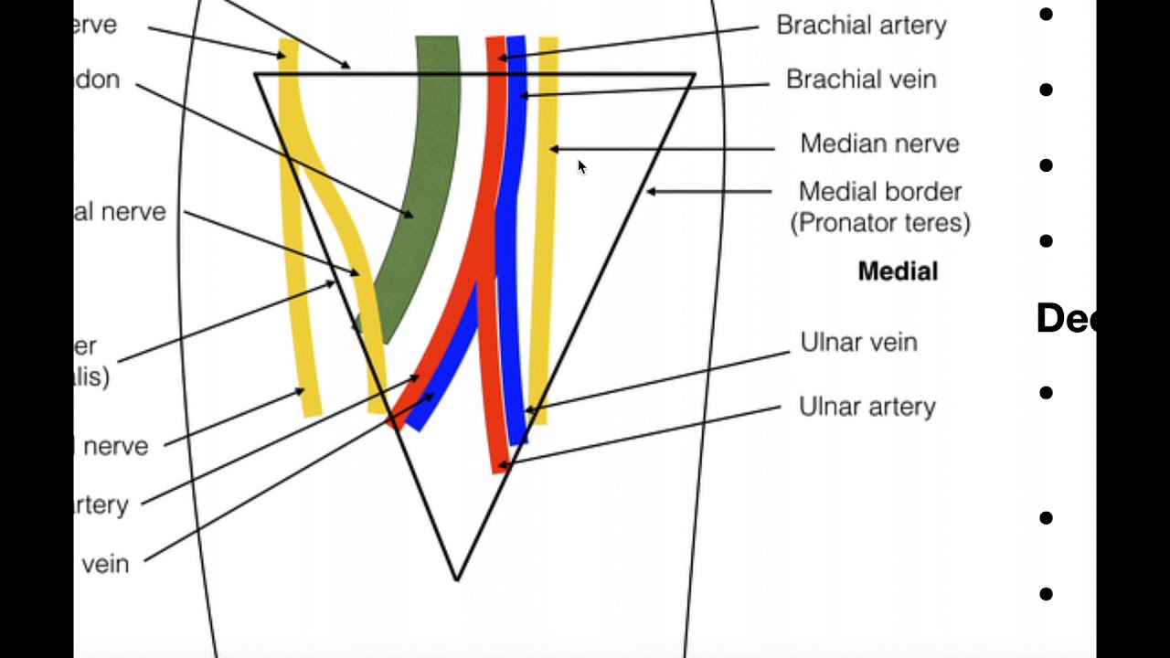
other boundary which is the medial boundary of the cubital fossa is formed by the lateral border of the pronator teres muscle and then the third border or the third edge of this cubital fossa being a triangular space is formed by an imaginary line that runs across the epoch on dollars in the epoch Andra area of the humerus now this triangular area has certain key structures and we can look at those structures now the first one of these is a tendon it's the tendon of the biceps muscle which is the main muscle of the arm this
tendon here runs across the cubital fossa and goes on to its point of attachment on to the proximal radius it's a strong tendon and can be easily palpable in the cubital fossa area if one continues to palpate more approximately one can feel the pulsations of the brachial artery which is seen here this is another key structure that enters the cubital fossa and then exits from its inferior edge more medial to the brachial artery is another key structure in this time a nerve this is the median nerve which is one of the key branches from the
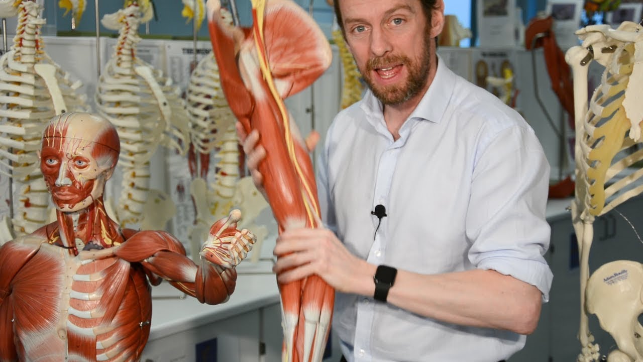
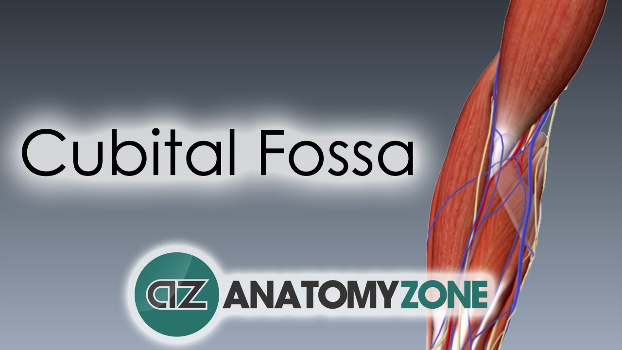
brachial plexus and is seen here the median nerve also enters the cubital fossa and exits out from its inferior or medial edge the medial nerve goes on to supply many of the key structures muscles are in the forearm and the hand thus we have reviewed the cubital fossa it's found formation and boundaries and some of the key structures that traverse this region let's now look at a photograph of a dissection of this region again to orient ourselves we see the biceps muscle in the more proximal part of the photograph here and the tendon of this
biceps muscle has been cut partly at least and so we see this part of the tendon going on its way to its point of attachment on to the proximal radius the other part of the biceps tendon which extends more medially is shown here and that has been cut away this is the bicipital aponeurosis which goes on to attach on to the medial side of the forearm specifically to some of the fascia and the ulna this has been cut away and removed in order to see some of the structures in the cubital fossa one of the
key structures in the cubital fossa is the brachial artery and we see this key structure running down into the cubital fossa over here so this is the brachial artery and note that it is on the medial side of the biceps tendon in exactly the same relationship if one were to palpate the cubital fossa in a patient on the medial side of the brachial artery is this important nerve known as the median nerve and it is seen here and to complete the picture of the cubital fossa we will put the two muscles that form the boundaries
the pronator teres which is seen here shaded in red this is all the pronator teres muscle and this forms the medial boundary of the cubital fossa the other muscle which is on the lateral side is seen here and this is the brachioradialis muscle the two muscles overlap and come close to each other in the inferior angle or inferior point of the cubital fossa [Music] you