[Music] in this lesson we will review the muscles that participate in the formation of the flexor group of forearms specifically we will look at the superficial muscles of this group let's start by looking at a simple diagram of the anterior part of the elbow joint and the proximal forearm as shown here the flexor muscles of the forearm can be thought of as being a position in layers and the superficial group is what we will look at in this lesson this group is made up of four muscles the first one of those muscles is known as
the pronator teres sometimes also abbreviated as PT and this muscle has its attachment on the medial epicondyle of the humerus this turns out to be the point where all of these muscles have a common attachment more proximally and the fibers of this muscle extend laterally and inferiorly and attach on to the mid shaft of the radius as shown in the diagram the name pronator signifies the function of this muscle and the function is pronation of the forearm Terri's refers to the shape of this muscle specifically in a cross-section Antares means round and this muscle indeed
is round in its cross section the second muscle in this group has a slightly longer name it's known as flexor carpi radialis often abbreviated as s gr and this is situated on the medial side of promote ares approximately it has the same attachment onto the medial epicondyle and then it extends down into the distal forearm on the radial side of the forearm this muscle has a action on the wrist because it crosses the wrist joint unlike the pronator teres muscle which does not cross the wrist joint the name of this muscle signify is exactly what
the function is and also gives some indication of its location flexor means that it has an action of flex Carpy relates to the carpus or the wrist and radialis signifies source suggests exactly where the location is in this case it is on the radial side of the forearm and the radial side of the wrist this muscle crosses the wrist and it attaches on to the base of two of the metacarpals the second and third specifically we have yet another muscle which is known as the palmaris longus muscle often abbreviated as PL and this is situated
on the medial side of the FCR this also has an attachment more approximately onto the medial epicondyle the common flexor attachment and then it extends distally down the middle of the forearm to cross the wrist joint and in fact attaches on to the palmer Eppler osis it's a small muscle it's a flimsy muscle doesn't generate too much power and is often described as being an accessory muscle or a vestigial muscle and in about 10 to 15 percent of the population depending on the series that you look at it is either absent on one side or
on both sides in individuals this is a very helpful muscle however in those who have it and is often used as a source for reconstructive surgeries it's an expendable tendon and is often used wherever reconstructive surgeries are required to recreate function that may have been lost at another location the fourth and final muscle in this region in the flexor group of for our muscles is known as the flexor carpi ulnaris and like the remaining muscles in this group it also has an attachment more approximately on to that medial epicondyle and then the fibers come down
the ulnar side of the forearm and crosses the wrist joint and it also flexes the wrist joint and it is attached onto one of the carpal bones specifically the pisiform bone and it produces the flexion movement at the wrist joint and that's reflected in its named flexor meaning it flexes carpi the wrist it is on the ulnar side and hence the name on eros so these are the four muscles that are described typically as being the superficial muscles of the forearm group they have a interesting nerve supply the pronator teres muscle along with the FCR
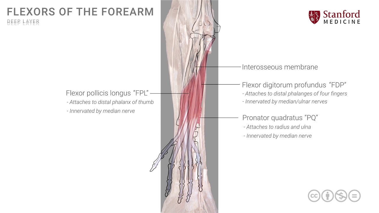
the flexor carpi radialis and the palmaris longus are all supplied by the median nerve the flexor carpi ulnaris is an exception and it is innervated by the ulnar nerve the flexor group of muscles of the forearm are divided into three layers and we have just looked at the superficial layer we will now look at the intermediate layer this layer has only one single muscle and by making this a simple line drawing I can put the muscle on it's called the flexor digitorum superficialis or FDS it has a more proximal attachment at the medial epicondyle which
is the common flexor attachment and then somewhere in the mid forearm it divides into four different segments these segments go into each of the four fingers the index finger the middle finger the ring finger and the little finger so this is the flexor digitorum superficialis and like all other naming conventions the name describes exactly what it does or where it goes the flexor suggests that it has an action of flexion the digitorum suggests the digits and superficialis means it is the superficial muscle and by convention one can guess that there will be a deeper muscle
as well and we'll look at that in a moment so the flexion here is across all joints that it crosses so it will have a little bit of flexion at the wrist joint but its primary flexion is on the small joints of the fingers or the digits so the flexor digitorum superficialis in each finger the tendons attach on to the middle phalanges and we'll look at the exact details in a subsequent lesson this muscle is entirely innervated by the median nerve let's now look at a photograph of a superficial dissection of the forearm muscles and
we'll put some names on here the first one is a muscle shown here on the radial side and this muscle is the brachioradialis muscle this muscles extends from the distal humerus and goes all the way up to the distal radius technically speaking and this is not a flexor muscle of the forearm it actually belongs to the extensor group but it is seen in an anterior vantage point so it's worth noting it here otherwise it tends to cause some confusion in the minds of learners the other muscle that is seen here is actually a muscle of
the flexor group of muscles and it is known as the pronator teres this is the first muscle and I'm going to color it in here between these two muscles the brachioradialis and the polarity aries is the cubital fossa and if we look in that cubital fossa for a moment we see in the depths of the cubital fossa a muscle that forms the floor of this cubital fossa which is known as the brachialis this is one of the key muscles on the anterior side of the arm the structures that are normally present in the cubital fossa
such as the biceps tendon and the neurovascular structures like brachial artery and median nerve have been removed in this dissection in order to clearly see the brachialis muscle so let's revert our attention back to the flexor group of muscles we have already named one which is the pronator teres the other muscle which is immediately adjacent and medial to it is this muscle here that I am going to now color in a slight purple shade this is the flexor carpi radialis muscle and you can see the tendon extending on to the wrist area and then crossing
the wrist to produce an action of flexion on the wrist the other muscle which is seen here is the most medial of this group which is seen here as the flexor carpi ulnaris this is the flexor carpi ulnaris muscle and it goes also and attaches distal to the wrist and has an action of flexion the rest there is a muscle that one can see between these two flexor carpi muscles the FCR and the FCU this is not the palmaris longus which would one would have expected to see here the palmer as long as actually it
was absent in this individual and therefore we don't see it what we do see is the muscle that is from the intermediate layer but it is visible here the flexor digitorum superficialis which is over here it's a large muscle and then divides into its multiple four four tendons that go into the four digits note that the flexor digitorum superficialis seems to be somewhat in the same layer or the same plane as the FCR and the FCU and as a result sometimes people describe authors will describe the flexor digitorum superficialis as being part of the superficial
group but it is a little deeper and when primaries longus is present then this muscle is deep to that muscle and partly covered by it so strictly speaking an intermediate group of muscle is a nice way to classify the flexor digitorum superficialis [Music] you