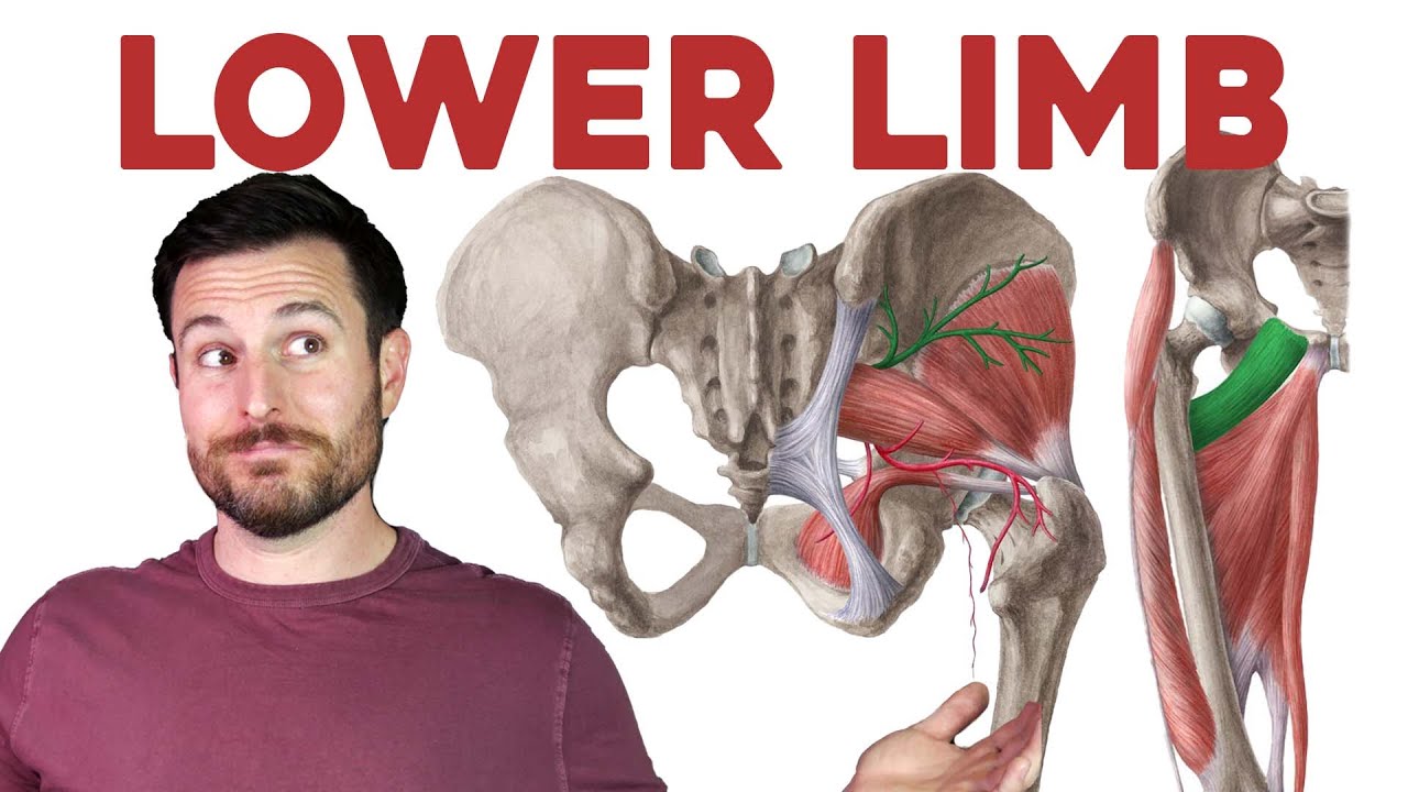
every now and then we need a chance to escape from our studies whether you do that by busting a move on the Dance Floor or taking a hike in the countryside to convene with nature you need those leg muscles today we're going to talk about one of the nerves that innervate the muscles of the lower limb which is the sciatic nerve and its branches before we begin I'd like to give you a quick overview of what we're going to talk about in this tutorial first of all we're going to discuss the origin and location of
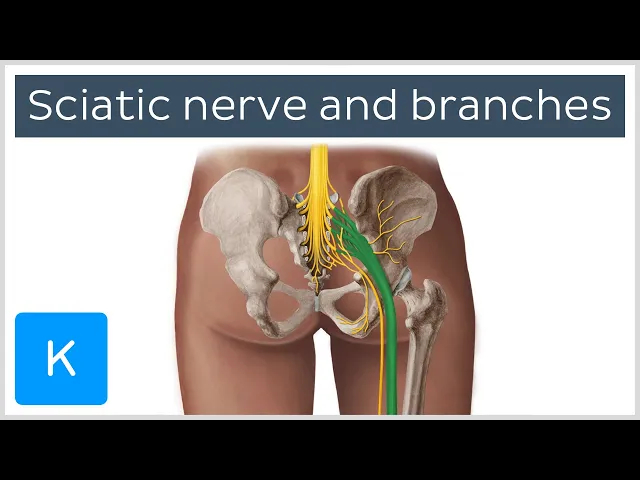
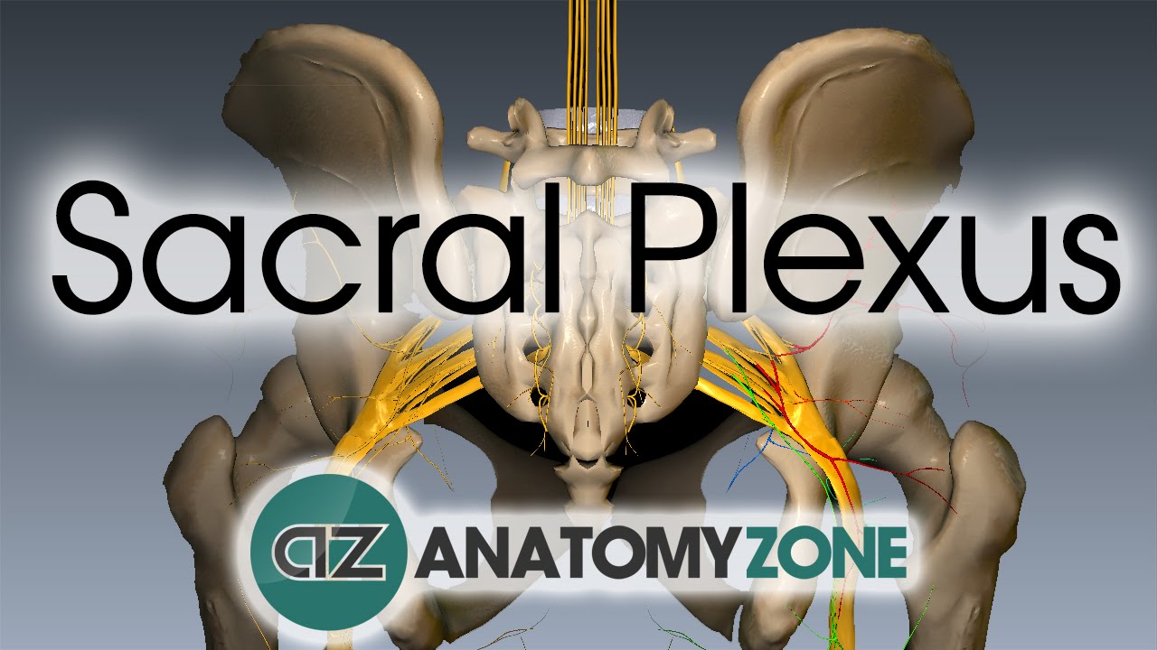
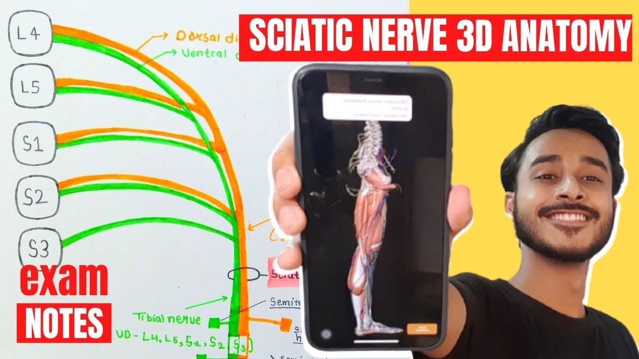
the sciatic nerve then we'll take a look at the branches of the sciatic nerve and some other nerves of the sacral plexus lastly we'll conclude our tutorial with some clinical notes about sciatica okay let's get started with the origin and location of the sciatic nerve the sciatic nerve originates from the sacral plexus which we can see here highlighted in green the sacral plexus is formed by the lumbosacral trunk which consists of contributions from spinal nerves L4 and L5 as well as the spinal nerves S1 2 3 and 4. here we can see S1 highlighted in
green and below it we can see S2 followed by S3 and finally S4 now we can see the sciatic nerve in all its Glory it is approximately two centimeters wide and is the longest and thickest nerve in the human body as I mentioned the sciatic nerve originates from the sacral plexus specifically it is formed by the anterior Rami of spinal nerves L4 to S3 the sciatic nerve is a key nerve of the lower limb as it supplies most muscles and skin of the posterior compartment of the thigh the leg and the foot okay let's take
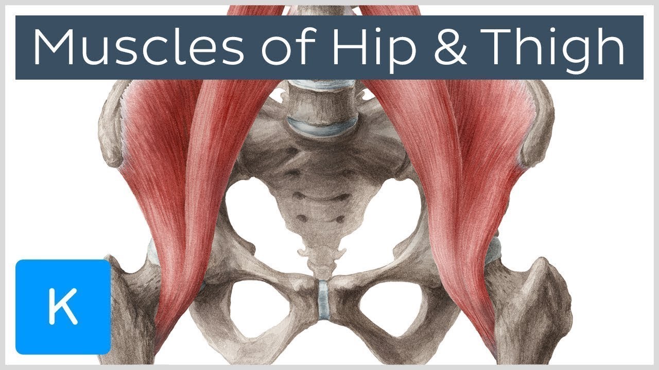
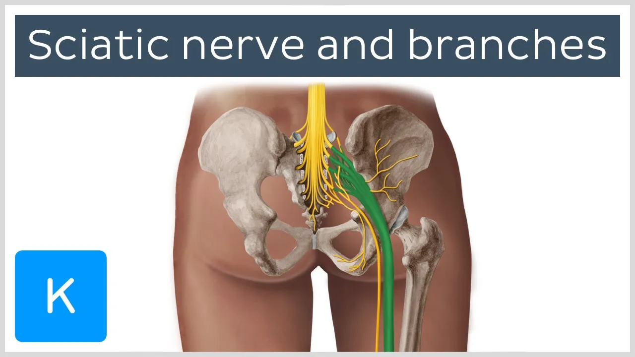
a look at the anatomical course of the sciatic nerve the sciatic nerve exits the pelvis to enter the posterior compartment of the thigh via the greater scientific foramen if we add some musculature we can see that the sciatic nerve emerges below the piriformis muscle this is an easy way to identify this nerve for example for if you're in the dissection lab and you see a chunky nerve emerging below the piriformis you're pretty sure to say that it's the sciatic nerve the sciatic nerve then courses inferiorly through the posterior aspect of the thigh and when it
reaches the apex of the popliteal fossa it terminates by bifurcating into the tibial and common fibular nerves okay now that we're familiar with the origin and location of the sciatic nerve let's move on to talk about its branches it's worth noting that the sciatic nerve can also be described as two individual nerves bundled together in the same connective tissue sheath as I mentioned previously the tibial and common fibular nerves usually separate at the apex of the popliteal fossa however in some individuals these nerves can separate as soon as they leave the pelvis based on this
principle we can see the tibial division of the sciatic nerve highlighted in green in this illustration we can see that the tibial division is formed by the anterior divisions of spinal nerves L4 to S3 the tibial division of the sciatic nerve innervates most muscles of the posterior compartment of the thigh including the semitendinosis the semimembranosus and the long head of the biceps femoris it also innovates the ischial part of the adductor Magnus which is a muscle of the medial compartment of the thigh in the next image we can see that the common fibular division of
the sciatic nerve which is formed by the posterior divisions of spinal nerves L4 to S2 in the posterior compartment of the thigh the common fibular division of the sciatic nerve innervates the short head of the biceps femoris okay so assuming that the sciatic nerve does bifurcate at the apex of the popliteal fossa let's take a look at its terminal branches here we can see the tibial nerve which runs down the medial side of the posterior compartment of the leg the tibial nerve innervates all muscles in the posterior compartment of the leg which are the gastrocnemius
muscle as well as the plantaris the Soleus the popliteus the flexor digitorum longus the tibialis posterior and the flexor hallucis longus muscle in our next illustration we can see the common fibular nerve which is located on the lateral side of the leg it's actually closely associated with the fibula which is where it gets its name from the common fibular nerve however is also known as the common peroneal nerve the common fibular nerve provides sensory innervation to the posterolateral leg alright so let's have a look at some terminal branches of the tibial nerve starting with the
serial nerve which is formed by fibers derived from spinal nerves S1 and S2 the serial nerve descends down the leg and is joined at a variable level by the serial communicating branch of the common fibular nerve which we can see here this nerve provides sensory innervation to the lower posterolateral aspect of the leg and foot in our illustration we can see two branches of the cereal nerve the first one that we can see is the lateral calcaneal Branch this Branch provides sensory innervation to the lateral aspect of the heel next we have the lateral dorsal
cutaneous nerve which is also a branch of the serial nerve this nerve provides sensory innervation to the lateral aspect of the foot we can also see how this nerve contributes to the innervation of the sole of the foot now back to the tibial nerve and its branches here we can see the medial plantar nerve highlighted in green which is a terminal branch of the tibial nerve this nerve provides motor innervation to the abductor hallucis muscle flexor digitor and brevis flexor hallucis brevis ends the first lumbrical muscle of the foot the medial plantar nerve also provides
sensory innervation to the plantar surface of the medial three and a half digits and the associated sole area as we can see in our illustration here if we have a medial plantar nerve we must also have a lateral plantar nerve this nerve is also a terminal branch of the tibial nerve and provides miter innervation to the abductor digital minimi muscle the flexor digit I mean in my brevis the interossei the adductor hallucis and the second to Fourth lumbricals it also provides sensory Innovation to the plantar surface of the lateral one and a half digits and
the associated Soul area the last branch of the tibial nerve we're going to talk about is the medial calcaneal Branch this Branch provides sensory innervation to the medial aspect of the heel and Associated Soul area now it's time to find out what happens to the common fibular nerve the common fibular nerve divides into two terminal branches we can see the first one in our illustration which is The Superficial fibular nerve this nerve provides motor innervation to muscles of the lateral compartment of the leg which includes the fibularis longus and the fibularis brevis muscles it also
provides sensory innervation to the skin of the lower leg and the dorsum of the foot we can't see the next branch in our featured illustration for this tutorial but it wouldn't feel right not to talk about it so here we have the Deep fibular nerve the Deep fibular nerve is a terminal branch of the common fibular nerve and as we can see it runs along the anterior aspect of the leg this nerve provides motor innervation to muscles of the anterior compartment of the leg including the tibialis anterior muscle the extensor hallucis longus the extensor digitorum
longus ends the extensor digitorum brevis and fibularis tertius it also provides a spot of sensory Innovation to the dorsum of the foot between the first and second toes all right it's time to leave the sciatic nerve now and to discuss some other branches of the sacral plexus it's worth noting that the sacral plexus gives off quite a few nerves but for today we're just going to focus on the ones that we can see in this illustration the first Branch we're going to talk about is the superior gluteal nerve this nerve is formed by the posterior
divisions of spinal nerves L4 to S1 and leaves the pelvis via the greater sciatic foramen to enter the gluteal region if we add some musculature we can see that the superior gluteal nerve emerges above the piriformis unlike the sciatic nerve which emerges below this muscle the superior gluteal nerve provides motor innervation to the gluteus medius the gluteus Minimus and the tensorflashy Lati muscle if we have a superior gluteal nerve then we must have an inferior gluteal nerve this nerve is formed by the posterior divisions of spinal nerves L5 to S2 and like its Superior sibling
leaves the pelvis via the greater sciatic foramen however like the sciatic nerve the inferior gluteal nerve emerges below the piriformis muscle this nerve provides motor innervation to the gluteus maximus muscle the next branch of the sacral plexus we're going to talk about is the pudendal nerve which is formed by the anterior divisions of spinal nerves S2 to S4 the root of the pudendal nerve is a little bit unusual it leaves the pelvis via the greater sciatic foramen and then enters the perineum via the Lesser sciatic foramen this nerve gives rise to branches that innervate the
external genitalia the skin of the perineum ends the muscles of the perineum which is this diamond-shaped region you can see here highlighted in green the last nerve of the sacral plexus we're going to talk about is the posterior femoral cutaneous nerve which is formed by the anterior divisions of spinal nerves S1 and S2 and the posterior divisions of spinal nerves S2 and S3 the posterior femoral cutaneous nerve leaves the pelvis by the greater circuit foramen and courses down the posterior aspect of the thigh it provides sensory innervation to the skin of the inferior half of
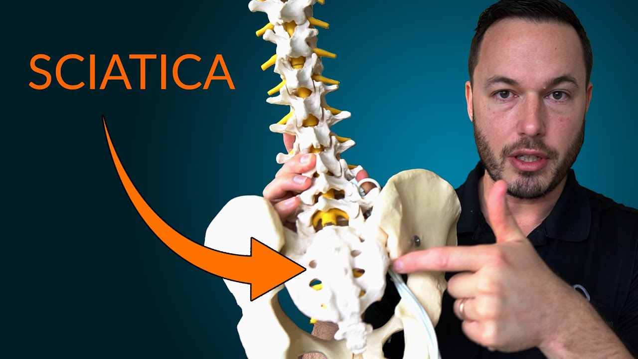
the buttock the skin of the perineum ends the skin of the posterior thigh and the proximal posterior leg interestingly IT Supplies the greatest surface area out of all the cutaneous nerves all right now that we're familiar with the sciatic nerve and its branches let's get clinical in today's clinical notes we're going to be talking about sciatica which is irritation of the sciatic nerve due to rubbing or increased pressure the most common causes of sciatica is lumbar disc herniation which can place increased pressure on its spinal nerves which form the sciatic nerve another cause is spinal
stenosis where the vertebral foramen and spaces between the vertebrae get smaller pinching the spinal cord and the nerves around it sciatica can also be caused by tumors within the spine or a back injury patients may present with pain tingling numbness weakness in their buttock the back of their legs their feet and even their toes it's worth noting that sciatica only affects one side of the body sciatica will usually resolve itself in four to six weeks but it can last longer symptoms can be managed with exercises and stretches as well as painkillers before we bring our
tutorial to a close let's quickly summarize what we've learned today we saw that the sciatic nerve originates from the sacral plexus which is formed by the lumbosacral trunk and the spinal nerves S1 S2 S3 and S4 we then looked at the sciatic nerve and its anatomical course we moved on to look at the branches of the sciatic nerve starting with the divisions which are the tibial division of the sciatic nerve and the common fibular division of the sciatic nerve at the apex of the popliteal fossa the sciatic nerve bifurcates into the tibial nerve and the
common fibular nerve the tibial nerve has several branches including the serial nerve which gives rise to the lateral calcaneal branch and the lateral dorsal cutaneous nerve the tibial nerve also gives rise to the medial plantar nerve the lateral plantar nerve ends the medial calcaneal branch whereas the common fibular nerve divides into two branches The Superficial fibular nerve and the Deep fibular nerve next we looked at some other nerves of the sacral plexus starting with the superior gluteal nerve and the inferior gluteal nerve then we looked at the pudendal nerve followed by the posterior femoral cutaneous
nerve we then concluded our tutorial with some clinical notes about sciatica and that brings us to the end of our tutorial on the sciatic nerve and its branches but don't let your learning stop there visit kenhub.com where you can read interesting articles test your knowledge with challenging quizzes explore our Atlas with beautiful anatomical images or watch more video tutorials like this one yes you'll find everything you need to master anatomy in no time go on click the button you know you want to