welcome to the da vinci academy histology video course the entire video course is available on youtube and covers all of the fundamental principles of histology and relevant cell biology you can find all the videos from the course by clicking the histology playlist link in the description below and then you can access the corresponding practice questions and histology lab videos by going to our website which is also linked in the description below welcome back in this video we'll investigate the histology of the vascular tunic the lens and the mechanisms of controlling the amount of light that's
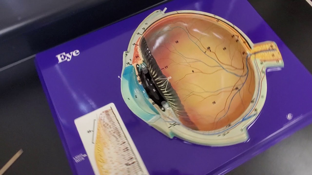
coming into the eye and the process of accommodation deep to the fibrous tunic we have the vascular tunic also known as the uvea so deep to the really pink layer which is the sclera in the back which transitions to cornea here we have now the yubia or the vascular tunic which has this really dark pigment especially in the back these pigments are attributable to the melanocytes that populate this layer so the vascular tunic is connective tissue layer that is rich with not only melanocytes but also the vasculature the melanocytes in the vascular tunic are a
little bit unique and different from those that are in the skin in that the melanocytes here produce their pigment melanosomes and hang onto them within their cytoplasm rather than giving them away to the surrounding cells as is the case in the skin so a lot of these dark pigment in the vascular layer or uvia is due to the melanocytes holding onto their pigments anteriorly speaking the vascular tunic comprises the anterior portion of the iris and the melanocytes here of course impart that particular color of the eye so if the melanocytes in the iris happen to
produce a lot of melanosomes or pigments then you would end up with a really dark eye color as opposed to melanocytes that produce much less pigments and holding onto them then we would end up with something like blue eye color or something like that iris has a central opening and this opening is not caught here the plane of section through this three-dimensional structure we just didn't catch the pupil in this plane so the pupil must be either behind or in front of this screen peripheral to the iris we have these kind of bulky structures that
are seemingly bulging into the interior of the globe this ring-like structure because we're talking about the three-dimensional structure is called a ciliary body it has to spoke and bunch of these in-foldings that are called the ciliary processes and although it's a little bit faint there are a bunch of these really really fine fibers that extend from these processes to the lens so these are all parts of the ciliary body the main bulk of the ciliary body is attributable to the ciliary muscles which play an important role in accommodation we'll talk about this in a little
bit here but in terms of the ciliary muscles we have to remember in three dimension the ciliary muscles are circular muscles comprised of smooth muscle tissue that's arranged in circular fashion like this and the ciliary muscles and the ciliary body are positioned kind of in the same plane as the lens kind of like this so what i just drew is a cross section through this black line so the ciliary muscle being a circular muscle imagine what would happen if this muscle contracted versus relaxed so let's say that the ciliary muscle the circular muscles are contracted
what that would do is it would reduce the diameter of this opening where the lens is positioned and the lens being held to those processes by the fiber ligaments like this and lens being kind of elastic in its nature it'll round up a little more and now let's imagine that circular ciliary muscles relaxing should that relax then we would have the ciliary muscles that have relaxed thereby kind of increasing the diameter of this aperture and what that would do then is it would pull on those ligaments attached to the rim of the lens and in
this manner the lens would actually flatten and that describes the process of accommodation where if we're looking at something really really close let's say an apple for example the light emitting from that apple is coming in at a greater angle and although the cornea bends that light the most as it hits the lens it needs to actually round up a little more in order to bend that light even further so that the focus of this apple is hitting right at this area where we have the greatest acuity now let's imagine that we're looking at the
same apple from the long distance here the light that's coming into the cornea or the eye is coming in in this manner so we don't need to bend that light quite as much because that angle of the light coming in is not as great so again the cornea bends the light the most but the lens actually can be flattened because it doesn't need to bend the light any further to focus that image onto the area of the retina with the greatest acuity like that so when we're looking at something far away we're actually relaxing the
ciliary muscles but when we're looking at something close by then we are contracting the ciliary muscles to allow our lens to round up so that's the process of accommodation and this is also the reason why when we are watching something really really close like watching this video several times then our ciliary muscles are in a contracted position for a long period of time which then fatigues this muscle and we experience our eyes getting kind of sore so it is certainly encouraged to look away occasionally and look at something far to relax our ciliary muscles now
before we move on one clinical pearl that's worth mentioning right now is the press biopia this is an age-related farsightedness that results from the loss of elasticity in our lens so despite the fact that our ciliary muscles are working properly when we look at something really close our lens has lost its elasticity so it cannot round up as much as as it used to and it remains kind of flattened so a lot of people as they get older they hold their phone or book or something like that farther and farther away from their eyes in
order to focus and read or use a magnifying glass so that we have a little more refraction in the light that is coming into our eyes so again presbyopia age-related farsightedness resulting from the loss of elasticity in our lens ciliary muscles are working just fine something to look forward to as we get older another important structure of the ciliary body are the ciliary processes these are these infoldings of tissues that's comprised in its core the vascular tunic but the interior is of course all lined by the extensions of the neural tunic so together ciliary processes
are comprised of these two layers and they're important in terms of producing the aqueous humor on a continuous basis lastly we have the suspensory ligaments these are these fine fibrous structures that extend from the ciliary processes to the peripheral rim of the lens so it goes all the way around so as you can imagine as the ciliary muscles contract and relax the suspensory ligaments in response become loosened or become more taut therefore contributing to the process of accommodation the choroid is the rest of the vascular tunic or the uvi it's the greatest component of the
vascular tunic highly vascular and heavily populated with melanocytes that are holding onto the melanosomes or the pigment particles to reduce the amount of scattering of light that does come in through the pupil now looking at the histology of the iris a little closer we'll zoom in on this boxed area and at higher magnification we can really see the two layered organization of the iris the exterior or the anterior component that is the vascular tunic and the interior posterior component that is the neural tunic extension this is the posterior chamber and this is the anterior chamber
so the thick layer of the connective tissue once again is the component of the vascular tunic and the anterior most aspect of this connective tissue actually does have a really thin but a lot of times incomplete epithelial lining there deep to that we have the stroma that is highly vascular so we can see some arterioles venules and capillary networks there as well as a lot of these spindly brown cells that are the melanocytes and it's these melanocytes that are holding onto the pigments that's mostly responsible for the color of the eye or the iris the
neural extension of the iris in the back are also heavily pigmented and they block out a lot of excess light coming in through the iris itself so that light coming into the eye is limited to the through the pupil within the stroma of the iris there are two types of muscles called the dilator pupillae and sphincter pupillae just based on the names can you guess the functions of these two muscles well that was a rhetorical question hopefully so when we look at the iris from the anterior aspect what we'll see is this circular structure with
a hole in the middle which is the pupil so this is the iris now the iris has a dilator pipilla muscle that are radially oriented going away from the pupil of course these are histologically comprised of smooth muscle cells and when the dilator pupillae muscles contract they pull away from the pupil so it actually opens or dilates the pupil size so that more lights are entering the eye these dilated pupillae muscles are actually innervated by the sympathetic innervation so when we are excited or emotionally aroused our dilated pupillae are contracting therefore our pupils are dilated
and we're trying to get a lot of light and information into our eyes the sphincter pupillae on the other hand are circular muscles again smooth muscle tissue that are at the center of the iris abutting the opening of the pupil so as we saw earlier whenever a circular muscle contracts it actually reduces the aperture or size of the hole so when sphincter pupila contracts it reduces the size of the pupil and when it relaxes then the pupil can then dilate along with the dilator pupillae contracting the sphincter pupillae are innervated by parasympathetic innervation so when
we are relaxing or digesting food or something like that our sphincter a would be contracted therefore our pupil would be constricted the posterior surface of the iris or the internal aspect of the iris is actually lined by the stratified cuboidal epithelium of the neural tunic and here the cells of the cuboidal epithelium are heavily pigmented once again to limit the light entering through the iris and in between this neural tunic and vascular tunic we can actually see a strip of this radial muscle smooth muscle cells that comprise the dilator pupillae and we would only see
the sphincter really close to the edge of that iris abutting the pupil now let's look at the ciliary body in a little more detail as well so this is a boxed area at a higher magnification we can see a little bit of the lamina propria of that conjunctiva right here and then the sclera over here in darker pink color and then we have a little bit of the last bit of the iris over here with the vascular tunic and the neural tunic and then we can start to see this bulk which forms the ciliary body
so anatomically the ciliary body is slightly posterior kind of peripherally to the iris while the bulk of the ciliary body is comprised of the vascular tunic we still need to remember that the inner aspect is lined by the extension of the neural tunic still the difference here is that although this inner lining is still stratified cuboidal epithelium just like in the lining of the iris the two layers are actually differently pigmented with the innermost layer having cuboidal cells that are not pigmented but the deeper layer or more appropriately since we're talking about a spherical structure
the exterior layer is comprised of the cuboidal cells that are still heavily pigmented more on that during the neural tunic discussion later now as for the actual bulk of the ciliary body as discussed earlier we have the ciliary muscle so it's comprised of the smooth muscles that are arranged in a circular fashion and in this cross section we can appreciate how the smooth muscle tissue is cut in a more transverse fashion so we can imagine the smooth muscle spindles coming in and out of the screen as they're forming this circle just posterior and peripheral to
the iris now these extensions are the ciliary processes of the ciliary body the stroma from the vascular tunic extends into the ciliary processes and form the core and true to the name the cores of these vascular tunic would be highly vascular and then the internal lining is the extension of the neural tunic and together with the vascular tunic core and the epithelial lining from the neural tunic produce the aqueous humor and secrete it into the posterior chamber from which that aqueous humor can go into the vitreous humor or into the anterior chamber via the pupil
and at a higher magnification we can also appreciate these really fine and wispy little fibers that are the suspensory ligaments so these are the ligaments that are extending from the ciliary processes in the body to the peripheral rim of the lens so together the ciliary muscles ciliary processes and the suspensory ligaments all function in the accommodation to review this process of accommodation it's a process of finely adjusting the roundness of the lens this lens has a natural elasticity so naturally it likes to stay in a rounded form so when the ciliary muscles are contracted we're
talking about from anterior view ciliary muscles when they contract they're reducing that size of the aperture in the middle with the lens over here which allows the suspensory ligaments to kind of loosen a little bit so that pooling force on the lens is kind of removed so that the lens can round up and bend the light a little more so this is particularly good for looking at something that is close by because the light that is emitted by the objects are coming in at a greater angle so the cornea will bend the light the most
as it comes in but in a fixed amount so the roundness of the lens will allow that light to bend even more just so to focus that image onto the area of the retina with the greatest acuity now what happens when the ciliary muscles are relaxed relaxation of the circular muscle allows that aperture to get larger if you will with the lens being kind of about the same size and that pulls on the suspensory ligament so the suspensory ligaments become taut so the pulling forces all the way around the periphery of the lens will force
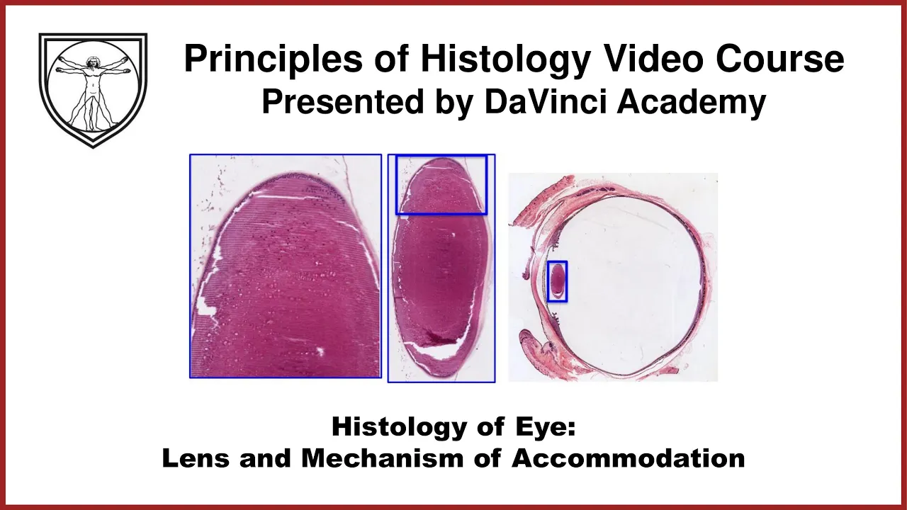
the lens to become flattened and that is good for looking at an object that is far away because the light that is emitted by an object is coming in at a lesser angle if you will so the lens does not need to be as round in order to refract a light so that it focuses right on that spot that we'll talk about with the neural tunic discussion now let's look at the lens histology a little bit we have to remember though that the lens is not a part of the three tunics of the eye kind
of hangs out there at the plane of the ciliary muscles and held in place by the suspensory ligaments this transparent structure is still comprised of the cells but these cells of the lens are also called the lens fibers which are elongated and a nucleated cells that are filled with this transparent crystalline proteins and these fibers are arranged once again in a precise alignment in order to make the lens transparent looking at the lens at a higher magnification we can see that this structure is comprised of number of these fibery looking stuff these are all the
lens fiber cells that for the most part are a nucleate and are just packed full of proteins at a higher magnification of the boxed area we can see the lens fibers clearly and they're all kind of precisely aligned here some of these nuclei that you are seeing belong to some of these cuboidal cells that line the anterior surface of the lens so indeed we have a simple cuboidal epithelium on the anterior surface but none on the posterior surface so when we're given an image like this without this big image for perspective based on the location
of the simple cuboidal epithelium in the lens we can still orient that this is the anterior surface and this is the posterior surface up top we're seeing an extension of that suspensory ligament fiber up there at the periphery we see a little more nuclear features here and that's because the simple cuboidal epithelial cells at the rim of the lens are the ones that are undergoing cell division in order to replace any lens fibers that may have degenerated or are getting older in order to replace them and as these processes occur should these lens fibers not
line up precisely or crystalline proteins not be produced in the just the right way we may end up acquiring some opacity or cloudiness of the lens which we well know as the cataract last feature of the lens histology is the fact that there is an acellular capsule that surrounds the entire lens on its most exterior surface now the last component of the vascular tunic is the choroid although we're discussing it the last this is the largest component of the vascular tunic at a higher magnification of this box area can really see what this layer is
comprised of externally we have the sclera and here with a lot of dark brown cells as well as vasculature is the choroid of the vascular tunic and internally we have the beginning of the retina right here so the delineation is located right here where the laser pointer is so again this is a very vascular component that supports the retina in terms of the vascular supply and all of these melanocytes that are just packed full in this vascular layer is quite important for limiting the light reflecting or scattering around once it has entered the globe and
one structure that may be of clinical importance is the brooks membrane so this is an acellular really thin basement membrane-like structure that delineates the division between the retina and the vascular tunic and the reason why the brook's membrane is of clinical significance is because of the melanocytes that are located here should any one of these melanocytes become cancerous and form a uvial melanoma the breach of brook's membrane has been associated with a significantly lower prognosis let's wrap up the discussion of the vascular tunic by reviewing some clinical pearls that we've discussed already presbyopia is an
age-related farsightedness that usually starts around fourth decade in one's life and that has to do with the loss of the lens elasticity cataract involves the developing opacity of the lens so we can't see things as clearly that either has to do with inappropriate crystalline proteins in the lens fibers or the imprecise arrangements of the lens fibers themselves and lastly we discussed the uvial melanoma which is the cancer of the uvul melanocytes thank you for watching this video from the da vinci academy histology video course which is completely available on youtube to access the corresponding practice
questions and histology lab videos go to our website using the link in the description below foreign [Music] foreign [Music] you
![Histology of the Retina [Special Senses Histology Part 3 of 4]](https://img.youtube.com/vi/zG4ZhMusF4Y/maxresdefault.jpg)
![Eye Anatomy and Histology of the Outer Layers of the Eye [Special Senses Histology Part 1 of 4]](https://img.youtube.com/vi/u1fUUr1A34Y/maxresdefault.jpg)

![Liver, Pancreas, Gallbladder Histology [GI Histology 4 of 4]](https://img.youtube.com/vi/_kA9x9FcjWA/maxresdefault.jpg)
![Ear Histology [Special Senses Histology Part 4 of 4]](https://img.youtube.com/vi/hTeIRe2cBDQ/maxresdefault.jpg)
![Histology of the Ovary and Ovarian Follicles [Female Reproductive Histology Part 1 of 2]](https://img.youtube.com/vi/cDs6goPOD1I/maxresdefault.jpg)
![Pituitary Gland Histology [Endocrine Histology 1 of 2]](https://img.youtube.com/vi/XZrS4zkrSps/maxresdefault.jpg)


![Arteries and Capillaries Histology [Cardiovascular Histology 1 of 4]](https://img.youtube.com/vi/Oi9rqymW6m8/maxresdefault.jpg)