um today we're going to um continue talking about um antibody structure as well as thinking about antibody function so when we left off last time I told you about this y-shaped protein that um scientists had discovered that was sort of able to lead to protection in certain um instances that was called the antibody and I told you some experiments last time where um bomarang and kado were transferring um different either Solutions or cells to try to find out what the protective thing was and people after them did things like use proteases or lipases or things
like that to say it was a protein um in times after that people did additional kinds of biochemistry experiments to try to understand some parts of the structure of that protein and so the next sort of thing that I want to tell you about are three different um processes or three different things that scientists did um to try to break this protein to help them understand its parts and so we've got three different degradation that were done that really helped us understand the structure of the antibody um one of them was that they uh treated
it with a compound called mercapto ethanol um the as you can see here um it's listed as mercaptoethanol reduction um but the thing that you should that I that you can know about mercapto ethanol um if you don't want to think about the chem the word reduction that chemistry word scares you the point of beta mapto ethanol or mapto ethanol is that it breaks disulfide bonds it breaks disulfide bonds and so they treated antibodies with mercapto ethanol to break disulfide bonds and when they did that this was you know the antibody that they started with
and they found out that they actually got four protein chains so this thing that looked like a y was actually four separate protein chains that had been held together by dooli bonds those four chains had were basically two chains that were identical and that were big and two that were identical and that were small and we called the the two that were identical big the heavy chain and we called the two small ones the light chain and so this y shaped uh protein is actually a complex of four different proteins two identical heavy chains two
identical light chains and you can see the disulfide bonds holding them together so you can see the the heavy chains are in dark blue um and they kind of make this main part of the they're held together by a couple disulfide bonds right there and then we've got a light chain on either side held together by a disulfide bond and so this is what it looks like in my antibody here the heavy chains are in yellow and the light chains are in Orange um and so this is sort of the overall structure they found by
doing um the bmer capol reduction um they then wanted to use some other types of um things to kind of learn about the structure of anti um and so now they use a protease to degrade the protein and to break it into different parts the proteas they use in this example is called papane um and when they did their papain digestion they found that they got three pieces one uh piece that was sort of the tail that you can see here and these other two identical arm parts and when they looked really closely they found
that the arm parts which you can see are made up of a heavy chain and a light chain each could bind to antigen and so they called those parts Fabs or fragment antigen binding so we get two identical Fabs with our Paine digestion um and they also got this tail piece that's made of the rest of the two heavy chains they named that fraction the FC region the thing that FC stands for actually is like oldfashioned and no one cares um but if you wanted to have something for it to think about you could say
that it was constant it is the fraction that was constant because we're often we we will frequently talk about a constant region and it happens to be basically the same part so we'll call it concentration yeah sure uh fraction or fragment with antigen binding yeah um and so you can see um the results of the papain digestion um here on the right um this is the version from your textbook you're seeing it from a different textbook on the left um your fun fact of the day um You probably have not in other classes heard of
the proteas papain um or thought a lot about the proteas papain but you actually probably know the proteas pine um you ever the the more common is pineapple if you eaten pineapple and you know how you get a tingly feeling in your mouth it's because it's full of a protease that's actually um starting to act in your mouth that produce is called papine it was originally just it was actually originally isolated from papaya so that's how it's got that name um and literally the only time I've ever heard of it in my entire life is
in a pinee digestion of an antibody and in fun facts about pineapples like I I honestly don't really hear about it very frequently otherwise um they also did a different kind of proteas digestion and now they used a proteas that you might have heard about um in other places which is pepsin so they also did a pepsin digestion um and when they did their pepsin digestion they basically got one part of the antibody and the reason why I say they got one part is cuz the rest was like like totally destroyed um and the one
part that they got you can see was like the two antigen binding arms connected together so the degradation happened below this disulfide Bond so the basically the whole FC was destroyed and they got this molecule called a Fab 2 um that again kind of helped them understand what this overall structure looks like there are situations where immunologists today will make either Fab fragments or Fab 2 fragments or XC fragments to use uh in experiments um and so and in fact I'll mention a little bit more about those um a little bit later in the lecture
um and so that sort of gave us this uh understanding of the antibody structure so your an so antibodies are made of two each individual antibody two identical heavy chains two identical light chains um that you can see here from your textbook uh shown in green and in yellow you can see your disulfide bonds between the heavy chains as well as the disulfide bond between the heavy and the light chain um and so that's going to be one of the key parts of this structure that we're going to think about when you um look at
this these images of the antibody structure you can also notice that the antibody is drawn as being made of some rectangles so you can see we've got rectangles depicted here that actually is um immunologist shorthand for a particular type of protein fold or protein domain and so after people had discovered the antibody and started to really look at the protein they found out that it has multiple copies of this same protein fold um that make up its structure if you remember on Wednesday I told you that sometimes we use this term of immune globuline or
imunoglobulin when we're talking about antibodies that fold that makes up each of those rectangles is called the imog globulin fold or the IG fold um and so um this is a kind of major protein fold that is found in many of these um types of proteins um what you can notice with the imunoglobulin fold is we've got these arrows so those are all beta sheets that are connected by Some Loops so we've got some Loops connecting some beta sheets um and we also have a dulfi bond kind of going through the middle and um you
can see this in its actual three-dimensional form here where we've got all these beta sheets kind of coming together like this and these little Loops coming off the top um I was actually taught to think about this as it being like one of those little fancy person sandwiches that has a toothpick in the middle and so you have two sheets of bread of beta sheets and a dulfi bond as toothpick holding it together in the middle so the disulfide bond is kind of like in here holding this whole thing together and otherwise it's like a
little sandwich made of two sets of beta sheets and we got those little Loops up at the top um at some different times in the semester I'm going to mention the loops so when I'm talking about the loops different times those are the those Loops particularly the ones that are labeled there up at the top are going to be the ones I mean um and you can see this sort of here as well so this is the Fab of an antibody so this would be like just this arm you can see there's like four different
locations four different little rectangles so this would be four different imunoglobulin Folds and here they are looking like two little sandwiches and you can see there's some Loops at the end that are going to be particularly important um this structure of the imunoglobulin fold um was first described in the antibody but now we found that tons of proteins have amog globulin folds um and you can see some examples of them here these are all really important proteins of the immune system system so we see this fold over and over and over and over again in
proteins of the immune system um and sometimes uh people draw it with this little like circular thing with the disulfide bond um in between so if I happen to draw something like that that might be what I'm trying to draw I will also say um once upon a time when I was a grad student taing I drew an imunoglobulin fold containing protein on the board um with little curves like this um I was drawing and kind of not looking like this to look at it and I realized I had just drawn the most phallic drawing
I'd ever drawn in my life giant on the board um and so I don't tend to draw the pro these proteins as much anymore um so that's kind of one aspect to understand about this antibody structure is that you can see that there are these different domains that are depicted here as different rectangles um we can also come up with some other ways of talking about different parts of the structure and one of the biggest things that we often talk about is shown on the right here um which is that the very last imunoglobulin domain
of both the heavy chain and the light chain so my little antibody here it's the darker colored ones here it's the red ones are actually the place where the antigen binds that amog globulin domain is the really important part and sometimes we refer to those parts as the variable region and so you can see they're listed here in red as the variable region the rest of the light chain and the rest of the heavy chain will be known as the constant region and so we can also talk about variable region and constant region um of
our antibodies um and I know this is going to be really shocking um but the variable region and constant region got those names based on how many diff like if I took all the antibodies of your whole body how many different variable region versions would I find and how many different constant region vergins would I find um I'm going to make give you estimate right now the numbers are totally made up I will never like ask you in both cases I'm lying but they'll give us a ideal um for variable regions the answer would be
about 10 to the 16th so that is a Quin 10 quintilian um pretty variable there's a lot of them um constant regions um for our purposes I'm going to say the answer is five so not not totally constant but a whole heck of a lot more constant than the variable region um we can have we have names for those different for those five different constant regions so we can we actually can sort of call those bodies that have different constant regions different things the different constant regions first of all you can notice from um this
image and other images the different constant regions the constant region is what binds to antigen the variable region is what binds to antigen the constant region is what allows the antibody to actually do something it's allow what allows the antibody to have its function and deal with a pathogen um and that's going to come up much more later um but when I think about my antibodies and their constant regions I think of five constant regions again there's actually sub types of some of them but I'm not really going to worry about those for us we
just got five and those five are shown here one of them is called IGM that means that that um this sort of the blue domains of um the heavy chain are present um there is actually you will see later a gene that's involved in IGM that's called the MU constant region mu is the Greek letter for m so they go together there's also one called igd which is encoded by Delta you can see it has these heavy chain constant regions um notice that when we're talking about this we're really talking about heavy chains we're leaving
light chains out there's reasons that's a later problem um you can see we have one called G we have one called e we have one called a um these are um this order of me telling you them um is actually an order that matters to us as immunologists it will not matter to you right now but later you will want to know MD G EA um and I like to remember this with with a pneumonic um you guys have um heard the phrase an apple a day keeps the doctor away so I learned to remember
this with MDS like doctors MDS give everyone apples um and that's how I remember mvg um in terms of the order um for now you can just use that to remember what the five are um going forward when we talk about these antibodies that um have that differ in their constant region we refer to them as different isotypes so this slide as you can see from the title is officially telling you the five isotypes that exist and so we can I might say what isotype is this antibody oh it's an IGG imunoglobulin is the IG
like we're just shortening that whole imunoglobulin word and then putting a letter at the end um so that is those are sort of the options and some of the details I'm going to tell you right at the moment for the constant region um but then I have to tell you some other aspects of some other parts of the structure um so just um really briefly I'm not going to say a ton about this here um but you can notice that there are some um additional parts that are not imunoglobulin domains particularly this region called the
hinge region it's sort of the I don't know shoulders of the Y um and what I want you to just know about the hinge region is that the hinge region is super flexible in fact antibodies can wave and change the distance between their Fabs they can rotate their Fab arms I don't really I can't really rotate my shoulder super well but they can rotate this way um they can bend the FC sort of like the elbow they can even like move the tail um and so we've got lots of flexibility the antibod is going to
be able to to of reach antigens really well by being really flexible at the hinge region there's also another piece of information that is really important about when we think about the structure of our sort of stereotypical antibody which is that we've got two sites that bind antigen to Fab sites two variable regions that are identical so we can actually bind the same antigen twice here one of the reasons why that is important is that it actually changes um some details of what the biochemical interaction between the antigen and the antibody look like so often
times biochemists think about like on- rate and off- rate how quickly a pro two proteins interact and how much they don't how quickly they come apart right we call that Affinity because the antibody and so you can see this is kind of what that would look like at the top because the antibody has the ability to do this B ve valent interaction to interact more than once it actually doesn't have a normal onoff rate because you can see both arms can come on if one arm happens to fall off it's doesn't it isn't like infinitely
able to potentially bind anywhere it's being held right close to its binding partner and it's going to bind on more quickly so in fact this has stronger binding than two times this um because you basically have reduced the ability of it to them to come off um as they are being held really close to other copies of the antigen um and so this is more than double as strong as this because of this um repetitive thing sometimes we refer to that that combined strength as the avidity of antibodies and so antibodies also have this thing
called High avidity um because of their um balent interaction um so the other sort of aspect of the antibody structure that I want to tell you about is um some details of the antigen binding sites and how we bind antigen one of the most important pieces of this is that um when we're binding to antigen with our antigen binding sites that antigen binding site is made of a combination of the heavy and light chain it's not just the heavy chain doing it alone it's not just the light chain doing it alone it's actually completely about
the combo of them and sort of the combined interface or the combined shape the combined structure that they make um and so um here in sort of grayish color I don't know what color that that oval is that's the antigen so in this you're the antigen and you're looking at the antibody um and when you're looking at the antibody you're actually making contact with this part of the heavy chain so there's a h right there and this part and this part as well as you're making contacts biochemically with parts of the light chain so it's
really about the combo of the two not just which heavy chain is there or which light chain is there um you can see the same thing kind of as a side view here if we look at the antigen you can see that the antigen is interacting with both the heavy chain and the light chain and so what this antibody is specific for is partially based on which heavy chain it has and also partially based on which light chain it has um so you can notice the antigen binds here every so often and I actually understand
why this is but every so often often when students try to draw an antibody they put the antigen here that's not where the antigen goes antigen goes here um making contact with both the heavy chain and the light chain um you can also notice um from my from this diagram that the antibody is actually um has you U sort of three different parts of the heavy chain three different parts of the light chain seem to be binding to that antigen those three different parts are actually those Loops I showed you before the three Loops from
this imunoglobulin domain the three Loops of this imunoglobulin domain so if I go back to my imunoglobulin domain you can see up at the top it's got three Loops that we're holding it together those three loops on these amog globulin domains are actually what are making the physical contact and so you can see Loop one Loop two loop three Loop one two loop three of the heavy chain and the light chain here um sometimes we even will refer to those Loops as cdrs or complementarity determining regions because they are the things that tell you which
antigen the antibody is complimentary to yes okay okay uhuh uhuh yeah so so what so what that means is that the light chain has a variable and a constant and the heavy chain has a variable and a constant so this is still a heavy chain this is still a light chain it's just that in this light chain there is a variable region and a constant region the red is variable the blue is constant here there's a variable region and a longer constant region yeah that's fine well so usually we see the we talk about the
light chain as being sort of the outside one and in a lot of ways that's just the convention of how we always draw it is the light chain is always it's smaller and it's we usually draw on the outside because the heavy chains kind of have to be next to each other to give you this tail okay all right yeah okay are they so I have only mentioned details that's a good question I've only mentioned details about the heavy chain constant region there is a light chain constant region that for the most part no one
cares about we there is legitimately like one spot in one lecture where I'm going to mention it and there's one place where sometimes I mean all just talk about it so there to if I am being fully truthful there are two kinds of constant regions of light chains one is called Kappa and one is called Lambda and sometimes you have a Kappa and sometimes you have a Lambda and no one cares um so I didn't really mention them but technically there are these there are specific um light chain constant regions Kaa or Lambda yeah yes
every light chain has a constant and a variable every heavy chain has a constant and a variable okay yes um I also want to just tell you guys um I have many times in the past told a story of one of the first one of the Early times I can't remember exactly which year it was when I taught it drew of a question that somebody asked in um my Immunology class um which I thought was like the most brilliant question I had ever heard and I was just like I can't believe it was basically like
he figured something out multiple lectures in advance and it was like this question I was just like I can't believe he figured that out and so I've always been like oh my God that question um the person who asked it is now a microbiology and neology professor at tcj um multiple times so far this semester I have been like man these are like Tyler level questions um so like good job I've been like super psyched um so I've already basically told you this on that previous slide um but but the heavy chain and the light
chain um actually uh combine to form the antigen and so this is just um another version of this showing you that really what happens is we have kind of the heavy chain and light chain coming together making some kind of three dimensional structure that is complementary to the antigen um when I was in when I was a senior um in grad or in college I was talking to one to my roommate who was a political science major and I told her that actually as an immunologist all I do is match shapes it's really like I'm
in preschool um is I I ask if shapes match or not um and so you can kind of see that it's the heavy chain and a light chain here this always shows it as the two of them being super symmetric and that's not totally true but whatever this works um when if I were to take all of the 10 to the 16th variable regions from all of your antibodies in your whole body and I was to do um protein sequencing on all of those antibodies or all of those variable regions I get a lot of
differences obviously they're variable regions but I would find out that the differences we're mostly in one part of the variable region so the variable region doesn't isn't sort of equally variable all the way across the imunoglobulin domain there are parts of it that are extra variable that are like the most variable we refer to those as the hyper variable regions or hvrs oh that's the wrong side this is the right side and when I actually look at that you can see I've got three areas that are super variable three hypervariable regions the three hypervariable regions
are the loops they actually encode the loop parts and the three hypervariable regions are the three low parts that are touching the antigen so in fact all the variation really between from antibody to antibody is in those Loops um and that's how they can each bind to a unique antigen and so sometimes when I think about an antibody I think about you know on each arm we've got heavy chain and a light chain and they each have three little Loops coming off like three little fingers that are actually the places where they vary and actually
change which antigen they bind and so here you can see kind of those three Loops shown at the end here you can see those three Loops shown in the structure here you can see this in a linear um amino acid sequence that there's three parts that vary way more than all the other parts yep Brena yes and so I didn't say that that's perfect and so it turns out that the CDR Loops the loops that are the complimentarity determining regions and the hypervariable regions are the same thing um and so those end up we now
know those are actually two words for the same same Loops so here you can see they're called both cdrs and they're called hvs or hypervariable regions um so what I hope you might notice from all of this is those Loops probably are kind of important um and as we move forward we're going to care about those Loops again um I told you earlier about antibody isotypes and isotypes being one way that we can divide up the many antibodies that a person has there are some other terms we can sometimes use when we're dividing up antibodies
and those are those are shown on a slide that maybe I can click on here um and so sometimes we'll talk about things like idiotype differences or Alo type differences um a little I will say there again this is one of those places where some of you who may there are a couple spots when we talk about antibodies where some of you who know a lot of biochemistry are going to look at me and be like no no no no or a lot of for molecular biology and the answer is I know what your problem
is and I promise you I will answer that question eventually um it's just that I haven't totally answered it yet but I have actually had some students get really confused here and so what I want you to notice first is that we we've got these antibodies from this person named Sherry I don't know why her name is Sherry but her name is Sherry and Sherry could have antibodies of different isotypes like IG versus IGG and so you can see that's the same difference I was showing you before which is the difference between isotypes if I
was drawing this slide I would have made this constant region a color that was like different green maybe it's darker maybe it's darker I would have made it like purple or something to indicate the difference between IG and IGG but they didn't do that what you can also notice is that um we can think about antibodies that have the same constant region but different variable regions so here you can see again from Sherry two different antibodies that are both IGG antibodies but that bind to different antigens so their constant region is the same but their
variable region is different now they did color code this time um and so you can see one of them is an IGG against antigen a so this might be against influenza and one of them is IGG against Anin B this might be against SARS K to so same isotype different um uh variable region gives you this difference in what's called idiotype so those are antibodies that are different idiotypes you can also see if I'm looking at two antibodies that are basically the same um but are from different people so here you can see an IGG
antibody against SARS K2 from Sherry and an IGG antibody against uh SARS K2 antigen B from John um we refer to those as different allotypes so two different individuals of the same spe species but they're same receptor and the idea here is that like there might be other genetic differences between Sherry and John maybe like Sherry has a mutation that changes the amino acid here and so her antibody isn't actually totally identical to Jon's because these are just two genetically different individuals and so we would call these antibodies that otherwise the same isotype same idiotype
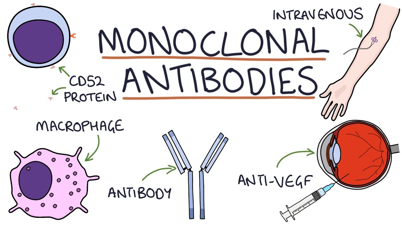
but just different from person to person um different in allotype yeah Jonah maybe um so there's one other kind of thing related to this I just want to mention um I told you guys last time about the difference between um polyclonal and monoclonal antibodies um some of you had heard the term monoclonal antibodies before if you remember the monoclonal antibodies were antibodies that were made especially in the lab where you have a whole population of antibodies that all bind the same epitope within an antigen so they're like exactly the same you had to do some
lab purification steps instead of the polyclonal response that you might get um we now use monoclonal antibodies for all sorts of medical uses we treat lots of diseases by giving people monoclonal antibodies that we've made in the lab um this is a super out ofd um list of some of the monoclonal antibodies that are currently in use um if you were to actually look at some details of um in the Pharma industry of kind of classes of drugs and how much money the Pharma industry makes off of them and how much the Pharma industry plans
to like do them in the future monoclonals win by far monoclonals are like where all the money is right now in Pharma and what everyone is sort of doing and so we know a lot about the so people in the pharmaceutical industry even if they're thinking about like heart disease end up thinking a lot about some of these sort of biochemical details of antibodies cuz a lot all the hot drugs are monoclonal anti anies Brena did you have a question so monal antibodies um so monoclonal antibodies would in fact all be an identical isotype idiotype
and allotype because we made it in the lab so it's like a whole bunch of super identical antibodies um and so what you can notice here is we've got some antibodies that um are used to treat non hodkin lymphoma some breast cancers some different types leukemias and lymphomas uh neuroblastoma other um other types of cancers um arthritis different types of arthritis croh disas also is colitis psoriasis um multiple sclerosis asthma you can see there's like a huge list of diseases where monoclonal antibodies are clinically in use um my guess I could be totally wrong on
this is that at some point over the weekend you guys some you guys might actually like have the TV on like see something on a on TV and you if and if you do that you might see an ad for some kind of medicine happens relatively frequently when that happens I want you to look at the name of the medicine um there are names listed here and there also will be on the next slide as well if the the name of the the drug has MB at the end that means it's a monoclonal antibody so
anything that ends in mab is a monoclonal antibody drug so yes for example um you know you might hear about Huma but and the official name for Humera is adelin RB so if you see a drug with any of those names um it that has mab at the end it's a monoclonal antibody um and now that you know that wherever you might see a drug ad look for the the name of the compound you will be shocked at how many drug ads you're seeing for monoclonal antibodies that have MBS at the end um we actually
also um can there's actually some other info in that naming because when we think about those antibodies we have to think about how we make them in a few different ways so once upon a time the way that all of our antibodies that were used for therapies were made is they were made in a mouse you had a you were like okay Mouse I need you to make an antibody against this protein and I'm going to use it for therapy and so we got an antibody that came out of a mouse and then we injected
that antibody that came out of a mouse into people to treat them and in many cases these drugs had like crazy positive effects like lifechanging effects but when sometimes when we did that they also had some side effects because you were pumping a person full of a mouse protein a Forin protein at some point your immune system is like what is this mouse protein doing that you put in um and so we eventually figured out ways to try to make the molecule more and more of a human antibody and not a mouse antibody so we
change first to have just a human constant region with the mouse variable regions we or to have the human everything but the cdrs and just the mouse cdrs or eventually to have the entire thing be a human antibody molecule it's kind of tricky you you might imagine that um I probably if I want to make a humanized antibody for a drug say I want to make grams of an antibody against SARS K2 I'm not going to inject you full of tons of SARS K2 and collect your antibodies I have to do a lot of lab
work not going to have a human patient who's making these antibodies for me um and so there's sort of a lot of biochemical um work that's been going on here but actually if you look at the name of the antibody the MB the part that comes before that tells you what kind of an body it is if it's a mouse antibody if it's a chimeric if it's partially humanized with just the mouse c3s or if it's a fully human antibody um and so you can see umab is a fully human antibody um zumab is humanized
um you can see uh usually it's um imab is the chimeric um and omab is the mouse and so again this is one of those things where you will start to see some of these um name these drug names now that you know that the thing if you see a drug ad look for that and you'll be surprised at actually how much information is given in that drug name and you'll and how many monocon antibodies um that are being used um one of the ways that we're actually able to do fully humanized antibodies now um
is someone actually generated a mouse that has the entire human antibody genetics section so we basically put the entire um human entire human antibody Gene region which is a massive region of the human genome into mice so that we can have mice make human antibodies for us um and so you can see um the these mice are known as the bosam mice um and this was a this was you know 10 years ago there were already 10 Therapeutics with the veloe mice and they were probably the most valuable mice ever engineered having already brought in
$2 billion at that point and they just keep on going um there are other situations where people have done things like say you know what using an antibody for therapy this protein that we're trying to use as a therapy for somebody is actually too big of a protein this protein actually like can't fit in the special anatomical location where we need to do treatment we need something even smaller than this protein and what they've ended up doing is they've ended up designing things like just Fabs or even just variable regions in the lab and injecting
people with those um some of this uh we figured out how to and sometimes we even do heavy chains alone without light chains um it turns out that there are some organisms that are super weird that only have heavy chains that don't have light chains the most famous organism that only has a heavy chain and no light chains and its antibodies can I tell you why no but it's a camel camels only have heavy chains a camel biologist I I do not know who found that out um and so based on looking at the structures
say those camel antibodies or otherwise trying to make like just Fabs or just FC's there are even times where people will make Fab 2s so you can get like cross binding but you don't have the tail in the way um will be used for a lot of different Therapeutics officially these are all called nanobodies um which are like these further modifications of antibodies based on all these different structural parts um so at this point we've seen the structure of an antibody but we haven't totally talked about the function of an antibody and if you read
online stuff especially kind of during covid and things you would not believe the verbs people use with antibodies that they think antibodies can do antibodies sound like freaking superheroes in a lot of cases like they're wearing a little cape and like they're doing all sorts of crazy things but really an antibody is a protein with four chains a protein cannot eat things right there there are kind of limits to what you can actually do to get rid of a microbe if you're just a protein made of 14 and so what we have to think about
is how in the world does this little protein actually help us get rid of a microb um there are a bunch of answers to that question which are known as the antibody effector functions um I always think of them as the five antibody affector functions this image from your textbook or from a different textbook says there's a sixth one I kind of get where they're going with the sixth one so I'm we can have six sure let's have six um different antibody effor functions and so we need to see these six different antibody effector functions
um the first one that I'm going to tell you about is pretty easy and the reason why I'm telling you about it first is because you actually already know it you just don't know you know it so one of the ways that antibodies can deal with a pathogen one of the affector functions of antibodies is that antibodies can start the compliment Cascade so one of the reasons why antibodies can be actually useful in getting rid of a microb is that they can start the classical pathway of compliment like we already learned about so fixing compliment
or starting the classical compliment Cascade is one of the ways antibody can get rid of stuff yay um there is a second one that you also kind of already know about um and that second one is opsonization so you have heard of opsonization when we talked about complement as well at that point you heard about how compliment can do this thing called opsonization and when I said talked about that I said that opsonization is when you coat something with something else and make it more likely to get phagocytosed I told you it was like butter
when you put a coating on something to make it more look more tasty and more likely to get eaten so you could opsonize or coat a microbe with compliment and that would make it more likely to get phagocytose and more likely to get eaten it turns out if you coat a microbe with antibodies it does the same thing it also opinize it also is a coating on that micro that makes that microb more likely to get eaten so it's a different kind of butter um and so the second way that antibodies can um deal with
microbes is that they can opsonize those microbes when we talked about um opsonization and complement we specifically I I had to tell you that there are um some recept ctors on the surface of some fago cells that were complement receptors and that was how the phagocytic cell knew there was compliments on the the pathogen surface and knew to do phagocytosis because there was a compliment receptor well now our coding is an antibody so complement receptors may not be the super helpful so you what you might notice is there probably should be an antibody receptor as
well there is an antibody receptor that anti there are multiple types of antibody receptors in fact and all of those receptors for antibodies bind to the FC portion of the antibody which we can kind I again remember I told you shorthand wise is constant it's this tail part down here these receptors that are binding to the FC portion of the antibody are called FC receptors um and so we can have um opsonization happen because fosic cells um have FC receptors on their surface um you can see a bunch of different FC receptors here um many
of them are imunoglobulin domain containing proteins and you can also notice that they Al some of their names have things like FC Epsilon FC Alpha FC gamma um FC Epsilon binds IG the the e one because e and Epsilon e is the Greek letter for Epsilon or Epsilon is Epson and E are the same and I don't know how I phrase that a second ago similarly the FC Alpha receptor binds IG the FC gamma receptor binds IG um and so on so that's two of our six functions um the third of our six functions is
also one that I find relatively straightforward which is called it's called neutralization um we often think about neutralization as being a really useful function when we are thinking about viruses and so so neutralization comes up a lot when we're thinking about viruses um I as an immunologist who thinks about Virus Infection think about neutralization pretty often um a lot in fact a lot of times um virologists like will look at their antibodies and they will do some experiments to see if their antibodies can do the function of neutralization and if they can't they're like these
are trash um and we're learning more and more over right the other ones matter for viruses too not just neutralization maybe those other one shouldn't maybe we shouldn't make the other one be trash um there there are those other five functions after all oops um this is a debate that's happening sometimes in the field but to understand neutralization um we can look at this slide um that's shown here and on the left we're seeing a situation where there is no antib body and there's no neutralization and what you can see happens is we've got this
virus it's going to bind to um something on the surface of the cell that's going to let the virus into the cell to infect and cause you know terrible destruction if however there are antibodies around those antibodies can bind to the virus and prevent that virus from being able to interact with our cells basically this antibody is blocking the virus from being able to interact with the receptor on our cell and so this virus can never get into cell and never cause problems so neutralization is this physical blocking the antibod physically blocking in this case
a virus from interacting with a receptor that's what what neutralization is um sometimes I have described neutralization in my life as a fancy word for steric hindrance because it's really just this antibodies getting in the way of these other two things binding um and you can see um this is a a sort of similar figure this is actually a um structural reconstruction of rhinovirus which is one of the things that causes the common cold and here you can in green you can actually see antibodies that are binding to that rhinovirus us and if the Rhino
if this little spot is what needs to interact with the receptor if you just put an antibody in the way then we can't have receptor binding we can't have vir replication we don't get a cold pray um and that binding can be to the exact same spot um or it actually can just be close enough that it's a structural structurally is in the way um one other place where um neutralization is really important is in the case where we have toxins so some bacteria make toxins that cause us a lot of harm some um other
situations involve toxins that can cause us a lot of harm and so usually so that toxin can interact with some receptor on our on the surface of our cell and then can cause us harm we can have an antibody that can neutralize that toxin so that the toxin cannot interact with its receptor and not cause us any harm um and so neutralization is a great way that antibodies are able to protect us against some of these microbes yes great question so um they're not they're generally not going to be necessarily alive so it's hard to
say that they would get killed but um generally they're going to get degraded so they're going to float around for a while and they're probably eventually going to get cleaned up as random trash yes um so the antibody just has the the two Fabs and their job is just to bind antigen and then there's the FC and so usually what we think about in a case like this is that the FC C is in the way is is really what the idea is is just as FC is in the way of this other kind of
binding okay so of think of the FC is sort of this bulky annoying thing in the way um so that's three of our antibody functions um this is the fourth one no one likes it it's really lame um the base basic idea here is that if you have many antigen molecules and many antibody molecules because often times those antigens will have repetitive epitopes and the antibody has multiple arms you can sometimes get these giant clumps of multiple antigens that are bound to multiple antibodies and so you can see a nice example here these are known
as antigen antibody complexes these these clumps and when those are made either in blood or in tissues they basically like precipitate out they basically become a whole bunch more trash to get rid of and so it's sort of like we're taking this antigen and we're pulling it out of the blood we're pulling it out of the solution by putting it in this big trash pile of making this antigen antibody complex um and so um sort of as was mentioned before usually what happens if you have this big trash antigen antibody complex it usually gets cleared
by phagocytosis um as trash so that's the fourth one no no what do you mean no benefit it it's basically just getting rid of antigen is the only benefit of it um then there are two other ones that uh to mention to you um one and they both have something in common in fact they have this thing in common with opsonization and this this is that they this as well as the one I'm going to show you on the next slide also requires an FC receptor so you also have to have a cell in both
cases that has a receptor on it that's binding to the FC portion of the antibody so the antibody is kind of signaling to some cell with an FC receptor here the cell with an FC receptor is called an NK cell or a natural killer cell if we have antibodies bound to something and um we bring in an NK cell that NK cell cell as you might guess from its name of natural killer will kill and so basically the antibody is sort of tagging whatever as a thing that it wants the enk cell to come kill
um sometimes we refer to this as antibod dependent cellular cytotoxicity or abcc um it's also if if you wanted if you were going to write a definition of ADCC you would say it's activating NK cells um and the final uh function is really the same thing it's just that we aren't going to have it ink cell at this point it's uh now we're going to be seeing a mass cell get activated so we can also have mass cells that have FC receptors on their surface and if they bind to they're binding to antibody and to
a microbe that Mass cell can be activated and so you can see the um title of this one it says activation of mass cells and the definition of activation of mass cells is activation of mass cells um so it's really tricky um as you will see on Monday each of these different isotypes has different kind of uses each one is like really good at some function and kind of meh at other functions so the iso so there are different times when you might want different isotypes