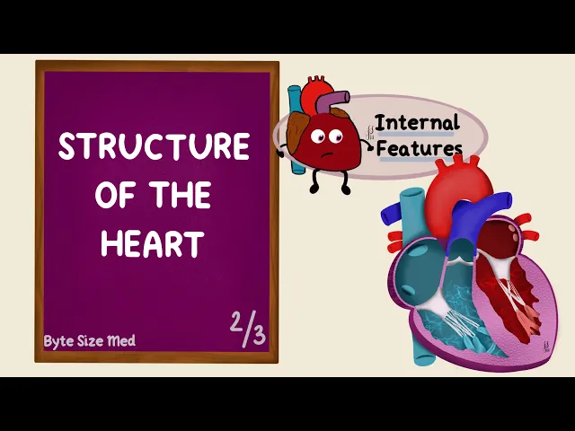
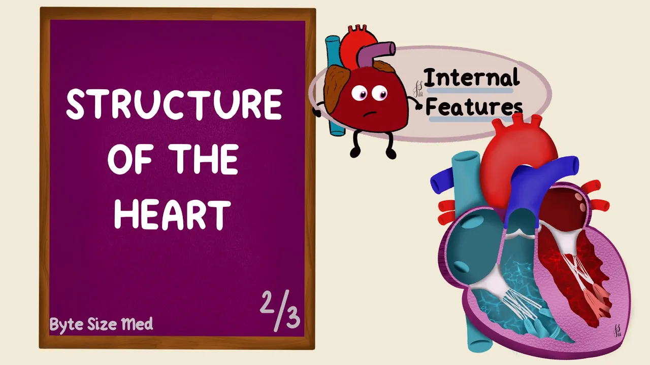
Hello. Welcome to Byte Size Med. This video is on the internal structure of the human heart.
First let's look at the path that blood takes through the heart. The oxygenated blood, which is used blood, from the whole body, reaches the right heart through the superior and inferior vena cava. They open into the right atrium.
That blood then moves into the right ventricle. The right ventricle pumps it out through the pulmonary trunk, which divides into the right and left pulmonary arteries, going to the right and left lungs. The lungs do their job of oxygenating that blood.
Oxygenated blood returns to the left heart. The four pulmonary veins bring that blood back to the left atrium. Blood then moves into the left ventricle.
The left ventricle pumps it through the aorta, which then leads into the arterial system, distributing it to the whole body. Once the tissues use what they want, the deoxygenated blood comes back to the right heart through veins, which ultimately lead into the superior and inferior vena cava. So right now we're in the right atrium, and in this video, we're gonna see what's inside all four chambers.
First up the right atrium. Now almost all the blood from the body drains into the right atrium through these two big vessels, the superior and inferior vena cava. The superior vena cava enters in the upper portion and the inferior vena cava in the lower portion, but both enter on the posterior wall.
Now that space is called the Sinus Venarum, and it's smooth, versus the anterior space which is called The Atrium Proper. Now this is rough. The anterior wall has got ridges and these are called the Musculi Pectinati.
Now that's why it's rough. The difference between these two is because of their embryological origins. The Sinus Venarum is actually derived from the Right Horn of the Sinus Venous, while the Atrium Proper is from the Primitive Atrium.
I'll be talking about this in my embryology video on the heart. The rough and the smooth portions are delineated by a muscular ridge, and this is called the Crista Terminalis. On the outside, that creates a groove.
That's called the Sulcus Terminalis. So Crista Terminalis on the inside, Sulcus Terminalis on the outside. The musculi pectinati are also seen in the right auricle.
That's a little ear-shaped extension, which overlaps the ascending aorta. That's why it's called the auricle, because it's ear shaped. Now I said almost all the blood comes in through the superior and inferior vena cava, and that's because there are other conduits as well, like the coronary sinus.
Now this brings back venous blood from the heart itself, because the tissues of the heart also need blood. That opens right next to the inferior vena caval opening. These two openings are actually guarded by valves.
The valve of the inferior vena cava is called the Eustachian valve, and that of the coronary sinus is called at the Thebasian valve. The two atria are separated by a septum, and that's called the interatrial septum. During foetal life, there is actually a space in the septum.
That's called the foramen ovale. It's an oval foramen, which helps blood move from the right atrium to the left. That's a shunt, and that's normal during foetal life.
But it closes at birth. So in adults what remains is an oval depression, called the Fossa Ovalis or the oval fossa, with its raised margin, and that's called the Limbus of the Fossa Ovalis, also called the Annulus Ovalis. In an atlas or a textbook, you'd see the right atrium usually from an angle like this.
So the heart is rotated and this is the right atrium, the right ventricle and this projection up here is the right auricle. And if we open up the right atrium, here we have the musculi pectinati. We've got the superior vena caval opening, the inferior vena caval opening, guarded by the eustachian valve; the coronary sinus opening guarded by the thebasian valve; the oval fossa, that's the Fossa Ovalis, with the Limbus of the Fossa Ovalis.
The right atrium leads into the right ventricle. That's through the Atrioventricular valve. Blood from the right atrium flows into the right ventricle.
That's through an Atrioventricular opening, guarded by a valve called a tricuspid valve. The tricuspid valve has three cusps or leaflets, hence the name. If we look at the valve from above, we can see the three leaflets.
There's the anterior, posterior and septal leaflets. The right ventricle has muscular ridges and bridges called trabeculae carneae. They form the rough inflow walls of the right ventricle.
There are papillary muscles which are like little pillars attached to the wall of the ventricle at one end, and the other end is attached to the tendinous cords. Now these tendinous cords are called the chordae tendineae. The other end of these cords is attached to the free margins of the leaflets of the valve.
The right ventricle has three papillary muscles. The anterior, which is the largest, the posterior, and the septal. So what do they do?
When the right ventricle fills up, the tricuspid valve is obviously open. But once it's done filling, that valve will close. When the ventricle contracts, the pressure forces the valve upward.
Now without the papillary muscles and the Chordae Tendineae, the pressure would push blood back into the right atrium and we don't want that. So when the ventricle contracts, the papillary muscles contract as well, tensing the Chordae Tendineae, keeping the valve shut. So blood can go the right way which is through the infundibulum This is the outflow tract of the right ventricle, and it leads into the pulmonary trunk.
Again this is guarded by a valve, the pulmonary valve. The pulmonary valve is a semilunar valve, and it has three semilunar cusps: right, left and anterior. Now unlike the Atrioventricular valve, the semilunar valves are not attached to chordae tendinae or papillary muscles.
So how do they stay closed? After ventricular contraction, the pressure change results in blood recoiling and filling up the pockets that the cusps form with the wall. These are the pulmonary sinuses.
This keeps the valve shut and blood doesn't flow back into the right ventricle. Instead it flows forward into the pulmonary trunk. The pulmonary trunk then divides into the right and left pulmonary arteries to go to the lungs, where oxygenation happens.
The oxygenated blood is brought back to the left atrium by the pulmonary veins. Four pulmonary veins open into the left atrium, and just like the right atrium, the left atrium also has a smooth posterior wall and a rough anterior wall. The anterior portion is derived from the Primitive Atrium, just like in the right atrium.
But the posterior wall is formed by the absorption of pulmonary veins. So in both the atria, the posterior walls are formed from veins. The pulmonary veins which bring back oxygenated blood from the lungs, open on the posterior wall of the left atrium, while the anterior wall has the musculi pectinati.
Blood from the left atrium enters the left ventricle through the bicuspid valve. Tricuspid on the right side of the heart, bicuspid is on the left side. It's also called the mitral valve.
Bicuspid, so it has two cusps, anterior and posterior, versus the tricuspid, which also has a septal leaflet. But just like the tricuspid valve, the free ends of these valve leaflets are attached to the chordae tendineae, which in turn are attached to papillary muscles. So they work similar to the tricuspid valves.
During ventricular contraction, the papillary muscles contract as well tensing the chordae tendineae keeping the valve shut. The closure of these two atrioventricular valves sends vibrations through the chambers. That creates an audible sound, called the first heart sound.
So S1 is because of closure of the Atrioventricular valves. The left and the right ventricles are separated by an interventricular septum and this has two parts. There's an upper membranous part and a lower muscular part.
The left ventricle like the right ventricle has trabeculae carnae, chordae tendineae and papillary muscles. But here there are two large papillary muscles, anterior and posterior. The wall of the left ventricle has more muscle than the right which makes sense functionally, because the left ventricle has to pump blood to reach all parts of the body against systemic vascular resistance, while the right ventricle just has to pump blood to the lungs against the lower resistance through pulmonary circulation.
But unlike the right ventricle, the left ventricle does not have a moderator band. The moderator band is a specialized trabeculum in the right ventricle. It's also called the septomarginal trabecula, because it's a bridge between the interventricular septum and the base of the anterior papillary muscle.
Now this is important in cardiac conduction. There are four important components to the cardiac conduction system, which are responsible for the heart contracting. The first is the pacemaker of the heart.
That's called the Sinoatrial Node. This is located in the right atrium, on that Crista terminalis, at the junction between the superior vena cava and the right atrium. The second structure is the Atrioventricular node.
This is located near the opening of the coronary sinus. The Atrioventricular Node continues towards the septum as a bundle that divides into two. That's the Atrioventricular Bundle, and at the junction between the membranous and the muscular parts of the interventricular septum, it divides into right and left bundle branches.
These branches then continue to form the last component. That's the Subendocardial plexus of conducting cells, called the Purkinje Fibres. This is schematic, but remember that the right ventricle has that moderator band, which contains a portion of the right bundle branch, while the left ventricle does not have this band.
The Purkinje Fibres spread through the muscles of the ventricles, and together this whole system initiates and coordinates cardiac contraction. Blood from the left ventricle gets pumped into the aorta through another semilunar valve called the aortic valve. Again it's got three semilunar cusps: right, left and posterior, similar to the pulmonary valve, except there is anterior, here its posterior.
They also form pocket-like sinuses. So the recoil of blood filling the sinuses keeps the valve shut. The closure of these two semilunar valves again creates a sound.
That's the second heart sound, S2. The blood can then move forward through systemic circulation, supplying the whole body along with the heart itself. And that is from the right and left coronary arteries.
From the right and left aortic sinuses, two vessels arise. The right and left coronary arteries. When blood fills the pockets after the ventricle contracts, it gets forced into the coronary arteries, forming coronary circulation.
And I'll go over that in Part 3, when we look at the blood supply of the heart. And that's what's inside the heart. I hope this video helped you.
If it did, you can give it a like and subscribe to my channel for more. Thanks for watching and I'll see you in the next one!