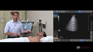
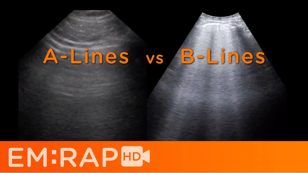
hello this is Jacob Aila and what we're going to talk about today is how you can use ultrasound to identify pulmonary edema so for this exam you can use any of the three main ultrasound transducers but my favorite probe for this is going to be the curv linear probe if you don't have the Cur linear probe the phase array will work just fine let's talk about what we're going to be looking for when asking yourself the question does this patient have pulmonary edema there are specific patterns of artifacts you need to look for and they're
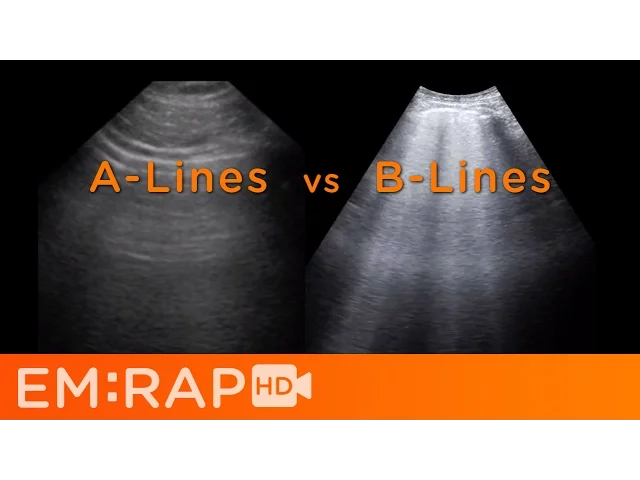
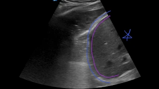
called a lines and B lines there are theories as to exactly how these sonographic artifacts are formed but I don't believe you need to know how they're formed to be able to identify them and to use them clinically so let's look at this super high-tech drawing here so this is a rib and a rib and this is the plural line right here these things down here these are called aines they are horizontal reverberation artifacts that are generated when there is just air beneath the plura a equals air an aine means not pulmonary edema in contrast
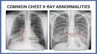
these vertical artifacts here are beines they mean that there is extra fluid in the lungs now for it to be considered a beline it's got to be vertical it's got to extend from the plural line all the way down to the bottom of the ultrasound image and it has to move with respirations sonographic beines have the same meaning as the curly beines that you see on chess X-Ray when you have pulmonary edema here's an example of an area of the lung without pulmonary edema so imagine a a rib rib Shadow rib rib Shadow this is
the pl line right here and this guy right here this horizontal artifact is called an a line a stands for air it means not pulmonary edema so these are beines so this is the plural line right here and you can see these vertical artifacts starting from the plural line extending all the way down this implies that the patient has pulmonary edema need those side by side here are two other examples the clip on the left side of the screen doesn't have pulmonary edema the one on the right has pulmonary edema here you have horizontal artifacts
a lines and here you have vertical artifacts these are called bines the presence or absence of be lines in of themselves do not necessarily mean anything in order to be able to D someone with pulmonary edema you need to see a certain pattern of beines in a specific distribution firstly you need to divide each hemithorax into four zones now the original study that described the zones supplied this picture that you see here now in the sead coddle orientation or up and down you use the posterior and anterior axillary lines then in the transverse plane you're
supposed to draw an imaginary line dividing each hemithorax into four equal zones the only thing I want to mention is that the picture that most people use which is this one is a bit confusing to me because in the text of the article they state that the transverse line should be placed in the third intercostal space which is a bit higher than how it sits in the picture it's kind of semantics I just divide the anterior and lateral chest into four equal zones now the posterior region while it's extremely important when you're looking for types
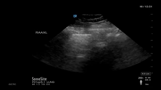
of pulmonary pathology other than pulmonary edema isn't really that important when you're looking for pulmonary edema itself after you divided the chest of the patient into these zones you should place a probe on the chest and look for either a lines or B lines if you see B lines in two zones per hemithorax then you have pulmonary edema now a single beline essentially has no real clinical significance two beines still essentially have no real clinical significance it isn't until you have three or more beines in a zone is that considered to be pathologic personally I
start looking in the anterior lung Fields bilaterally because that area is the most specific for pulmonary edema so to recap to have a positive zone for pulmonary edema you have to have three or more beines so three or more Bel lines per Zone equals a positive zone for the patient to be considered to have pulmonary edema so it's diffuse pulmonary edema you have to have two positive zones per side so three or more beines in two or more zones per side gives you a patient with pulmonary edema now the thing that I have to mention
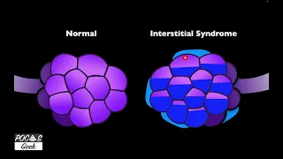
here is that the presence of a positive Zone doesn't necessarily mean that the patient has cardiogenic pulmonary edema beines technically are caused by any kind of increase in density in the lungs increase in density definitely happens with cogenic pulmonary edema the lungs get soggy but you can also have that with interstitial lung disease ARS pneumonia ectasis pulmonary contusions all these things can cause beines so you have to make sure to look at the patient in front of you and don't use that ultrasound in isolation