arando huran biology and Medicine videos please make sure to subscribe join the foran group for the latest videos please visit Facebook atand again please like and here you can also ask questions answer questions and please post some interesting things including artworks and you can also change the quality settings to the highest one for better graphics and in this video we're going to talk about skeletal muscle contraction and so with this we begin with a neuron a nerve cell and this neuron is specifically what's called a somatic neuron and here we have a muscle fiber from
a muscle and so a skeletal muscle will contract only when stimulated by a sematic motor neuron and so if we have a signal here that comes towards a neuron this will this can cause the muscle fiber to contract now a somatic motor neuron can inate between 3 to up to 1 th000 muscle fibers and so when a neuron the neuron's nerve fibers connects to a muscle fiber this is this complex is what we call a motor unit so it's where we have a nerve fiber and a muscle fiber so if you remember what a muscle
fiber is if we have a bone here with a muscle attaching on it we have the tendon which attaches to the to the Bone and here we have the actual muscle now the muscle contains many structures within it uh portions of the muscle known as as fases and these facles then contain many muscle fibers and so here is where our muscle fiber is so now let's zoom into one motor unit here and see what kind of structures are around in the muscle fiber and also the end of this neuron that assists that assists in muscle
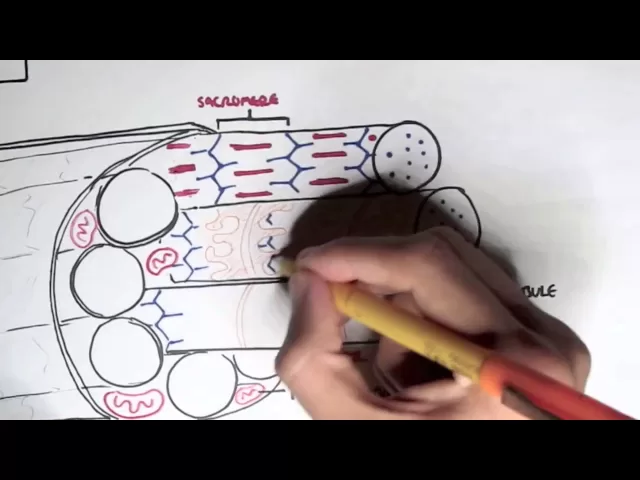
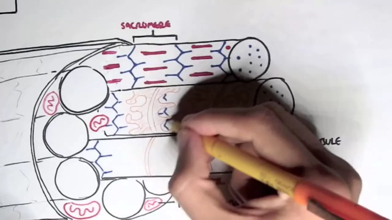
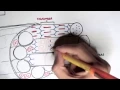
contraction and so here we have the N bulb the Aon terminal of the motor neuron and the Aon terminal of the motor neuron the endbulb has many vesicles here and these vesicles contain neurotransmitters known as ayic Coline so now we'll just stop there for now and look at the muscle fibers and so here we have the muscle fiber one muscle fiber and the muscle fiber contains many myof fibral so here we have the myof fibral and around this area within the muscle fiber we have also mitochondria which can provide ATP and we also have many
nucleus around the muscle fiber because the muscle fiber is multinucleated the muscle fiber has a membrane called the sacma which contains many receptors and ion channels now let's stop there for a bit and look specifically at the myof fibral so if you remember the myofibrils contain Thin and Thick filaments here in blue is the thin filaments and in red the thick filaments from one Z line to another Z line of the thin filaments is what we call a sarcomere and so we have this within each myofibril however in each myofibril you can say we also
have uh what's called the T tubal which runs around the myofibril and the T tubul surrounding it we have What's called the sacroplasmic reticulum abbreviated Sr and these sacroplasmic reticulum contain calcium ions let's have a closer look at the T tub t tubu because a t Tule has an important role in skeletal muscle contraction the T tubal essentially connects with the outer membrane and wraps around the myof fial and the tach will contain special ion channels for calcium here shown in Orange surrounding the CH tubil we have the sacroplasmic reticulum which contains calcium ions so
surrounding it here we have the sacroplasmic reticulum specifically uh what's called the terminal sister uh which is part of the cyop psychop plasm reticulum and the terminal cister contains these calcium ions but these calcium ions they cannot go into the T tubu because the ion channels the calcium ion channels are blocked and therefore they need to be opened for calcium to come out and here of course um beneath this T tubal or or within the myof fibro we have the thin filaments and thick filaments and the thin filaments requires calcium in order to initiate muscle
contraction but remember the calciums are still within the sacroplasmic reticulum so how do these calcium ions come out well let's have a look let's begin at the neuron the neurons nbob the axon terminal where an action potential has arrived this action potential will cause these vesicles containing ailine to release ayic Coline out these ayic Coline will then bind on to The receptors in the sacma let's have a closer look at this interaction between a silicol and The receptors in the sacma so here we have the N bulb the Aon terminal of the motor neuron and
here we have a vesicle containing ayic Coline here is our receptor for ayic Coline on the sacma and a point to make which is pretty important is out outside in the extracell fluid we have high concentrations of sodium and within the muscle the the within the Saca inside the cell we have high concentrations of potassium ions so what happens is when the vesicle containing ayic Coline releases acyline out these acolin will then bind onto this receptor which will cause the receptor to open when this receptor opens sodium ions can then come inside and when sodium
comes inside this will open the voltage gated channels which will then create an action potential these Action potentials will then travel across the sacma where the action potential can then propagate down the t t tubal when the action potential travels down the T tubal it will cause these um calcium ion channels which were previously closed to open up and it opens up because there's a change in voltage in the T tubal which will cause the calcium channels to open and so when these calcium channels open calcium uh will then move out from the terminal syst
into the T tubule we will travel down and essentially go and bind onto the thin filaments and so essentially calcium and also ATP will help um with muscle contraction and to understand how they help in muscle contraction please click on the link for the sliding filament theory um which will look at the contraction at a molecular level at a um sarom level please like comment and subscribe and share