hi my name is Louise Murray I'm one of the clinical oncology consultants in Leeds and my job today is to give you a brief introduction to radiotherapy the end of the session is to provide a fund of an overview of the fundamental principles behind radiotherapy and it's delivery and I hope that by the end of the session you can firstly explain the basic mechanism of radiotherapy and it's indications for use describe the basic radiotherapy process and lastly understand some of the common expected side effects from radiotherapy so how are we going to do that well
brief introduction that's what we've just done then we're going to do a brief quiz then I'll move on to talk about the basic science of radiotherapy the radiotherapy process and lastly some side effects so first of all you leave there got a copy of the quiz or I'll read the questions out now so it's basically true or false this is just to assess what you know before I give you the rest of the talk I'm hopeful that by the end of it you'll get more of these right so first of all is it true or
false 40% of all patients cured of cancer will have received radiotherapy true or false in the UK in 2011 approximately 1 million doses of radiotherapy were given is that true or is it false radiotherapy is given as an outpatient true or false radiotherapy can be given as an emergency treatment is that true or false radiotherapy is available at most hospitals is that true or false radiotherapy is delivered accurately to within 2 millimetres of the target is that true or is it false radiotherapy is visible true or false radiotherapy causes burns is that true or false
radiotherapy can cause cancer is that true or is it false and lastly radiotherapy and chemotherapy can be given at the same time is that true or is it false don't worry we're going to answer these questions as well as others as we go through the talk so what is radiotherapy so radiotherapy is the treatment of disease mainly cancer using ionizing radiation now if you say to a patient I'm going to give you radiotherapy and they say to you what's that doctor and you see it's ionizing radiation you'll start to look pale and quite unsettled what
I tell patients and what you should tell patients is radiotherapy is high-energy x-ray treatment all of our patients have had scans and x-rays before and so that concept they can understand so radiotherapy is high-energy x-ray treatments and the whole crux of radiotherapy if you remember nothing else is the aim is to get the optimal dose of radiotherapy to the tumor and the minimum amount of radiotherapy to the surrounding normal tissues because it's when we treat the normal tissues that we run into problems with side effects so as with most things on college in oncology radiotherapy
can be given with four different intents and you may have come across some of these before sometimes we can do curative radiotherapy otherwise known as radical radiotherapy which as the name suggests is when we're trying to cure the patient get rid of the cancer for the patient and we've got common examples of that often when we're giving radiotherapy we give with curative intent we give chemotherapy around about the same time so common examples are head and neck cancer and uncommon ik another common example is cervical cancer we're giving the radiotherapy plus or minus chemotherapy with
the aim of cure in fact radiotherapy is a very effective treatment for cancer 40% of patients who are cured of their cancer will have had radiotherapy at least as a component of their treatment so that's radical or curative radiotherapy adjuvant radiotherapy adjuvant radiotherapy follows a definitive procedure so for example patients might have surgery and there may give radiotherapy after that a good example is a woman who has breast cancer and she has the lump removed or a wide local excision and then we give radiotherapy to the rest of the breast just in case there are
microscopic cells left behind and we know that by adding radiotherapy Orange having had surgery for breast cancer effectively we achieved the same result as having a mastectomy and the lady gets to keep her breast as well so that's a different radiotherapy following a definitive procedure and then we do lots and lots of palliative radiotherapy as well this is in patients in whom we've accepted we cannot cure the cancer and in whom they have symptoms related to the disease so a common example might be a patient with prostate cancer that's to the bones or breast cancer
that spread to the bones and the patient has symptoms often pain related to the spread so supposing somebody had a bone metastasis in their leg from prostate cancer and it was causing them pain we should give them some radiotherapy and we've got a better seventy eight out of ten chance of improving their pain after about one to week one to two weeks and that benefit can last for several months so that's palliative radiotherapy given with the aim of improving the patient's symptoms and then sometimes we do neoadjuvant radiotherapy that's prior to definitive procedure and quite
a good example would be locally advanced rectal cancer the patient has a rectal cancer it's spread a little bit locally if the surgeon went in and did an operation there'd be a very high chance of leaving sales at the margin of where he cuts we know that if we give five weeks of radiotherapy along with some chemotherapy beforehand we can shrink the cancer down a bit and when the surgeon goes in a few weeks later we increase the chances of him getting in and doing an operation and not leaving any cells behind which we know
increases our chance of controlling that cancer so for different indications for radiotherapy radiotherapy is not available in all hospitals and it's given in what we call tertiary hospitals so for those of you who are based in leads for your oncology placement you will be in a hospital that has a radiotherapy department radiotherapy is an excellent treatment one of the things that makes it very good is we can give it as an outpatient patients don't need to stay in hospital the treatment only takes a few minutes and so there's no value in patients being in hospital
for 24 hours a day several days a week for such a short treatment so most treatment is governor's and outpatients and we generally do radiotherapy Monday to Friday with the weekends off and when patients have radiotherapy they don't see it they don't feel it they don't hear it and it only takes a few minutes to deliver there are different types of radiotherapy and there are two main types of radiotherapy that are used in the UK the main one that we're going to think about is photon radiotherapy that's the bulk of the radiotherapy that we deliver
and four times as I've touched on our high-energy x-rays and what they do is they penetrate deep into the body's tissue but at the same time spear the overlying skin and what happens is the four tines produce secondary electrons and it's all secondary electrons that damage DNA single strand breaks and double strand breaks and that's what can kill cancer cells the important thing about radiotherapy is it can kill cancer cells and it can kill normal cells the reason that radiotherapy works is that normal cells up to a point can repair radiotherapy and juice damaged damaged
tumor cells often have defective DNA repair mechanisms and that means that they can't repair radiation and just damaged so when they come to divide they realize that they're damaged and those cells can die I'll touch on that again in a second another type of radiotherapy that we sometimes use our electrons and those are electrons directly and they damage DNA directly and and we can use electrons to deliver treatment to relatively superficial lesions so maybe skin metastasis is near the surface we don't penetrate very deeply with electrons and so it's only for superficial lesion skin metastasis
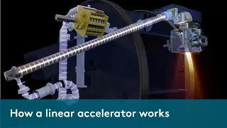
is a good example both photons and electrons are delivered by a machine called a linear accelerator and I'll show you an example of that in a second and protons you may have heard of those in the news protons are a relatively new treatment to the UK we've been around for several decades protons are a different form of radiotherapy their particle radiotherapy and we generally use them in very specific circumstances and generally we use protons for young people who've got brain tumors children have so got a growing brain and we want to deliver a treatment that
delivers a very low dose to the normal brain and that's where protons have a potential rule if you think about a proton compared to photo and if you think about Angry Birds on your iPhone the red angry bird is like a photon it comes along hits the pig and keeps going that's like a photon we get this exit of the Angry Birds or the exit of the photon protons are more like the black Angry Bird and they travels into the tissue you press the Angry Bird and it drops down and delivers all its doors in
one place and it doesn't travel on that's a bit like a proton there's no exit doors and so if you're a three-year-old or a five-year-old who's still got a growing and developing brain we want to deliver little radiotherapy as possible to the normal tissue beyond the tumor and so that's where we might use protons but they are new to the UK and they're not going to be a big focus of any exams that you're going to get I only mention them because you may have heard of them already as I've touched on how does radiotherapy
work it's what we get what we call a differential sale kill so normal cells have got effective DNA repair mechanisms and so up to a point they can repair the damage that radiotherapy causes cancer cells normally have defective DNA repair mechanisms and so when they get damaged by radiotherapy when the next come to divide they realize that they're damaged and they can undergo cell death sometimes that's a pop ptosis sometimes it's what we call the mitotic catastrophe and mitotic cell death but regardless we get this differential cell kill normal cells can repair themselves up to
a point tumor cells generally can't repair radiotherapy and just damage however if you give enough radiotherapy to normal cell eventually you will damage it to the point that it can't repair itself and that's when we get into trouble with late side effects which we'll come to but also that's when we can run into trouble with second cancers radiotherapy as well as being used to treat cancers can cause second cancers the risk is low but we know that it's real and that's why there are very strict radiation protection laws governing where radiotherapy can be delivered who
can deliver radiotherapy and how much exposure different patient groups and professional groups can have to radiotherapy when we talk about radiotherapy doses we talk about the doors in gray so radiotherapy doors is great GRE Y and radiotherapy is often given in a series of small doses and we call each individual radiotherapy treatment a fraction so we might give our doors of C fifth degree in twenty five fractions that's twenty five usually daily treatments because we deliver treatment and lots of lots of fractions we deliver a lot of fractions per patient in fact we deliver about
two million fractions of radiotherapy per year in the UK so each fraction form are inaccurate tumor often not exclusively but often each fraction is around about to agree and to grieve just about enough radiotherapy to kill some cancer cells but is less likely to cause long-term normal tissue damage and that's why when we're doing radical treatments it's often a lot of small approximately two great refraction treatments so for example for some head and neck cancers we might give a dose of 70 gray and 35 fractions so that's two gray per fraction and remember we treat
Monday to Friday once a day weekends off and so 7 degree and 35 fractions takes about seven weeks to deliver palliative radiotherapy for patients who've got incurable cancer we're just wanting to control symptoms we want patients to have relatively straightforward treatments with minimal hospital visits we tend to do larger doses per fraction and part of the reason for that is we're less worried about late side-effects in that patient group and so for palliative treatments we might give eighth grade in a single fraction or we might give 20 degree and five fractions or thirty great and
ten fractions so a bigger dose per fraction short course of treatment in fact for every single fractions patients can come to the department have their treatment designed have their treatment all in one visit and it's done so we have a whole radiotherapy process that I'm going to talk through this is just a kind of overview of the process we've got the pretreatment phases the treatment planning phases and then the treatment itself and we'll focus on various aspects of that pathway the things we're going to focus on most of all are immediately and are the consent
process immobilization CT simulation tattoo volume definition treatment itself and then follow-up during treatment so let's go to the beginning of that process so the consent form all patients will sign a consent form for radiotherapy a lot of what we do is about communication tell the patient why we're going to do it tell them what the benefits are what the intended benefit is tell them what the alternatives are and tell them what the side effects of treatment are we've got standard forms that we use that we individualized the treatment and the patient that we're dealing with
so always consent your patients tell them what you're going to do and why you're going to do it the next step in the process is immobilization if somebody has a tumor let's see on they're cheek I want to make sure that every time I turn the radiotherapy on maybe for 35 treatments I want to be confident I'm definitely going to treat the cheek if I go too high I will miss the tumor and I'll put some radiotherapy through the eye that could meet the person blind that could give them a cataract equally every time I
turn on I want to hit the cheek I don't accidentally want to I don't accidentally want to miss the tumor I go to law and then I'd cause a soror reaction in the mouth so we need to hit the right spot every time that's the crux of radiotherapy and if patients have a brain tumor or a head and neck tumor one of the ways to help us ensure that we always treat the right spot is to keep patients still and so immobilization is the area of radiotherapy where we make sure the patient is kept in
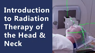
the same position so it's reproducible between designing the treatment and delivering the treatments so head and neck cancer patients and brain tumor patients get a mask made like you can see here this is a perspex mask essentially the patient comes to the mold room we use like plaster of Paris to create a mold of their face we cover that in perspex and get an individualized patient shell and it is designed to hold you still it's not tight tight tight but it is snug because that's the point of the treatment and you can see somebody wearing
an example there that's the patient wearing the mask as I see it snug but it's not tight tight tight and then the other sort of masks the other sort of masks that we use or what we call thermoplastic shells these are like quite sheets of plastic and they feel rigid we put them in a hot water then they go all bendy you can shape that over the patient's face and then that six heart and again we've got an individualized mold of that patient's head or their head and neck area so we've got a couple of
examples here adult patients we just tend to use the plain white patients our younger patients which we a lot of young patients with brain tumors sadly and we individualize the mask to make it even more bearable so we turn that young patient into a superhero so I believe that's Darth whoa we've got other sorts of immobilization depending on which parts of the body we're treating so this is for a patient we're treating their torso and we've got them up with their arms above the head and that blue mold just gives keeps things in the right
place this is called a vacuum bag patient lies and what's a bit like a blue bean bag we suck the ear out and effectively that sets hard to giving us a mold of the particular part of the body that we're interested in so the first one that we can see we're concentrating on the upper part of the body and down below you've got a patient in a whole body mold and that we're trying to keep still over their whole body this is a CT simulator so this is just like a normal CT scan with a
few important differences and first of all the bed is hard and flat everything's to do with keeping the position patient in the correct position and the same position for treatment to treatment and so this is a hard bed so they can't sink down and change the position and also there are marks along the side of the bed so that we can always line the patient up with reference to these marks otherwise it's very like a normal CT scanner so the patient has a mobilization as necessary and they have a CT scan on which we design
the radiotherapy it's different to the diagnostic scans they have this is a specific scan to design the patient's radiotherapy well they come for their CT scan they often have a tattoo in fact they often get three tattoos one in the middle or one on either side and that's to allow us to line the patient up on the couch in reference to these CT scans we often use the laser lights you can just see the corner of a laser light on that slide and we use these laser lights to line up to their tattoos and then
we know we've got the right position on the outside we can then do scans to check the position on the inside so we give patients tattoos they're absolutely tiny you can see a picture of a dime with a tattoo next to it so they are there forever can sometimes remove them but they're there for a long time but they're very very small so the first step immobilization CT scan and tattoos the next thing that involves the clinical oncologist or the doctor is designing where we want the treatment to go and there are three key volumes
when it comes to designing where we want the radiotherapy to go first of all we draw tumor that we can see that's called the gross tumor volume or the GTV so that's tumor that we can see who would obviously want to treat that however we all know that on a scan tumors don't stop right at the edge of where we can see them we know that there's liable to be microscopic spread and so in most cases we don't just stop at the edge of the GTV we've put on a margin to take account of that
microscopic spread and that margin we call the clinical target volume and then we also know that tumors can move about a little bit and they're never in the same position from day to day even though we've got good immobilization and so we need a bit of a margin to take account of potential variation set up from day to day and in tumor motion and we call that margin the planning target volume and just about all tumors and in fact all tumors need some sort of planning target volume because things can vary from D to D
let's think of the prostate for example if somebody has a full bladder the prostate might be a little bit lower down and so we need a margin to take account of that if somebody's passing gas through the rectum the prostate might be pushed forward and if we don't have a margin to take account of that then we could miss the prostate for a part of that patients treatment so we have to have this planning target volume as well so the gross trimmer volume trimmer that we can see the clinical target volume margin for microscopic spread
and the planning target volume our margin to take account of day-to-day variability and potential tumor motion we can see an example of this here this is a patient who's got a lung tumor on the left side you can see we've drawn around the tumor that we can see in red persona margin added in green to take account of microscopic spread and then there's another margin in blue to take account of day-to-day variation also patients breathe and so you get more emotion and in a super elf direction than you do site to site and so you
can see on the coronal view that we have a larger margin sipping them for our planning target volume than we do side to site we draw the tumor that we want the tissue that we want to treat that's what we've just discussed I also spend a lot of time trying areas that I don't want too much radiotherapy to go to so that's your normal tissue so for example in this patient with the lung cancer we would draw the spinal cord we can't give it too much radiotherapy or will cause paralysis and we draw the heart
we don't want it to get too much radiotherapy or we could cause cardiovascular damage and we don't want the normal lung to get too much radiotherapy or we could cause fibrosis and therefore lots of breathlessness so we draw for everyone the radiotherapy to go and we draw where we don't want too much radio therapy to go after we've done that our design on the patient planning CT scan passes to physics and they do lots of sums and calculations to work out how to get the optimal amount of radiotherapy to the tumor but at the same
time minimize the doors to all those normal tissues that we've drawn on the scan now in the case of a palliative radiotherapy treatment we just want something relatively simple with low toxicity that's easy for the patient to manage and so this is an example of a patient who has a large pale that born metastasis I've drawn round out in red that's where we want to treat there's a bit of a gap to take account of the fact that we have set up variability in a clinical target volume and this is a very simple beam arrangement
we're putting one beam in from the front and one beam in from the back and essentially that meets in the middle which is where the tumor is so that's a simple palliative treatment beam from the front and a beam from the back nice and easy here's another example this is a patient who's had a lumpectomy rar wide local excision for breast cancer and now we want to deliver adjuvant radiotherapy to the remaining breast and what we've got our two beams going across the breast one is coming in from the left on one is coming in
from the right we call this a glancing pair they glance across the chest wall therefore encompassing all of the breast tissue but minimizing the doors to the lung underneath and to the underlying heart we use this arrangement because if we just did one beam that came in from the front we would cover the breast but we get lots of exit doors through the normal lung and through the heart with this way you can see there's a small sliver of lung that will get radiotherapy but it's only a small sliver and a very small proportion of
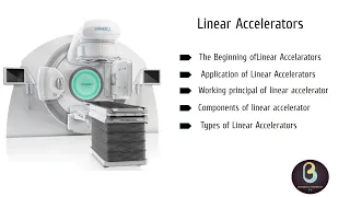
the heart might get some radiotherapy nowadays we can treat these patients in breath-hold and that means that we fill their lungs before we turn the radiotherapy on and that actually moves the heart back or effectively fills the lungs with air to minimize the amount of radiotherapy received by the heart because we know that long term that can put patients at risk of cardiovascular disease and we obviously want to minimize that and here's the machine that does a business this is our radiotherapy linear accelerator you can see it's got a black couch the same as the
one on the planning CT and again it's got those marks on the side so we can line the patient up in reference to the tattoos that they've got using the laser lights so that we get the same position on the as we had when we did the planning CT scan the radiotherapy comes out the round head of the linear accelerator and that gray circle is essentially a gantry so that head can move round in a circle and so radiotherapy can be delivered from 360 degrees and so we typically use multiple beams to angle around the
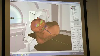
patient so that they all Center and on the chip but again minimizing the doors to the individual normal tissues so as an example here's a bit of a mock-up the patients on the bed the radiotherapy linear accelerator moves around the patient radiotherapy is delivered from multiple different angles there by focusing in when the tumor and a roughly central point so that maximum dose goes to the tumor and the doors elsewhere is spread out or minimized also to try and protect the normal tissues and make sure the radiotherapy is going exactly where we want it we
shape each radiotherapy beam and that's achieved by virtue of lay leaves of tungsten shown on the right hand picture which are in the head of the machine so that picture on the right is in the head of the feet machine and effectively these are like shutters are effectively like shutters and what they do is they can move in and out and create an aperture so for example if we were treating a perfectly circular aperture we would position those leaves of tungsten so that they were in a circle if we've got a square tumor we would
position them so the aperture was a square etc we can create any shape and those are automatically moved to match to the shape of the target that we're trying to treat here's a mock-up of radiotherapy treatment delivery and patients lying on the bed this is a patient who's got two lung tumors being treated at the same time on this occasion they're really the Linear Accelerator is on all the time and the machine is moving around a patient in an arc radiotherapy has been delivered all the time and the beam is constantly being shaped by those
leaves us tungsten that sit in the head of the machine you can see they're moving down the bottom left corner so all the time that beam which is mocked up in yellow it's constantly changing shape to ensure that we get the optimal dose to the tumor and the minimum doze elsewhere from each angle that we're delivering the radiotherapy as I said patients don't see it don't he don't feel it and it only takes about two to two five minutes to deliver a treatment so that's how we do it what about toxicity well it depends on
what part of the body we're treating and acute toxicities are caused by transient damage to the normal tip sales that will repair usually takes a couple of weeks for the acute side effects to start and then this settle down a couple of weeks or begin to settle down a couple of weeks after the treatments finished those are the local side effects fatigue is quite common and a skin reaction can be common as well depending on which bit of the body we're treating when it comes to read your therapy side-effects the way that I think about
it is think in the short-term the tissue gets inflamed you get an itis and the longer term the tissue can become Phi bruised and then you get fibrosis so think osis the short term itis long term OSIS and we'll talk through these as we go through it think about which bit of the body you're treating think about what normal tissues the radiotherapy passes through onyx we into the tumor and think about what normal tissues the radiotherapy passes through beyond the tumor and then think short term itis long term ulcers so let's think about a head
and neck cancer patient let's say somebody who's getting their tonsil treated in the short term they may well get a skin reaction the skin becomes red and inflamed that's radiation dermatitis and that causes pain we don't call it a burn it's a radiotherapy skin reaction skin reactions can be very sore in radiotherapy just as the skin on the outside can get red in and flew in from radiotherapy so can the mucous membranes on the inflate and that gives us some mucositis when we give patients mucositis and the saliva glands don't work quite as normal we
don't produce as much watery saliva and you get left with this thick sticky useless oral secretions which are quite unpleasant for the patient unsurprisingly and if you've got six sticky oral secretions and your mouse or your teeth doesn't normal either patients can get very fatigued with head and neck radiotherapy and long-term we can cause permanent fibrosis of some of the saliva tissues and that means they don't work and patients are left with a long term dry mouth so let's have an example of that this is a patient who's maybe about ten days into a course
of Atika radiotherapy and the skin is ever so slightly red you almost have to appear to convince yourself there's some redness there so this is a very mild radiotherapy radiation dermatitis at the end of treatment maybe in the final week of seven weeks of treatment the skin can look like this we see it less actually with our modern art treatments but we do see skin reactions like this so this is right at the end of treatment you can see the skin has peeled off the surface it's denuded it looks really red and angry that will
continue for two weeks after the treatments finished and then it will gradually begin to heal and cosmetically we'll look essentially normal the skin might be a slightly darker pigment longer-term just slightly deeper red and if you felt that patients neck years down the line it would feel more firm and that's because of fibrosis of some of the normal tissues in the skin leaving the patient with less elasticity than they had before but as a general passing them Street you may well not notice any difference once that's had time to heal as I said just as
it goes red and angry on the outside it can also go red and angry on the inside so this is a patient with mucositis so all the linings on the inside of the mouth are red and sore and peeling and you can understand why it's therefore difficult for these patients to enjoy food in fact a lot of patients can lose weight during head in radiotherapy and to try and prevent that these patients see a dietician every week who could advise them on supplements and if we're very concerned that they're losing a lot of weight or
if we know that they're going to lose a lot of weight during treatments then we can pass an NG tube or we can put in a peg prophylactically to try and keep their weight up as we know that that's better for patients so how do we manage these side effects should they happen so patients having had an ex radiotherapy you need a very good mouth care so we get some new guide we get some gel clear which is like got clear and a clear gel that they can put on for some soothing and mouthwashes aspirin
gargles mucus got some anaesthetic and some lubricant in it to help with pain and to help patients swallow things over and normal feeling nebulizers are good for these thick secretions helps patients get them up and get them clear lots and lots of emollients for skin care and we've got special preparations ship the skin breakdown analgesia because this is very sore patients are often and often end up on opiates and the short-term and often towards the end of a radiotherapy course they can end up coming into hospital for a few days to have their nutrition licked
up by our dieticians but also in terms of optimizing their analgesia and sometimes patients into syringe driver to deliver optimal opiate analgesia let's think about prostate cancer so prostate at the other end of the body you've got the bladder up above and you've got the rectum behind and so if we treat the prostate it in itself might become inflamed during treatment in the short term so that you get a prostatitis and they can get inflammation of the bladder which is a cystitis and if you get so status patients might find they've got burning when they
pass water frequency and urinary urgency as well as I said the back passage is immediately behind the prostate and so the rectum can become inflamed and with that patients can get some diarrhea and they can pass some mucus or Shlaim from the back passage they can even pass some blood from the back passage just because the rectum has become very inflamed if the prostate becomes inflamed prostatitis then it comes hard to pass water and the urinary flow is reduced and if we're treating a lot of bowel patients may get some abdominal pain most of those
things will settle down longer-term we can cause damage to the small blood small blood vessels in the bladder and in the rectum and that can give later bleeding so patients can have in hematuria or they can have we call it proctitis but it's really a late side effect we can cause telangiectasia in the bladder causing hematuria which lands it easier and the back passage which we call prosper and proctitis which can cause bleeding in the future especially a lot of barrel and short term patients can get nausea as well so how do we manage these
side-effects well in the first instance of somebody's got urinary symptoms we make sure they don't have a urinary infection so we send a sample analgesia can help with pain if people are having urinary symptoms a good fluid intake keeps the urine nice and dilute if the floor becomes pure then time to loss and relaxes the muscles in the middle of the prostate to improve your only floor and if people are having a lot of diarrhea then fiber gel helps bulk out the stools and makes them more satisfying to pass last example let's think about lung
cancer so let's think about a tumor in the lung and close and involving some lymph nodes as well so we're treating the lung and the mediastinum in the short term the gullet or the esophagus will become red and inflamed that can cause esophagitis and therefore its source to swallow so Dana feed you and essentially that's mucositis in the esophagus and we can irritate the lungs which can cause coughing shortness of breath and and then patients can also get chest pain particularly with more peripheral tumours when we're treating those and catching some of the pleural tissue
and patients can get pain if we're treating a low down lung cancer and we catch some of the stomach or the bowel patients can feel a bit sick and they're pneumonitis inflammation of the lungs that can happen about six to eight weeks after radiotherapy patient becomes breathless has a dry cough and and we use steroids and oxygen to treat that that can be very serious if it involves a large amount of lung how do we manage these side effects well lung cancer patients are so breathless and are coughing we need to make sure they don't
have a chest infection so we'll get an x-ray in here there's a nebulizers are also important and remember a significant proportion of these patients will have smoking related damage as well so they're often already on up inhalers and nebulizers and so we need to optimize those analgesics for pain and miu clean is very good for esophagitis so that's some lubrication and some analgesia which helps patients swallow despite the fact they've got neko scientist for pneumonitis as I've said occasionally we need to use steroids to settle down that inflammation just to mention chemoradiotherapy we touched on
that before that's when chemo a chemotherapy and radiation therapy are given around about the same time often in the curative or neoadjuvant setting to try and increase our chances of cure it makes radiotherapy more effective we know that but it comes at the cost of increased toxicity and one of the things that we didn't used to see often with radiotherapy but can see much more with chemotherapy and must be aware of it's the risk of pneumonitis just to see because it's in the quiz and I haven't mentioned it yet when we turn the radiotherapy machine
on we want to be within two millimeters of our target to make sure that we're super accurate so now we're going to go back to those questions that we looked at at the beginning so 40% of patients cured of cancer will have received radiotherapy is that true or is it false so that's in fact true at radiotherapy is very important for the cure of cancers it's also very important as a palliative treatment and improving symptoms in patients who've got incurable cancers as well question 2 in the UK in 2011 approximately 1 million doses of radiotherapy
were given is that true or is it false so that's false in the UK usually we give about two million fractions pair you that's all those little treatments that are given that divide up the total dose question three radiotherapy is given as an outpatient is that true or is it false so radio therapy's mainly given as an outpatient because it's quite quick to receive and there's no benefit for patients staying in hospital for a prolonged length of time when we can come and go every day occasionally for emergency treatments or when patients are true unwell
to travel we do keep them in hospital or sometimes if patients are coming from a long way away we keep them in like hostel wards to save the journey every day question 4 radiotherapy can be given as an emergency treatment is that true or is it false so I didn't mention that specifically but radiotherapy can be given as an emergency treatment and sometimes we even do that at the weekend so the most common example is spinal cord compression I'm sorry I didn't mention that before hopefully you've come across radiotherapy emergencies before now question five radiotherapy
is available at most hospitals is that true or is it false so that is false radiotherapy is only available in tertiary centers which is why patients from peripheral hospitals like Huddersfield or Harrogate or York they have to travel to Leeds in order to have radiotherapy question 6 radiotherapy is delivered accurately to within 2 millimetres of the target is that true or is it false so that's true as a minimum we try to be within two millimeters of the target we often try to be more accurate than that and we achieve that by setting up patients
appropriately using the tattoos and the laser lights but also by doing CT scans before we turn the actual radiotherapy on just to make sure that the position is as accurate as possible unless it's not we can shift the radiotherapy couch by millimeters just to get the kids a patient in exactly the right position question 7 radiotherapy is is visible is that true or is it false so that's false radiotherapy is invisible you don't see it you don't feel it and you don't hear it at question eight radiotherapy causes burns is that true or is it
false so that's false radiotherapy does cause the skin reaction it's not the same as a burn it's a radiation dermatitis at question nine radiotherapy can cause cancer it's that true or is it false so that is true radiotherapy can cause cancer the risk is law it's when we damage DNA up to normal cell DNA up to the point that it can't repair but then it can start to get mutations and develop a second cancer that's very serious and that's why we have such strict laws regarding how much radiotherapy different professional groups can receive and lastly
radiotherapy and chemotherapy can be given at the same time is that true or is it false so that's true radiotherapy and chemotherapy can be given around the same time we can do it within an hour of each other and we know that as in chemotherapy to radiotherapy increases the effect of the radiotherapy at the price of increased toxicity those are our questions final couple of slides last couple of things we have a youtube video that shows a patient going through the radiotherapy departments and it's the voiceovers by Alan Bennett's a very famous yard Schurman so
I encourage you to have a look at that and secondly we have a video showing an adult patient having a mask made for immobilization which is what we use for the head and neck cancer patient so the brain tumor patient so I encourage you both I encourage you to have a look at both of those and lastly if you're so inspired there are lots of prizes available for people who are interested in radiotherapy and those can be accessed on the Royal College of radiotherapy website so those are for you at your at your at your
leisure any questions please feel free to get in touch at Jane freer can provide my contact details thank you you