Greetings to all anatomical fans, welcome to a new easy anatomy video by Juan José Sánchez. Today I bring you the second and final installment of the anatomy of the kidney. Before this one I uploaded a video talking about the first part of the kidney where we touched on the generalities, where we touched the kidney capsule, we touched the envelopes and we talked about the most important relationships.
of kidney and with this we would finish touching on the last topics of kidney and I invite you to watch the video of renal artery and vein after this so that you finally understand how I am linked to that kidney and in what arrangement the renal artery is placed and how it is drained venously by the renal vein. Let's start without further ado by first talking to you about the internal structure of the kidney, there are many people who have asked me for a video of the ultra renal structure, to talk about the loop of Henle, the straight tubules, convoluted tubules, but really that is more histological How anatomical, that's why here we are going to touch on things practically in a rough way, when we make a simple exception of a kidney without getting into the ultra structure, which, as I told you, would be histology, it has not been ruled out that at some point I will make that video but I don't have it. currently planned; Speaking about the internal structure of the kidney, we left in the last video the most external part of the kidney, which was the renal capsule.
This renal capsule is the part of the outermost envelope of the kidney and I told you that it was very easy to separate it from the kidney itself. when the kidney was healthy but in a diseased kidney it was quite complicated because this kidney capsule practically adhered to it, so in this image that I bring to you you can see a kidney cut, we are seeing it in a frontal view, this part What comes already in the internal structure of the kidney that would be practically below the capsule is what we call the cortex of the kidney, the kidney is basically made up of two large structures: a cortex and an internal structure that are these large triangles, I'll get ahead of myself. which are what we call the medulla, so this cortex apart from what we say is like the outermost envelope of the kidney, it is also found separating the medullary structures as we are going to see now, notice that the parts of the renal cortex , the cortical parts that are placed between the medulla are what we call the renal septum, before when anatomy was accepted the eponymous ones were called Bertin septa or Bertini septa, today we simply do not call it renal septum; So that is basically the cortex, when we study the arterial part in the kidney arteries you will see that there are some arteries that are arteries that penetrate the renal cortex as you can see there, but well it is no longer part of this video, Studying the spinal cord, which is a little more complex, even though it is shorter in length, it is more complex in anatomy.
We are going to start talking about these spinal structures that are inverted pyramids and in fact are called pyramids, these renal pyramids. They have an external base which is a base that is practically in contact with the cortex and a vertex that is lower, go towards the internal part, towards the central part of the kidney, approximately in each kidney we will find 10 to 12 pyramids although some kidneys we can even find more than 15 pyramids, but the most common thing is that there are 12 of 10 to 12 renal pyramids, before we knew these renal pyramids with the name of Malpighian pyramids, as well as with gh gh and finally today in Today, taking out that eponym, we only call it renal pyramids. You see that the tip of the pyramids becomes a little pale and that tip is leading into these structures that are already the urine collecting system.
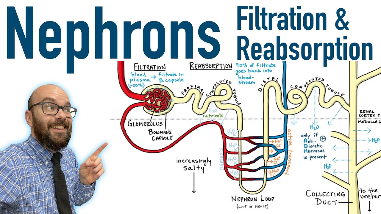
These are the famous minor calyces that we are going to study. Right now, what I want you to know is that the medulla is embedded, that is, it basically fits into those minor calyces through the papilla, so that pale one is the papilla and what is specifically in that papilla? Well, that papilla is where the final duct of the nephron , which in this case would be the renal collecting duct, is the one that finally carries the urine droplet or the urine and takes it to the minor calyces and they will then take it to the major ones and the major ones to the renal pelvis and that is where the urine begins to be poured or excreted as such, in any case we are going to see this image a little larger, Notice how these are a nephron, this is one, then what I was telling you about the ultra structure of the kidney, but in the end it will reach the collecting duct and those collecting ducts all perforate the renal papilla, which is this, this is like the tip of the medulla so that you can imagine and see that each collecting tubule or many collecting tubules pierce each stack.
In fact, it is said that for each kidney there may be up to 500 collecting tubules. So this area of the papilla, which is the one that is tucked inside the minor calyx, has many small holes or looks like a sieve, which is why anatomists gave the name cribriform area to the papilla or cribriform area because cribriform means sieve-shaped. , see it is the shape of a strainer as such.
Very well, you already know that these are the collecting tubules and this would be the then cribriform or cribriform area of the kidney, now we are going to quickly talk about the regulations of the kidney, perhaps you did not know this term but it turns out that embryologically the kidney is formed by the union of many right lobes, of these lobes there are more or less between 11 and between 17 lobes, each lobe can be even more than 17 to 19. Each lobe is made up of a Malpighian pyramid with the cortex surrounding it , then if you see that there are 10 to 12 pyramids for each kidney, then more or less those are the set of lobes that surely formed that kidney; This is a child's kidney, so that they more or less understand the concept of ovulation. These are those blood cells that begin to unite until, in the adult stage, there is practically no point, a lobulated kidney as such, but rather we can see vestiges of that perhaps there were or existed lobes that were united but it is a single structure in the adult, we are going to continue talking about the kidneys, we are going to talk about the area of the pedicle and the area of the renal sinus but first we are not going to detach ourselves from this video [Music] I invite you to subscribe here in the lower right corner by clicking [Music] and don't forget to like the video.
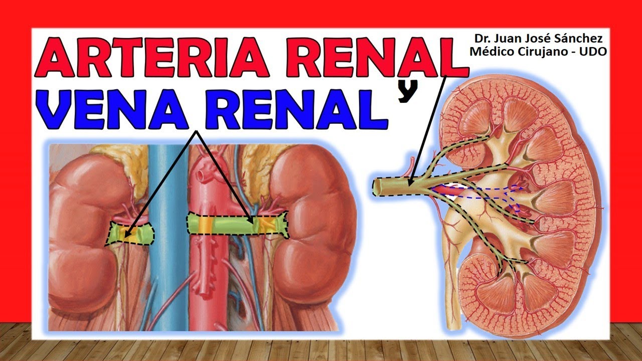
Very well, it is important that you subscribe to the channel below where you see that it says subscribe, I don't know how many times they told you about the videos that you have to subscribe so that you can access all the content of the channel and you can see the more than 162 of anatomy; We have anatomy of the kidneys, adrenal gland, ureters and, well, we continue making videos, we still need many, if there is any anatomy video that I don't see here but that you want to ask me for, you can tell me about it below in this same video, I will surely read it and At some point I will make the video that you are requesting, but sometimes I don't have time to respond to everyone in the comments or make the videos that everyone requests, but I always take them into account. Speaking of the renal pedicle, remember that in the previous video we talked about the hilum and the hilum was the area of the inner face of the kidney or the inner edge through which the structures entered or left the kidney, so those structures that enter and leave the kidney are what we call the renal pedicle, basically lymphatics enter the kidney, as well as nerves but they are structures that very few of us study, when we talk about the renal pedicle we almost always study a classic triad of three things, we talk about a renal vein, which is The most anterior structure that can enter at the level of the pedicle, behind it we find the renal pelvis, which perhaps you do not know that term, we are going to know it right now, better known as the pelvis of the ureter, it is very incorrectly called the renal pelvis, but like this They call it the anatomical list but the ideal is the pelvis of the ureter, this is posterior to the renal vein and the renal artery if it has a disposition that apart from being variable, its terminal branches are intertwined with a renal vein and with the renal pelvis that is the specific arrangement of this pedicle, if we see it in this image we see the anterior renal vein again, remember that there is a video only dedicated to the renal vein, behind it to the renal pelvis or pelvis of the ureter and you will see the even more posterior disposition of the renal artery and with its terminal branches that are penetrating at the level of that renal hilum; Speaking now of the renal sinus, remember that the entrance to, let's say, the kidney, to the internal part of the kidney, was the hilum that was located on its internal edge or medial edge, well that takes me to a space, a hole literally that is a recess really what is the renal sinus, in that renal sinus we are going to see that it is also lined, look at the same renal capsule, it is lined by the renal capsule at the level of that renal sinus is that we find the famous one, see here in this cut before explaining to you in this cross section all this space that is there is the renal sinus and notice how the entire sinus is also covered by that renal capsule, as such there we also find adipose tissue that is part of the perirenal tissue or tissues perinephro; very well there where we find the famous renal pelvis or pelvis of the ureter, I am going to explain the ureter in another video that we are going to see, then I am going to explain to you how that ureter was previously the renal pelvis, how that renal pelvis was major calyces, how The major calyces were minor calyces, I am going to start from distal to proximal, this renal pelvis has a vessel shape, in fact that is what they call it a funnel shape, all the urine produced by the kidneys is captured by the renal pelvis and in the renal pelvis is carried through the ureters to the bladder, then this renal pelvis in its highest portion will measure 2 to 3 centimeters or 20 to 30 millimeters, as the latarjer text says, and in its widest part it measures 1. 5 at 2 centimeters, we are going to divide it into two portions, a portion that remains inside the renal sinus, which would be the portion that is in green here, which is the intrarenal, and a portion that remains outside the renal sinus, which is the one we see.
which would be the extra renal portion, very well apart from those portions we are going to see a base, the base looks into the renal sinus, the base is the place where the major calyces empty, then it narrows in an area that It is called the neck until finally the vertex comes, which is the most final part of that renal pelvis, when it becomes the ureter, I was trying to find the anatomical point where I stopped calling it the renal pelvis and started calling it the ureter and I really couldn't find it. , I didn't get it in the books and I couldn't get it on the internet, if any of you know where theoretically I should call it the renal pelvis and I'm starting to call it the ureter, let me know in this video so I can also say learn it, these are the structures So what make me up or into which this renal pelvis is divided, it is also made up of two faces: an anterior face that we are seeing here and a similar one but which is behind the posterior face, it is said that in fact it It is flattened when there is no urine at this level of the renal pelvis, but it literally bulges. It bulges when the production of urine that passes through the renal pelvis increases.
Then we would have the edges: an upper edge, see that it is rather convex upwards. and a lower edge that is concave downwards and that is much shorter than the upper edge, speaking then the major calyxes, these major calyxes are 2 to 3 remember that calyx means that it has the shape of a cup, so these three calyxes The larger ones will be the ones that bring the urine from the minor calyces and take it towards the pelvis of the ureter, then it would be made up of an upper major calyx, which is the one we are seeing here that is oblique downwards, a middle major calyx, this It would be the upper major calyx of the middle major calyx, which is said to be horizontal and that generally drains two or a maximum of three minor calyxes from the middle portion of the kidney, the upper major calyx drains between four to five minor calyxes from the upper portion of the kidney. , while lastly the lower major calyx is oblique upward and also drains three to four minor calyxes from the lower portion of the kidney; then these are the famous major calyxes, those major calyxes before they were formed had to join the calyxes minors, remember that we came from distal to proximal, so the minor calyxes will be approximately 7 to 14 for each kidney, they have an upper extremity that is the part of the calyx or the cup that fits on the renal pyramid where This famous structure is located that I explained to you at the end of the last slide what they were, you remember that I told you that it was somewhat whitish, very well, then those collecting tubules that penetrate or leave this structure that was the vertex of this Malpighian pyramid end At the level of the minor calyx, specifically the upper extremity, then the upper extremity is the one that is going to join the rest of the lower extremities and is the one that is going to lead to the subsequent formation of the major calyxes, this structure here that I had The name has gone away, specifically the renal papilla, which fits on the upper extremity of those minor calyces.