This is Randy shell and I'm making a podcast entitled valvular heart disease in the anesthesia it's part of our University of Kentucky College of Medicine department of anesthesiology didactic series let's first look at the ABA keywords related to valvular heart disease from the dates shown here 2005 to 2016 under aortic stenosis aortic stenosis and dysrhythmias one Specific dysrhythmia atrial fibrillation if a patient goes into acute onset afib and they have existing aortic stenosis you need that atrial Kick to fill that stiff thick left ventricle and maybe they may need immediate cardioversion if they're hemodynamically unstable
being able to recognize pressure volume loops in a patient with aortic stenosis and we'll go over those a Artic insufficiency treatment if they have your token Sufficiency medically that is afterload reduction drugs like nitroprusside increasing contractility and along with afterload reduction dobutamine would be an example of that and not putting in a balloon pump because a balloon pump inflates during diastole and can worsen the insufficiency especially in a patient with moderate to severe AI mitral valve disease Insufficiency we can drop the vascular resistance some and decrease the regurgitant volume micro stenosis can raise pulmonary artery
pressures and we will go over both mitral regurgitation and mitral stenosis HSS or idiopathic hypertrophic subaortic stenosis thickening of the left ventricular septum resulting in awful tract obstruction to the lvl 4 tract this is Especially important in the perioperative period because anti any reduction in systemic vascular resistance can worsen that awful tract obstruction and we treat it the hypotension resulting from it in a page with IHSS with fennel Efrain with the volume and even occasionally beta-blockers rather than giving drugs that increase contractility such as ephedrine or epinephrine and lastly there's a couple miscellaneous topics Like
bacterial endocarditis prophylaxis in patients with valvular heart disease and some new ways to go about putting valves and patients that don't require sternotomy our learning objectives first are to go over some of the pathophysiology of valve the heart disease to be able to differentiate what happens to the heart in regurge dn't tends to be big dilated hearts versus stenotic tends to be a thick heart ie a or texture gnosis that left ventricle And to recognize some of the pressure volume curves that are associated with these lesions and understand a little bit about how to determine
whether a valve lesion is severe or not and to discuss the anesthetic goals for patients who have valvular heart disease who are undergoing non cardiac surgery up at the top right shows a patient with mitral stenosis looking down from the left atrium Now management of patients with valvular heart disease depends on the severity of the lesion and whether it's a stenotic or regurgitant lesion stenotic lesion the classic example being a or Tech's nosis requires the heart to create an increased pressure inside the left ventricle to get blood out through that reduced orifice the aortic valve
stenosis a regurgitant lesion example would be a Aortic insufficiency where the heart is forced to eject large amounts of volume because during diastole blood can flow back from the aorta through that insufficient erratic valve and fill the left ventricle and so it's like blood is going back and forth back and forth and regurgitant lesions often result in very dilated heart so stenotic lesions often result in thick left ventricle a s regurgitant lesions both mitral regurge and erratic regurge Result in dilated hearts if they have to pump a lot of blood and they dilate surgical procedure
wealth of patients undergoing something that can be done under regional versus major fluid shifts it will determine what we're going to do with regards to monitoring and anesthetic management and how well the patient has adapted to their valve their heart disease will determine how we monitor and manage them for example do They have good left ventricular function do they have pulmonary hypertension is their stroke volume maintained which depends upon preload afterload and contractility what's the natural progression of a patient with valve disease who is left untreated and this graphic shows survivors percentage on the y-axis
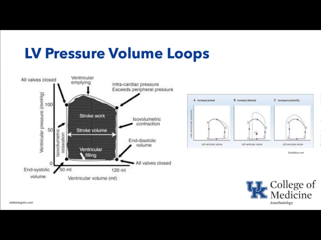
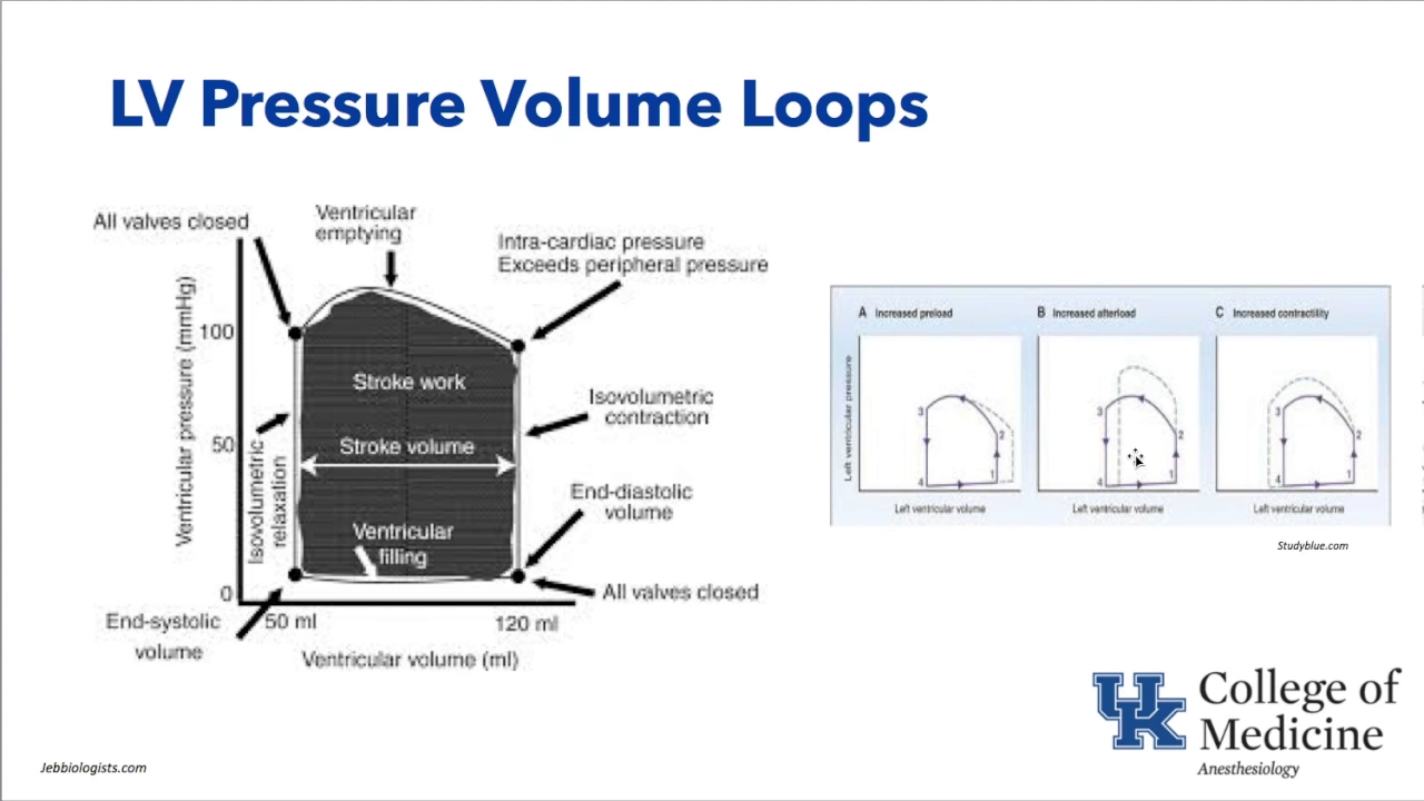
and on the x-axis time since diagnosis one thing that's interesting see here is that the Lesions a or tick insufficiency mitral insufficiency and mitral stenosis the yellow green and blues here have a pretty similar decrement in survivors over time since diagnosis but the or text analysis look at how bad it is the number of survivors after diagnosis at ten years is less than 25 percent so aortic stenosis is especially bad if left untreated let's look at normal left ventricular pressure volume curve so that when we Look at abnormal ones we'll understand them better pressure volume
curves characteristically show pressure on the y axis here and on the x axis LV volume and as we go through the steps 1 2 3 4 of what's going on into the heart let's start at the bottom left where the mitral valve opens and diastolic filling of the left ventricle occurs that's at 1 diastolic filling of the left ventricle blood is flowing from the left atrium into the left ventricle filling it and Then at n diastole as the heart starts into systole it starts to build up pressure inside the mitral valve closes and you have
isovolumetric and for action at two - the pressure builds up and opens the Artic valve an ejection through the aortic valve occurs at three and then the Artic valve closes as the pressure drops isovolumetric relaxation is occurring and we go back through the same cycle again a filling of that left ventricle so in diastole bottom right The difference between end diastole and end systole gives us an idea of stroke volume so the height of this curve gives us an idea about pressure inside the left ventricle the width of this curve gives us an idea about
the volume in the left ventricle looking at it in a little more detail here again starting at the bottom left and systole the heart is as a little residual volume left in it your the Mitral valve opens it fills the ventricle when the ventricle is full in the heart starts to eject so this is the end diastole at the bottom right eisah volumetric contraction is occurring the heart builds up pressure and blood is ejected out through the aortic valve and the aortic valve closes again isovolumetric relaxation we go through the cycle again and the width
tells us about stroke volume the bottom right volume tells us about end diastolic Volume so if preload goes up if you dumped in a couple liters of crystalloid or colloid the preload would go up to this heart and would be shown in the right side as the very first graphic a in the dotted line represents increased left ventricular volume notice that there's a shift in the end diastolic volume to the right as opposed to increased after load which is represented by the middle graphic here You can see that the heart has to create a higher
pressure inside the left ventricle and so the height of this pressure bomb curve is mainly affected and then increased contractility would be for the same amount of volume in the heart you generate more pressure inside that heart so let's look at pathophysiological e some pressure versus volume overload effects on the heart what it does pressure overload first if there's a Pressure overload to the left ventricle luck occurs in aortic stenosis the peak systolic wall tension goes up and the muscle sarcomeres tend to replicate in parallel and what happens is you get a very thick left
ventricle which then functions to reduce wall tension but what you see is concentric hypertrophy so this is the classic patient who has a Artic stenosis or long-standing hypertension who gets concentric thick left ventricle now if You have volume overload on the heart and diastolic wall tension goes up then heart is dilated during diastole lots of volume in it there's serial replication of sarcomeres and to end chamber enlargement like lve d the dilation and you get what's called eccentric hypertrophy AI would be the classic example of eccentric hypertrophy as opposed to a ser ex gnosis as
the classic example of concentric Hypertrophy let's look at now the path of physiologic pressure-volume curve associated with the valvular lesions again on the y axis you see left ventricular pressure on the x axis you see left ventricular volume normal is represented by a B represents mitral stenosis and you can instantly see that the left ventricular volume is less in this patient Micra stenosis under Filling of the left ventricle so think of a small left ventricle with mitral stenosis C represents aortic stenosis lack ventral has to generate huge pressures inside to get the blood out through
that small orifice and so the height of this pressure-volume curve goes up so this tall relatively narrow curve is a or text analysis and then if you look at DNA you see these huge volume hearts where e represents chronic aortic Regurgitation as opposed to D represents chronic mitral regurgitation both dilated hearts in the case of D during systole you can see this sloping quite steep sloping back because blood is being ejected not only to the systemic vascular Chur but also back to the mitral regurg attend valve and you don't have a nice all you isovolumetric
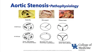
contraction because as the ventricle starts to contract blood just leaks right back through that mitral valve so You can kind of recognize some of these patterns of stenotic versus regurgitant lesions if we look at a visual picture of what a or text analysis looks like depending upon the cause at the far left is calcific your text analysis this would be the classic eighty-year-old with aortic stenosis who you pick up a systolic murmur and you can see during systole and diastole here the aortic valve doesn't open very far it's got Heavy calcification as opposed to a
patient with a bicuspid pathophysiology of aortic stenosis they often present early or 40 50 maybe 60 but definitely earlier than calcific eratic stenosis in this case they're Bournemouth can just with a bicuspid valve and you can see that over time that fish mouth like your attic valve becomes calcified and thickened and doesn't open as well and your texture gnosis pathophysiology occurs secondary To congenital bicuspid aortic valve rheumatic disease is associated with inflammation in both the mitral and aortic valve are often involved in this case showing a picture of the Artic valve with hamish oral fusion
from the inflammation the valve becomes very inflamed it scars down and only the little central part opens Thomas real fusion a characteristic of rheumatic a or text enosis Eric stenosis as you know is a systolic Murmur and this is showing it here in the yellow triangle a crescendo decrescendo systolic murmur and if we look at the natural history of these lesions aortic stenosis from the time of onset of symptoms shown up at the top right you can see that there are three major symptoms that we talk about heart failure and syncope and angina and from
the time of onset of symptoms like heart failure syncope or angina if you Don't have a retic valve replacement you're not going to live very long a little bit longer after heart failure symptoms then syncope an antigen oh maybe but all three of them are associated with from the onset of those symptoms to death without valve replacement not very long period of time why do they get heart failure well because they're pumping against the resistance why do they get syncope well let's say you exercise and Vaso dilate distal to that they already valve you your
heart can't get enough blood out there to fill that dilated vasculature your blood pressure drops significantly and you faint and you're not because you have a big thick left ventricle that's trying to get blood from the epicardium into the endocardium the endocardium is at risk and even without flow limiting coronary artery disease ie lesions in the coronary artery you can still get the engine in Patients that have a or text analysis so let's look kind of in a algorithm based format the pathophysiology of your neck stenosis will start at the top your stenosis is present
obstruction to outflow through the aortic valve the left ventricle has to pump against that it starts to grow when thickened left our mass goes up and early on the heart's not very compliant because it's like the thicker muscle and you have some form of diastolic dysfunction But contractility is maintained the ejection fraction is maintained and diastolic pressure goes up inside the heart and you're much more dependent upon the H or contraction which may be in a healthy state contributes 20 25% or so to the filling of the left ventricle and in the case of aortic
stenosis system a far exceed 50% such that if a patient loses their atrial contraction who has a Artic stenosis they may become very human and I'm glad stable they need That atrial contraction to fill that stiff high-pressured left ventricle and onset of afib may require immediate cardioversion in these patients now late the left ventricular mass itself the muscle can fibrosis the contractility can start to decrease ejection fraction goes from normal now less than 60 percent to fifty to forty percent and if nothing is done this patients going to die in a short period of time
so valve replacement we hope is done before you Start seeing these Decker mints in LV function eventually the left ventricle left ventricle dilates the mitral valve apparatus can be pulled apart and get mitral regurgitation stroke volume goes down and terminal events occur the aortic stenosis creates again an outflow tract obstruction or an LV outflow obstruction that is and the systolic pressure the inside the left ventricle goes up that's the reason for the increase LV mass it uses more Oxygen because it has more muscle the time and ejection the left ventricular ejection time goes up it's
ejecting for a longer period of time needs more oxygen to to perform that muscular ejection there's an increase in left ventricular end diastolic pressure so the pressure inside the heart at end diastole is actually higher instead of being you know 10 or 12 it might be 18 or 20 and the combination of less diastolic time And higher diastolic pressure is a bad thing because diastolic times when coronary blood flow to the left ventricle occurs and that's when the myocardium is getting its oxygen so it's using more oxygen getting less oxygen and can have myocardial ischemia
even without flow limiting coronary artery lesions now if we look at valve areas of the aortic valve versus flow flow being on the y-axis in this graphic on the left Here two valve pressure on the bottom on the x axis this is showing a normal valve area up at the top and then as it decreases the valve area all the way down to this magical point eight to one range which most books will tell you is the critical severe aortic stenosis or by definition usually most use one centimeter squared valve area as severe aortic stenosis
some books will tell you point eight and critical means that no matter what Pressure you generate in the left ventricle you can't get any more flow across that aortic valve its flow limited and so at that point if systemic vascular resistance drops there's nothing that patient can do they cannot generate more pressure in the left ventricle to get more blood to go through it and again up at the top right you can see the pressure-volume curve that characteristic one of erratic stenosis That shows very high pressures inside the left ventricle and initially as long as
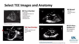
it's not a failing dilated heart it is not a big heart so high pressures is the characteristic finding inside that heart with aortic stenosis aortic stenosis creates turbulent flow across through the little orifice and that's one of the things we look for on echo the presence of turbulent flow and there's several views and we'll just Touch on some of these views as we go through to introduce them to you if you haven't been before but the first one is the mid esophageal long axis view meaning that the probe of the t'ee machine is sitting in
the middle cos and long axis being through the aortic valve and long axis as opposed to the mid esophageal air two valve short axis which is 90 degrees from the long axis you can see the three leaflets assuming it's a trial occluded artic valve with The non coronary cost sitting next to the atrial septum the left corner in cuffs which the left main comes off of and the right corner costs the most anterior so the aortic valve in cross-section the Artic valve in long section their long axis we do some measurements we can measure the
annulus we can measure the aorta aortic can often dilate posta notic dilation after long standing Urich stenosis if you stop the picture and Trace out around the opening which isn't a very accurate way of doing it but can be done this is called plenum 'try and we can get the opening size of the Artic valve in this case you can see it is 0.9 centimeters squared which is severe aortic stenosis another view that we use is what's called the deep trans gastric long axis view where the probe of the t'ee is sitting in the stomach
and shooting up through the apex towards the aortic Valve and in this case la stands for left atrium left LV stands for left ventricle and AV stands for a aortic valve the aortic root is the part from the aortic annulus up to the sino tubular Junction right here so when you hear about a root replacement this is what they're replacing but why do we get this view we get this view because we can line up a continuous wave Doppler here through the aortic valve and get a Trace that looks like yes and put a little
cursor on it and find out the peak gradients and if we trace over this we can actually get mean gradients also so continuous wave Doppler is used to measure the severity of your text analysis and if we look at the mean gradient in the valve area when you get over about a 40 millimeter mercury mean gradient that correlates in many patients with a valve area of about a centimeter squared so if the gradients High the valve area calculated is less than centimeters squared the jet velocity is very high we start thinking this is bad chaotic
stenosis in the cath lab they'll occasionally measure the gradient and there's a difference between the gradient that's measured in the cath lab versus measured on echo let me just touch on this very briefly on the top lap shows what it would look like in the cath lab if you had a Catheter inside the left ventricle which would be the LV trace you're the highest one and one out in the aorta so you're measuring pressures across the Artic valve both yeah before and after it and the maximum pressure gradient is shown here as well as what's
called the peak to peak gradient what you can see is not occurring at the same time the left ventricle creates a high pressure and there's a sometime after that that the highest pressure is created in new york Because obviously the Artic valve has to open and blood has to eject so that's called peak to peak gradient they're not at the same time as opposed to using continuous wave Doppler to look at the maximal velocity of the jet going across this is called v-max and that's the maximal grading by echo which tends to exceed the peak
gradient in the cat lab and there's some big equations that can be used to estimate the ik valve area in the cath lab and There's something called the continuity equation which can be used to better quantify the aortic valve area but we will not go into that detail in this podcast treatment of aortic stenosis to get severe aortic stenosis the classic area being less than one centimeter squared as we mentioned with the mean gradient greater than 40 millimeters of mercury if they're already undergoing coronary bypass graft surgery that's a class 1 Indication you better just
replace the Artic valve at the same time if the patient has symptoms you're gonna go right to aortic valve replacement with those measurements of 1 centimeter squared or less and mean gradient 40 or more if they're symptomatic maybe maybe not you're not sure you can do an exercise test blood pressure goes down you're going to take them to area 2 valve replacement probably and If these things are equivocal and their ventricle is good and you may wait for some time before you take them to valve replacement but the presence of heart failure or angina or
syncope symptoms these patients need here at valve replacement cuz we showed you before what happens if you don't do anything to the valve over time now valve replacement can be done with bile prosthetic valves which are shown on the bottom right here an example and Prosthetic valves made out of materials such as shown here carbon materials metal metallic materials and so these artificial heart valves two major types up at the top right but the little star is like a tilting disc bileaflet a tilting disc and at the bottom right is destructed notice the struts your
bio prosthetic valve now airtight valve replacement also can be done percutaneously in patients that are extremely high-risk is what we used To do in the past older patients decreased ejection fraction percutaneous aortic valve replacement has been done these indications are becoming wider now for replacing valves not through a sternotomy but through other approaches such as trans femoral transaxillary trans apical up at the top left a through D is showing a balloon going in across the aortic valve The catheter is is present across their attic valve with the the artificial valve which as the balloon goes
up and spreads that artificial valve and its little struts open in the position of the aortic valve you now have your new valve that's open with a strutted or with a supporting wire mesh apparatus like that's holding that dirty valve open with aortic valve in the middle of it And just the simple deployment of this puts an aortic valve in place without having to open the sternum open the chest etc in some cases not opening the chest now one of the questions key words that the ABA asked in the last couple years with what are
some of the complications of this form of aortic valve replacement well one is if you're going through major vessels like the femoral vessel and get vascular dissection perforation of vessels Retroperitoneal bleeding aneurysm formation gut ischemia and there's multiple ways that you can put catheters into the femoral vessels to the trans axillary through the apex of the heart the he's sending he ordered south and each of them are associated with some complications more than others but stroke myocardial ischemia rupture dissection etc are some of the complications of aortic valve replacement Done in this method so some
treatment goals for patient with aortic stenosis big red arrows pointing to the aortic stenosis the two major points are one afterload you do not want afterload to go down the heart cannot get blood to that small amorphous to fill the dilated vasculature so turning up your inhaled agent high concentrations turning out a night to process those would be very bad things to do phenol Efrain treatment of Hypotension would be a good thing rhythm we really want that patient in sinus because we remember pointing out the importance of the atrial kick to filling of that thick
non-compliant left ventricle so keeping the afterload up keeping the rhythm sinus and preload maintained and contractility maintained that's the best things that you can do in a patient with aortic stenosis a couple clinical pearls here One spinal anesthesia an epidural if you put in a spinal anesthetic and have a rapid onset of sympathectomy t1 through l2 your sympathetic nervous system and you decrease the preload and you decrease the after load this is not going to be good for that thick non-compliant left ventricle that needs to be filled and needs after load up and patients can
become severely hypotensive so putting in a spinal anesthetic in a patient with the Artic stenosis that's Severe probably not a wise thing to do goals for general anesthesia keep their SVR up sometimes fel efferent infusions are used during induction to keep that SVR maintained keeping preload up because that thick non-compliant left ventricle needs to be filled and if they went into a rhythm outside of sinus trying to get them out of that and back into the atrial kick sinus rhythm that they need Tom made opioids are especially useful in these patients remi Fentanyl etomidate for
example patients who have a us can have angina even without flow limiting lesions as mentioned earlier and that's mainly because they have this big thick left ventricle and epicardial coronary arteries are trying to go all the way from the outside of the heart to the inside of the heart at endocardium is especially at risk for ski Mia because not only is it a long distance to a thick left ventricular Muscle but also the pressure inside the heart left ventricular end diastolic pressure is high and remember that coronary perfusion pressure is diastolic blood pressure - LV
e DP and if L V DP is high that can negatively impact corner perfusion pressure we don't want the patient get tachycardia tachycardic because that will increase oxygen demands of the heart also another pearl is this atrial Fibrillation concept of needing the atrial kick if a patient with with aortic stenosis loses their a chicken becomes hypotensive remember cardiovert early rather than waiting CPR if needed in a patient of the our external says chest compressions are not going to be very effective in generating adequate pressures across that air tube valve so early instigation of therapy before
they get to this needing CPR The Artic insufficiency let's move on from a s up at the top right shows a picture of blood leaking back through an aortic valve during diastole and on echo you can see the picture at the top right that blue jet from the aorta through the aortic valve into the lv o full tract into the left ventricle the ideology of Eirik insufficiency could be rheumatic fever could be destruction of the valve from bacterial endocarditis trauma like chest trauma After a motor vehicle accident dissection of the ordered dissect down interrupts the
competency of that aortic valve when you have a Artic insufficiency there's a volume that's overloading the left ventricle during diastole that blood is leaking back in the left ventricle filling it up and it dilates over time you get a massively big dilated heart eccentric hypertrophy remember Compensation is large stroke vine yeah you fill that heart with a lot of blood it's got to get rid of it so it's got a huge stroke volume the peripheral vasodilation will help reduce the aortic insufficiency and the heart rate goes up because the the more time you spend in
diastole the more time there is for blood to leak back into that left ventricle and fill it so we want the heart rate a little fast days are dilated And we'll talk about some of that in the clinical pearls discussion in just a couple slides let's go over the pathophysiology of AI now it's a volume overload of the left ventricle fiber length goes up there's dilation if the heart dilates a lot it can pull apart the mitral apparatus and pull apart the mitral valve annulus and you can get mitral regurgitation blood leaking back into the
atria and into the pulmonary system and causing pulmonary Hypertension and eventually right heart failure the heart dilates over time and reduces the oxygen cost but a heart overtime can get huge the pressure inside not only the left ventricle but the left atrium can get high also contributing to the pulmonary hypertension that may be seen in long-standing eratic insufficiency and eventually you get a fail failure of the heart with reduced stroke volume they Just can't keep that up over time this pressure volume curve is attempting to show the difference between acute a high versus chronic a
high first thing you can see is chronically I has a huge heart look at end diastole over here 300 350 400 cc's of blood in this heart as opposed to acute AR it's not as big of a heart it hasn't had time to dilate but look at n diastole the pressure because the heart hasn't had a chance to dilate and take up the pressure from all this Blood coming back yeah if a cute AI occurs such as a healthy patient motor-vehicle axon test crunch the aortic valve becomes incompetent all that blood backs up and the
LV DP goes up very high and the patient could go into pulmonary edema quickly as opposed to the patient with chronic eye over time the left ventricle dilates so much bigger heart if the heart has had time to dilate how do we quantify AI what's a little Bit more difficult some aspects than a s but one of the things that is looked at is the width of what's called the vena contract if you look at the place where bloods coming through the aortic valve rate almost at the annulus that little width of that jet as
opposed to the width of the LV awful tract gives us an idea how severe the eye is for example if the width of this jet fills the whole LV awful tract that's pretty bad if it's just a teeny little narrow jet Suggest that it's not as bad so the width of the jet is called the vena contracta and it's one of the things that's used to estimate Yodok insufficiency other things like cougar jet and fraction regurgitant volume effective regurgitant orifice area are complicated in quantitative ways that echo is used to assess AI so what do
we want to do in patients with AI clinically well if the after load is decreased there'll be less AI the Pressure in the aorta will be less and blood will not go back as much through that the aortic valve so turning on nitroprusside turning out inhaled agent which would basil dilate the patient promotes forward flow so keep the afterload down you can't put a balloon pump in these patients remember because in moderate to severe AI that a or DIC bloom pump will inflate during diastole create higher pressures during diastole and force Blood back into the
left ventricle so you need a competently Artic valve or at least only mild AI if you needed to put a balloon pump in to support the left ventricle the rate it would be nice if it's a little bit faster an AI why because less time and diastole less time for it to fill during that diastolic phase where insufficiency is occurring some clinical pearls related to AI again the graphic on the top right is showing pressure volume and You remember that AI is a regurgitant lesion and therefore has a big dilated heart and chronically would be
the e in this picture at the top right now if someone has a eye and you want to put a Spile or an epidural in it's not contraindicated as long as you keep the preload maintained dropping the afterload actually can be good and reduce the regurgitant amount through the aortic valve for general anesthesia you really don't Want there afterload to go up so intubating someone lightly for example and their pressure jumps from 120 up to 200 and they get tachycardic via that sudden increase in afterload can raise Eric pressure and make more blood go back
through that a or a bow so we want to decrease after older than our inhaled agents Evo Dez and I so do that and we want to avoid bradycardia because with bradycardia there's more diastolic time and it's during diastole that blood Is leaking back into the left ventricle medical management of acute AI was multiple keywords between the years 2005 and 2016 from the american board of anesthesia let's touch on that here a patient has a disrupted area valve for whatever reason and it's acutely leaking they don't have time to dilate and therefore there's very high
left ventricular end diastolic pressures and it backs up into the lung and they often have pulmonary edema require intubation A mechanical venatori support but then we want to reduce the amount of blood that goes through that air incompetent Eric valve so turning on a vasodilator to decrease systemic vascular resistance nitroprusside would be an example turn I know dilator like dobutamine which via it's a beta wanna fact increases contractility the its beta to affect results in dilation therefore the final dilation and also remember that intra attic Balloon pump is contraindicated because of the fact that it
raises diastolic blood pressure and will increase flow into the left ventricle let's look at the mitral valve now since we've touched on a or Texas New York insufficiency will now look at micro stenosis and mitral insufficiency the valve at the top left is a mitral valve taken from a heart removed and the Coeur D cut and you can see the anterior leaflet at the top the posterior leaflet At the bottom at the top right is a four chamber view of the mitral valve and in that case is a mite roastin onic valve notice the big
left atrium the mitral valve that isn't opening very much and has a small orifice at the bottom left graphic is showing the anterior leaflet and the posterior leaflet and there are designations two portions of these leaflet called anterior one in tier 2 and tier 3 and p1 p2 and p3 not an echo that can be Used to determine exactly where the leaflet pathology is so if someone said they had a p2 flail that would mean that the flail of a leaflet was in the p2 segment again that's beyond the discussion at this point but just
to introduce you to some of the concepts of echo used to help diagnose the location as well as the severity of valve disease and if you look at the mitral valve it involves not just the leaflet but the valve apparatus being the chordae Tendineae the papillary muscles attached to the left ventricle any part of these that become disrupted or damaged ie papillary muscle disruption from ischemia a flail leaflet from accordi rupture a endocarditis leaflet that has holes in it was destroyed or the wall the left ventricle that becomes ischemic or infarcted all those can disrupt
mitral valve function that caused problems Let's look at mitral stenosis first rheumatic fever severe calcification Mixel modest lesions in the left atrium not real common but can also cause mitral stenosis and endocarditis can cause regurgitation and stenosis so at the top right shows a graphic of what the artist gnosis would look like the stenotic and that is mitral stenosis would look like the static mitral valve is here the left atrium is big there's a hockey stick Like appearance of that mitral valve and restricted opening of it such that there's a very small orifice and if
we looked at that in long axis the graphic at the bottom shows that left atrial enlargement in those very poor opening of the tips of the mitral valve leaflet commercial fusion and if you looked at in short actions you can see that teeny little opening so mitral stenosis reduced mitral valve opening and mitral stenosis causes a diastolic Murmur as blood is flowing from the left atrium through a stenosis mitral valve into the left ventricle during diastole you hear that diastolic murmur and it also often is a decrescendo and then pre systolic accentuation crescendo murdering bastard
but the main point is it is a diastolic murmur again remember a artx ptosis is a systolic murmur of aortic insufficiency is a diastolic murmur and now we have a mitral stenosis which is a diastolic Murmur let's look at an algorithm for mitral valve disease pathophysiology at the top you can see stenosis or regurgitation can back up pressures into the pulmonary artery system and if the pulmonary pressures go up you can get right ventricular hypertrophy and right ventricular failure you can get dilation of the annulus in the right side of the heart tricuspid valve can
become regurgitant And it can back up such that the liver becomes huge and eventually right heart failure can occur along with left heart failure mitral stenosis causes an increased grading across the mitral valve such that the left atrial pressure goes up as the left H of pressure goes up the left atrium tends to enlarge over time going from the normal four centimeters or so and many times you can see six and seven and eight centimeter left atria that are In atrial fibrillation commonly because of their dilation and when they're huge and eight really fibrillating little
clots can form especially in the appendage such that thromboembolic can occur and these patients are anticoagulated and if you're going to cardiovert them you need to make sure that there's not a clot stuck in the atrial appendage now as left atrial pressure goes up pulmonary venous pressure goes up and Pulmonary hypertension and again right-sided failure right-sided dilation tricuspid annular dilation and tricuspid regurgitation can occur so left-sided mitral stenosis can cause right-sided failure and pulmonary hypertension now that mitral valve when it gets really small in fact when it gets down to about one centimeter squared that's
critical nitrous and also it's critical and the fact that no matter what pressure you generate Across that mitral valve that's on the x axis mitral valve pressure gradient no matter what that grading is you can crank it all the way up into 50 here and no more flow goes across the mitral valve so it's critical at once in a meter squared approximately and the flow characteristics and oncotic characteristics and hydrostatic characters of blood vessels in the pulmonary system are such that if the Pressure gets too high it's going to exceed oncotic pressure and you're going
to force fluid through the alveolar capillaries right into the Ovilus itself and you're gonna get pulmonary edema so pulmonary edema can occur with mitral stenosis and up at the top right is showing a mitral stenosis pressure-volume curve pressure on the y-axis volume on the x axis and you have an under loaded left ventricle is the Classic appearance of such on t ee there's several views that we look at one is a short axis view of the mitral valve which shows the fish month appearance and the other one that we classically look at is the mid
esophageal for chamber where you see the left atrium and left ventricle in the mitral valve here and there's some other ones like the Commish oral view we can look at to see whether those coma shares are fused or not and the mid esophageal 2 chamber which is really good for looking at the left atrial appendage to see if clots are present there and when we look at it the mitral valve under echo te at the bottom left is a great way to look at the left atrium left atrial pendant size the left atrium and the
mitral valve because it's right near the probe which sits just behind the left atrium in the esophagus and here's that hockey stick appearance of a my Tristana Crowl reduced opening and oftentimes these leaflets are restricted in a mobility and calcified very white for example here's a transthoracic echo picture on the top showing again dilated left atrium hockey stick appearance of the leaflets of the mitral valve and reduced orifice opening and if you looked at in short axis you can see that the little fish mouth is small indicating that the orifice size is small One of
the things that we can do to quantify a mitral stenosis is to put a pulse wave Doppler rate at the annulus or at the tips of the mitral valve and here it is during diastole blood is flowing through a normal mitral valve in this case the blue flow away from the probe across the mitral valve and if the sample volume was right at the mitral valve leaflets tips or so what you see is during diastole notice on the EKG diastole Being from the T wave all the way to the QRS complex so this is diastole
you get an E and an A wave in a wave refers to the passive filling that occurs when the atrium starts to empty when the mitral valve opens and blood flows across and then diastasis when the pressures equalize buds in left atrium and left ventricle and then the atrial kick assuming that they are in a sinus rhythm and so there's a P wave down below you'd see that atrial kick now when we look at Mitral stenosis patient with echo and put that little Doppler signal between the atria in the ventricle at the tips of the
mitral valve leaflet we can look at the peak gradient the higher the velocity across that little orifice the worse the mitral stenosis so the peak and mean gradient can be based on the velocity of blood flow across this mitral valve and another thing that we look at is how long does it take for a decrease in Velocity across the mitral valve and that's called the pressure half time and basically to make a long story short if you have a wide open mitral valve this velocity falls off very quickly as opposed to a small little orifice
it's small it falls off over longer periods of time when we say there's a long pressure half time and we can use that to estimate mitral valve area so the pressure gradient across it and the pressure Halftime accosted are used to quantify mitral stenosis and if we have an area less than one centimeter squared remember the aortic valve area less than somewhere around one centimeter squared also was considered quite severe the mean gradient for a Artic valve was 40 in this case a mean gradient from left atrium to left ventricle of greater than 12 is
considered severe and if you have concomitant pulmonary artery mean pressures greater than 50 associated With it that's also severe what do we do with a patient with mitral stenosis clinically well we need to keep up the preload maintained because that left atrial atrium has to be filled to try to get blood squeezed across that small mitral orifice but if you give a whole bunch of fluid or put a person in head down position it has mitral stenosis you can actually put them in acute pulmonary edema now you think well why don't I Just decrease the
afterload a little bit and that's a bad thing to try to decrease left-sided afterload why because blood is trying to get to that orifice of the mitral valve and if you dilate the left side it can't get enough blood through that orifice to fill the your body and your blood pressure tends to drop so turning it on a drug like nitroprusside would be a really bad idea but remember blood is backing up from that stenotic mitral valve into the Atrium into the pulmonary system so they often have pulmonary hypertension and so reducing right-sided afterload is
something we should do so don't let them get hypercar Bacoor hypoxic or acidotic things that constrict the pulmonary vasculature the right ventricle may not be working too well because the pressures have gone back through the pulmonary system and pulmonary hypertension and right heart failure can be occurring and we really Really want the atria to be contracting on its own that would be nice to push blood across that stenotic mitral valve but often there in atrial fibrillation we often just have to control their ventricular response with drugs that slow AV conduction so some clinical pearls for
mitral stenosis what about the pregnant patient with mitral stenosis remember during pregnancy your interactive volume goes up and as your intravascular volume goes Up left atrial pressures can go up and symptoms pulmonary edema can get worse especially immediately after delivery where you get all that increased volume from the uterus contracting down and the blood volume goes up and cardiac output goes up shortly after delivery that is the time where they can go into failure spinal anesthesia is not a great idea why because you will drop after load and You can't get enough blood across thats
not mitral valve to fill the dilated vasculature if a patient will happen to be in sinus rhythm and then went into new onset atrial fibrillation and lost their atrial kick which was so good for squeezing blood across that stenotic valve that's the patient who often a severely hypertensive and cardioversion could be indicated remembering again that cardioversion for someone who's in chronic afib you really need to make Sure that there's not a clot sitting in the atrial appendage some pre medication issues with severe ms if you have a patient with mitral stenosis and you give them
let's say fentanyl or midazolam and they hyperventilate their co2 goes from normal 40 up to 50 pco2 a 50 hypercarbia is gonna cause an increase in pulmonary vascular resistance and if they already have pulmonary hypertension and right heart issues you're gonna make those Worse so be very careful about pre medicating a patient with mitral stenosis and in most cases we avoid premedicate so some factors that might worsen pulmonary hypertension in a patient who has severe MS and blood backing up already and pressures backing up into the pulmonary system it's all the bad things hypoxia hypercarbia
acidosis light anesthesia we're norepinephrine levels go up and cause vasoconstriction through alpha-1 Mechanisms in exogenous alpha agonist like norepinephrine and phenol Efrain can raise pulmonary vascular resistance vasopressin may not have quite so much effect on the pulmonary vasculature as does norepinephrine and phenol Ephraim and then if you let a patient get cold they often will get evasive constricted in the pulmonary vasculature so if you already have on my hypertension don't let him get hypoxic let their co2 go up Let them get acidotic if they're catecholamines rise because you stimulate them ie learn gossipy in a
light anesthetic state pulmonary vascular resistance can go up dramatically and then don't don't let them get cold let's finish up with mitral regurgitation and IHSS and then some miscellaneous topics but mitral regurgitation how can it occur well mitral valve prolapse could be one where the leaflets just billow into the left Atrium that could have disruption of the chordal apparatus that holds that mitral valve in place like the papillary muscle or the chordae tendineae from ischemia or just old patients sometimes can have degeneration of the korte and just rupture and LV dilation of a patient has
family left ventral it's dilating and pull the annulus apart and cause a central often jet going back into the left atrium During systole and endocarditis can eat away at that leaflet and cause mitral regurgitation now mitral regurgitation pressure-volume curve is shown at the left here and you can see that it is a systolic murmur so during left ventricular systole Bloods going back through the mitral valve and that's when you hear the murmur acute versus chronic is shown here big left ventricle during regurgitation lesions like AI and M R And so you can recognize it by
on the x-axis the large volume one and then during isovolumetric contraction where the volume should stay the same go straight up and down blood is going back into the right atrium and left ventricular volume is getting smaller when during isovolumetric contraction it should stay the same until they are take valve opens that's because it's leaking back through the mitral valve the pathophysiology Is discussed here whether it's acute or chronic and let's do it cute first you have a sudden disruption of a papillary muscle or a chordae tendineae for a stroke line goes down backward stroke
volume goes up so they're going to be hypotensive and their body responds with high sympathetic tone the LV edv is gonna go up and diastolic volume and diastolic pressure goes up Blood is backing up into the pulmonary system hydrostatic pulmonary edema can occur pulmonary hypertension and right heart failure so this is the patient who has for example a papillary muscle disruption from coronary heart disease myocardial infarction a patient with chronic M R has time to dilate their left atrium and oftentimes it will go from that for up to five and six and seven and eight
Centimeters huge left atrium that is more compliant can accept that blood over time and and preserve the low pulmonary venous pressures now it's dilating to compensate for the mitral regurgitation and and try to keep those PA pressures down as opposed to on the top picture here you can see that acute mitral regurg the LV doesn't have time to dilate and the LA pressures are are high The LA pressures are normalized as they dilate in the chronic state how do we check the severity of my kroger tation one is the size that big jet going back
into the left atrium the size of the left atrium itself the bigger it is over time probably the worst of yet the leaflet pathology is it a flail flipping up a leaflet into the left atrium with each systolic contraction and one of the things that we look at is Does the blood go all the way back and go into the pulmonary vein such that during systole the contraction of the ventricle and blood going back through the mitral valve goes all the way up into the pulmonary vein and causes what we call systolic reversal of flow
in the pulmonary vein and if they do have that that's considered severe mitral regurgitation so this is just showing a brief example of the area that the jet fills the bigger the area it fills the Worse it tends to be and then if it goes all the way back and reverses flow in the pulmonary vein and in this case if you see a QRS that obviously assistant Li and blood should be going from the pulmonary vein into the left atrium during both systole and diastole but in this case systole it reversed diastole is still going
forward and then atrial reversal as the atria contracts so this waveform is below the line when it really should be above the line blood Should be going through the pulmonary vein into the left atrium rather than the left atrium into the pulmonary vein during assistant Li that's called systolic reversal pulmonary venous flow indicator of severe Ammar so mitral valve replacement or repair if it's a intact mitral valve with let's say a quarter rupture but not heavily calcified or thickened you might repair it or in some cases put a ring and do another form of repair
but this is Just showing an example of a replacement with a st. Jude valve or an annular ring as part of a repair the annular ring is shown here extending around the posterior towards the anterior often open in the anterior part here in this case and that helps keep the annular size intact and the mitral valve leaflets pulled together and in the bottom is a st. Jude valve a tilting disc bileaflet a tilting disk valve put in the mitral Position for a replacement if you have someone with severe mr moderate-to-severe mr for non cardiac surgery
what are some of our goals were one if we can keep their after load read down down more we'll go forward and less backwards so vasodilation is good in the case of mitral regurgitation the rate be nice to a little bit faster rate to improve the cardiac output and it's not so critical that they maintain their atrial kick but they're often those Patients if they've been in chronic regurgitation have dilated the left atrium and maybe an atrial fibrillation so one of the big things you can do is reduce after load and in this case if
you needed to support the left ventricle an introverted blood pump could be placed because if the aortic valve is intact it will increase diastolic blood pressure and flow through the coronary arteries and reduce afterload systolic blood pressure and maintain more forward Flow and decrease mr so a bloom pump is not contraindicated in mitral regurgitation so some clinical pearls one putting in a spinal or epidural not a big deal usually decreasing after a little may actually improve the mr reduce it in our general anesthesia goals we want the after load to go down and our volatile
anesthetics do that there vasodilators and we want to avoid anything that increases pulmonary vascular resistance if blood is already Going backwards into the pulmonary system with a high pressures the to raise the pulmonary pressures higher with hypoxia hypercarbia hypothermia light anesthetic state vasoconstrictors etc and keep the heart rate relatively fast one of the important points about echo an ejection fraction might regurge is that when someone has an ejection fraction reported let's say of 60% but has severe mr you say hey that's a good ejection Fraction 60% normal right well they're each acting some forward and
some backward and so that ventricles has it a little bit easier job in some ways because it's not having to make all the blood go forward if you suddenly put in a competent mitral valve and make that ventricle pump everything forward suddenly that LV dysfunction that's the present but you couldn't see it may become evident and their ejection fraction instead of 60% now looks like More like 20% and it may uncover a baseline poorly functioning left ventricle and require ionotropic support some pearls continued if you have acute mr such as a ruptured chordae patients in
the IDI with myocardial infarction and a ruptured chordae large amount of Bloods going back into the left atrium they haven't had time to dilate their pulmonary pressures go up dramatically what you can do is drop their SVR Vascular resistance with a drug like nitroprusside make more go forwards and backwards you can put in a balloon pump and you can add high note ropes to support them until you can get them to the operating room so that's called medical management of acute mitral regurgitation don't forget back to undercut itis prophylaxis in patients with valvular heart disease
and we'll touch on that at the very end about making sure that we give antibiotics to These patients tricuspid regurgitation about 80 plus percent of us have some amount of tr and so if you do a echo on most patients there's a little bit of tricuspid regurgitation in the classic view from transesophageal echo is the mid esophageal for chamber view which you see the left H and the left ventricle right hm right ventricle the tricuspid valve and the mitral valve so we're focusing on the right side the Tricuspid valve and we said that most people
have a little bit of tr one of the most common causes of tricuspid regurgitation is left heart failure where blood backs up and causes right heart failure and the right heart dilates and pulls apart the tricuspid annulus and you get TR another cause is the valve being eaten away by bacteria in patients that use drugs IV drug use and right-sided valve lesions another view for TE and tricuspid valve Other than the for chamber which we showed you before is this what's called mid esophageal RV inflow outflow right atrium right ventricle tricuspid and valve in between
a good view to line up a Doppler across it now why would I want to line a Doppler across tit to look at the tricuspid valve regurgitation velocity this is a somewhat complicated idea but I'm gonna try to make it as simple as possible when you are looking preoperatively for example at a patient Who's had a transthoracic echo and somewhere on the report the cardiologist says the estimated RV systolic pressure is 28 or whatever they say you know where did they get that how do they know what the RV systolic pressure is how do they
know what the PA system pressure is because those are usually equal if the pulmonary valve is not abnormal so they're giving you an estimate of the pulmonary systolic pressure Non-invasively and the way they do that is they line up a Doppler across this tricuspid regurge it and jet and this jet will have a higher velocity if the pressure on the right side right ventricle is much much much higher than the right atrial side and the greater the differential in pressure between the right ventricle in the right atrium the greater will be that velocity of jet
during systole and there's an equation that can be simplified down to the one In red at the top that the RV systolic pressure equals four times the velocity squared of the tricuspid regurge inject plus the central venous pressure so I gave you an example at the bottom here where a continuous wave Doppler was placed across a tricuspid regurge in jet we get a trace like this the velocity in this case is about three centimeters per second or three meters per second that is and so we can put that into the Equation and if it was
three it would be four times three squared or four times nine would be 36 Plus you add the CVP to it and if it was four in the example below the jet velocity four meters per second which is very high velocity it say 4 times 4 squared or 4 times 16 is 64 and let's say the CVP was estimated at 10 the cardiology would say that's a 74 estimated millimeters of mercury our V systolic pressure or PA systolic pressure that's pretty high and so this Is how the cardiologist gets our V systolic pressure from the
TR jet on your preoperative echo 4 times the velocity squared plus C V P so in tricuspid regurgitation really what you want to do is you want to maintain our V function and decrease the pulmonary vascular resistance so that more blood goes forward now how do you do these pulmonary vascular resistance oxygen don't let the co2 go up don't let them get cold don't let Them get acidotic don't let catecholamines be flying around and constricting the pulmonary artery all things that make the TR jet worse some clinical pearls oxygen and co2 and its effect on
tricuspid regurgitation if the patient's oxygen levels go down they're going to constrict hypoxic pulmonary vasoconstriction hypercarbia constricts the pulmonary vasculature pressure's going to go up in the right ventricle And worsen tricuspid regurgitation nitrous oxide probably not a great idea and some with severe TR and pulmonary hypertension because nitric oxide increases pulmonary vascular resistance positive pressure ventilation and peep if you have high peak pressures inspiratory and peep intrathoracic pressure can increase pulmonary artery pressure worsening the tr jet and but it also decreases venous return so be careful About peak inspiratory pressures and peep an ablation with
a TR if a patient who has dr has a sudden decrease in SP o2 saturation unresponsive to increasing your fi to don't forget that there could be flow across the PF o there could be so much so much blood and pressure in the right atrium that exceeds left atrial pressure and flow couldn't could occur a cost that PF fall from right to left that's a right-to-left shunt decreasing the spo2 up at the top shows A annular ring that can be put in in a patient with TR to pull those leaflets together pull the annuals together
to make it non regurge done so that's a tricuspid regurge attend repair hypertrophic cardiomyopathy the last major topic in this case on the left you're seeing a hypertrophied septum and that hypertrophied septum can set up a scenario where the mitral valve leaflet Is actually pulled into the LV outflow tract creating mitral regurgitation also the thick septum along with the ante relief of being pulled out there can obstruct flow going out to the body so lvl flow tract obstruction mitral regurgitation characteristics of hypertrophic cardiomyopathy here's an echo pitcher thick interventricular septum the anti leaf of the
mitral valve being pulled out into the left ventricular Outflow tract during systole if you look at the muscle fibers of a normal patient in this nice laid out fibers as opposed to the hypertrophic cardiomyopathy they have disordered muscle fibers and the septum is thickened the proximal acceleration of blood trying to get through this obstructed little orifice creates a venturi effect which sucks the inter leaflet out to it that's called systolic anterior motion of the mitral valve Leaflet or Sam and can cause severe decrease in blood flow going out into the aorta and hypotension and low
cardiac output state all from that lvova tract obstruction our management goals of Holcomb and this was one of the most common keywords related to valvular heart disease technically this isn't valve of heart disease but it's covered under this category if you have someone with Holcomb you want to keep their afterload Ripped up you want to do everything you can to keep their ventricle big not emptied okay so SVR up phenol Efrain you want to keep the preload up so give them volume you don't want their contractility up because that makes the ventricle small or during
systole so ephedrine and epinephrine and don't mean usually bad choices we want their heart rate relatively low so that's time to fill and pull apart that little a mitral Valve inter leaflet away from the only outflow tract so the main thing that you want to do is this if you have a patient who has severe hypotension we're going to jump to the third one here first who has IHSS remember that outflow tract is the path of physiology offal tract obstruction if they lose volume they bleed suddenly get behind on volume that outflow tract obstruction is
going to get worse final Afrin can increase Preload can increase after load can be given as your giving volume but do your best to avoid any drugs that increase contractility because don't mean epinephrine will make things worse in fact classically as you're adding more and more drugs like that the patient gets worse and worse and then someone comes in and recognizing that this is IHSS physiology they stop all the beta stimulants and give a little fennel offering a little volume and suddenly Things get better and even beta blockers you have given volume replacement so phenyl
efferent volume beta-blockers the management for Holcomb obviously the very first one putting in a spinal or an epidural could be bad for someone with IHSS sudden drops in preload sudden dropping after load worsens the LV outflow tract obstruction we want to avoid major sympathetic stimulation which would make the heart smaller from the Beta one effects and maintain our SVR and preload that's our general anesthetic goals one of the interesting things is there's a dissociation between the pulmonary artery catheter findings and te findings and patients with Holcomb and basically just think this that if you have
a pa catheter and what it's going to tell you is is that if you have LV outflow tract obstruction the pressure is high and the left ventricle during systole you're going to see that Pressure and you're gonna measure pretty high pulmonary artery pressure Junior say oh they're hypovolemic or there have a failing heart and you're probably gonna do the wrong thing you're gonna add I know Tropes would be a really bad choice as opposed to seeing it with te and seeing the LV outflow tract obstruction the acceleration of blood out the LV offal track the
mitral valve being sucked into The lvova tract in that situation you would say AHA this is IHSS physiology you're gonna give phenyl efferent and volume and maybe beta blockers the antibiotic prophylaxis new guidelines cardiac conditions that are associated with high risk of endocarditis infective endocarditis if someone has a prosthetic cardiac valve in place or has used prosthetic material for valve repair think need to give them antibiotic prophylaxis Conditions no longer requiring antibiotic prophylaxis mitral valve prolapse just because they've had rheumatic heart disease or have a bicuspid aortic valve or an old patient with calcific yodok
stenosis you don't have to give them in about a prophylaxis this ends a relatively long thank you for sticking with me on this podcast discussion of valvular heart disease here's a quote from Proverbs 17:22 a merry heart doeth good like a medicine I Hope you have a great day to day never stop learning and thank you very much