this is a short video on the various cardiac imaging modalities I'm going to be going over a brief overview of the clinical methods of visualizing the heart and the different modalities that we're gonna be talking about are listed down on the left here we're gonna start off talking about chest x-rays we're going to talk about echocardiograms the different ways to get echocardiograms such as T te and te e we're gonna go into myocardial perfusion imaging we're going to look at MRI that's specific for the heart catheterization for diagnostic and treatment purposes as well as cardiac
CT so this is just a preview of these six imaging modalities we're going to jump into each of them in more detail tell you what they're good for tell you some advantages some disadvantages and show you some examples of the pictures that could be obtained with each of these modalities so let's begin with chest x-ray often abbreviated as CX our four for billing purposes chest x-ray very commonly used dates back over a hundred years ago first used to diagnose TB so if you saw cloudiness in the lungs you knew there might be some some bacterial
infiltrate there it's used to differentiate or resolve objects based on density and this is just based on the physical principles of x rays passing through the chest so a high density object like a bone or metal appears white that's why you see a skeleton on an x-ray a low density object such as a gas or gas in your lungs appear black because the x-rays passed through those objects or that or that material with without without reflecting much so water based tissues which make up the majority of the bodies are so the majority of the body
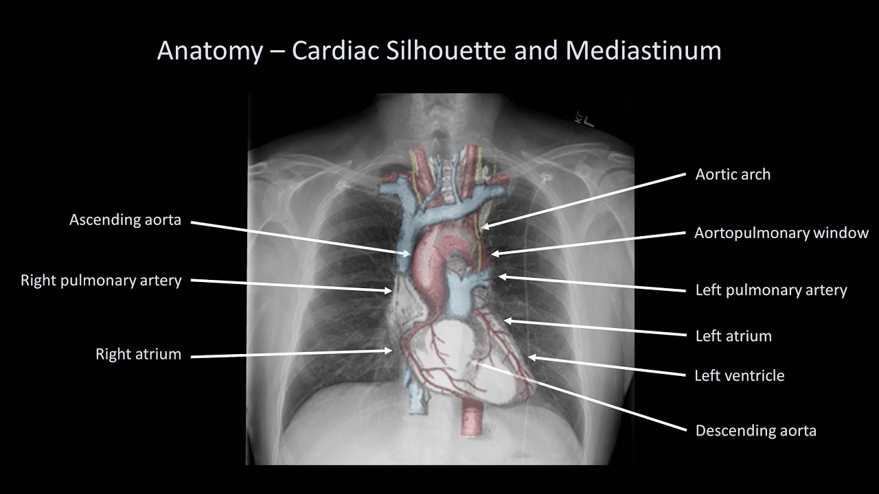
is somewhere in between those two somewhere between black and white one downside to the chest x-ray is that you cannot resolve cardiac structures but you do see a silhouette of the heart this means that if you know the general positioning of the heart and the general anatomy the heart you can still to do certain things based on this silhouette and you can still calculate cardiac ratio now cardiac ratio if you look over at this top right image you can see the image of heart here cardiac ratio is that horizontal distance that the heart takes up
divided by the horizontal distance of the entire chest cavity so in this top left image here we could see a normal-sized heart if we look down to this bottom image we see a heart that's much larger this heart on the bottom has a cardiac ratio that's larger than 50% meaning that the width of the heart is bigger than 50% of the width of the chest cavity so that's an example of cardiac megali an enlargement of the heart you can also see a pacemaker in that bottom right image chest x-ray can also be used to identify
manifestations of lung disease or of heart disease in the lungs this includes pleural edema and pulmonary effusion so oftentimes when we see cloudiness at the base of the lungs where that pointer is now this can be an indication of congestive heart failure of fluid in the lungs as a result of heart problems and just kind of in summary text x-ray is cheap there's a small radiation exposure it's just one quick x-ray and it's used frequently in the emergency room when people present with chest pain its moon move on over to echocardiograms there's a lot to
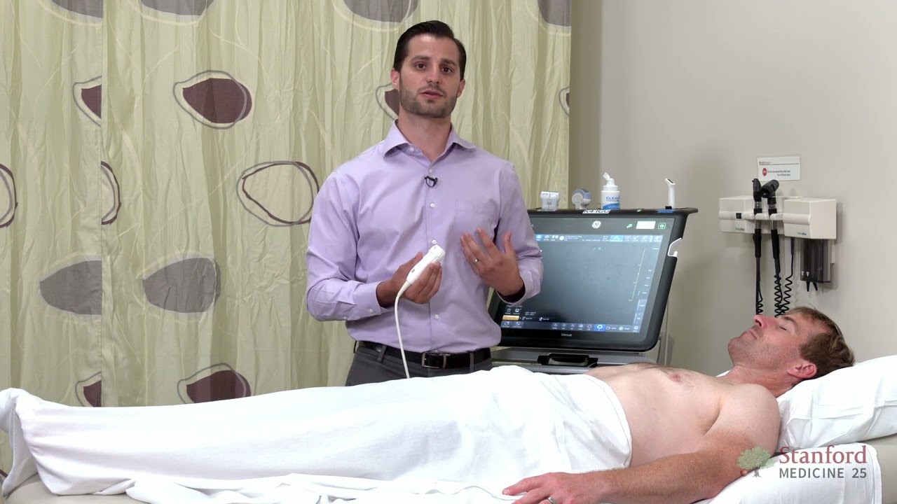
cover here a lot of different methods you can see some moving images here too basic principle is that ultrasound waves are emitted from piezoelectric transducers and those emitted ultrasound waves bounce off tissues the reflected ultrasound waves are then measured processed by a computer and made into an image because we're dealing with pressure waves here there's no big risk or toxicity like we had with x-ray and and like we'll see with with cardiac CT especially there's a low cost to echocardiograms so that's another benefit the two main types are transthoracic echocardiogram TT or transesophageal echocardiogram now
this picture at the top here that red one is an example of te e transesophageal and there's essentially a probe that goes down into the esophagus that probe has both the ultrasound transducers and the ultrasound receivers that can get a better angle at the heart otherwise the ultrasound transducer and receiver is placed directly onto the chest through the through the sternum and the ribs to access the heart and to get an image of the heart that way te e is specifically good for excluding intracardiac thrombus for ruling out the presence of an intracardiac clot now
when we do echocardiograms you can either have one beam which would give you one line of resolution or you could have multiple beams portrayed in an arc like these images here and get a 2-dimensional image of what's going on these images at the bottom just kind of show you the different orientations of the heart that you can get pictures of using echocardiograms and i believe that these were taken using a standard trends thoracic echocardiogram at ete they also have methods these days a finding of making 3-dimensional images and that's all with the image processing on
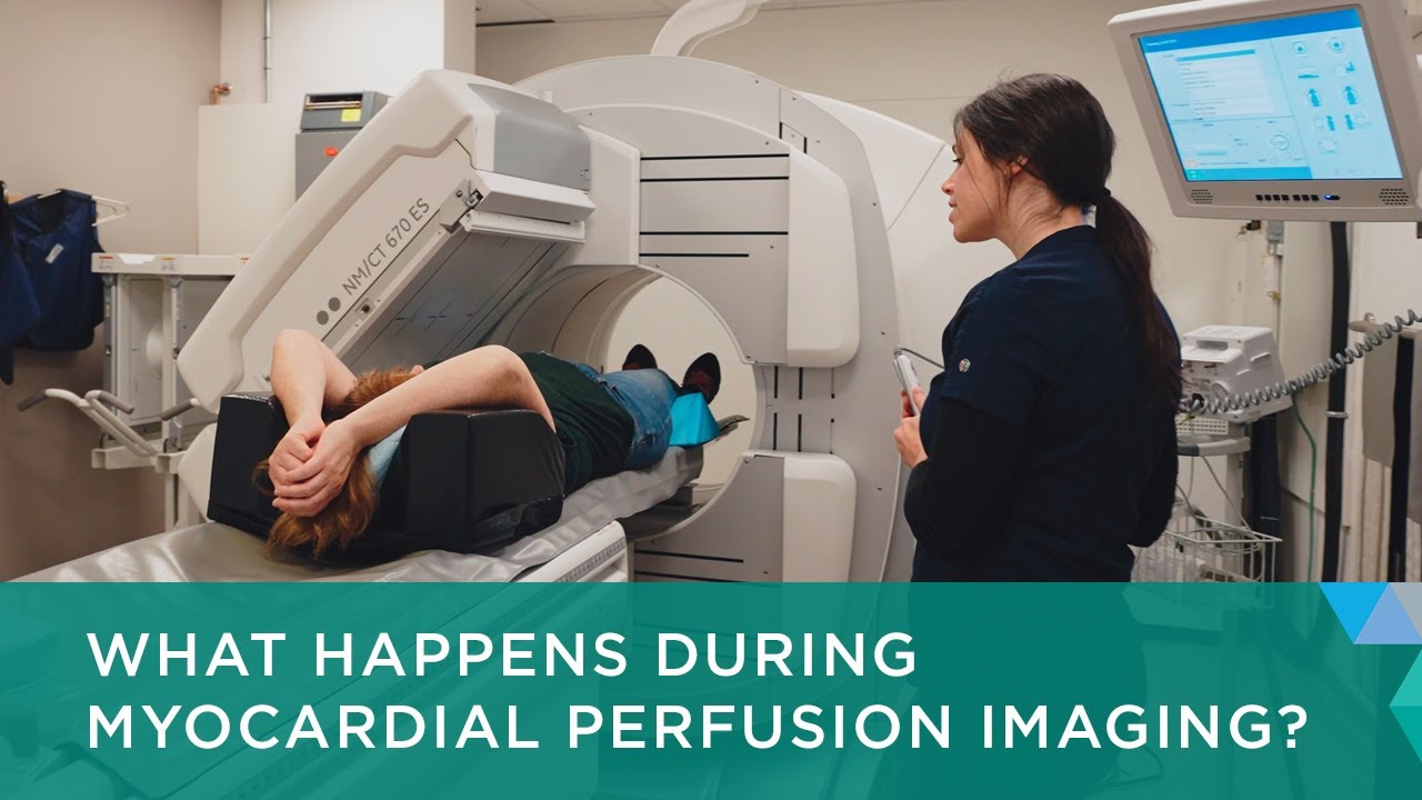
the computer Doppler echocardiography is specifically used to look at blood flow and its advantage is that you can identify turbulent regions I believe it's specifically used for looking at valvular diseases and valvular abnormalities so doppler should be associated with a valvular abnormalities next we have myocardial perfusion imaging this is essentially the use of a heavy element radioisotope there's a few of them that you could use to mark areas of myocardial perfusion not perfusion in the biological sense is the ability to deliver blood to the capillaries we want to be able to give blood to the
heart muscle and we kind of assess where cardiac or the coronary arteries are delivering blood with a heavy element radioisotope so we expose the we expose the heart muscle to stress after it's after it's perfused with these Radio isotopes and you can kind of see the results of a stress test at the bottom here the top row of images shows many heart muscles under stress and the bottom row shows the same heart muscle under arrest if we see a big discrepancy between these images we know that there is a there's a part of the heart
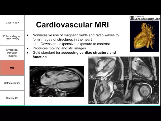
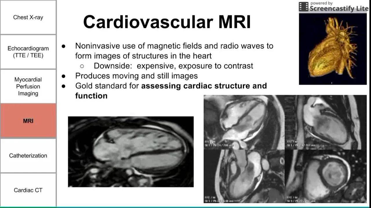
that is not being perfused that is not getting as much blood as it should and this could be of course a risk for a heart attack and could indicate ischemia which is the death of tissues usually due to the lack of blood and as we said this is a nuclear stress test because we're using a heavy element radioisotope and we have imaging before and after the stress to identify areas of the heart that are not being well perfused during stress we also have cardiovascular MRIs this applies the same principles of MRIs which is magnetic fields
in radio waves too to form images of a body it can be used to find us to make moving images and still images downside to MRI is as usual pretty expensive we have moving and still images as we said the best part about cardio or cardiovascular MRI is using it to assess cardiac structure and function and we could see some pretty good images of the heart beating here we have some small animations and there's pretty clear contrast between tissues which is something you don't get with CT or x-rays and that's one of the major benefits
of MRIs next we have cardiac catheterization it's a it's a it's a domain of procedures that involves insertion of the catheter into the chambers of the heart or the vessels of the heart we can use this for therapeutics or diagnostics one example of therapeutics inserting stents through cardiac catheterization when one of the downsides of cardiac catheterization that it is invasive it's not quite as invasive as surgery but it does involve putting a wire and a catheter it's it's it's done under anesthesia but the patient is still awake so it's a local anesthesia and there is
radiation exposure if you are using a contrast agent in the coronary arteries right heart catheterization involves the insertion of a catheter through the vein usually the femoral vein and it's used to measure the right heart pressure or the pulmonary artery pressure and this makes sense because if we're going through the veins and we're going up to the heart we're going to enter through the inferior vena cava into the right atrium and into the right ventricle and we can even keep going into the pulmonary artery to measure pressures there so that's the main use of right
heart catheterization so as we said right heart catheterization is best for assessing intracardiac pressures and it can also be used to look at cardiac output left heart catheterization is is performed by inserting a catheter through an artery usually in the arm and of course if we go through the artery back up to the heart we're gonna go backwards retrograde is the term they use through the aorta into the left ventricle and the left atrium now if we go into the left ventricle and release a dye a dye that can be seen using an x-ray that
dye is then going to be pumped out of the left ventricle into the aorta and into the coronary arteries the coronary arteries will then profuse around the heart and we can get these nice and geography images as you see on the right here these angiography images show the blood vessels the artery is the coronary arteries going around the heart muscle and feeding the tissue of the heart muscle so coronary angiography using left heart catheterization is the best way to visualize the coronary arteries and to identify stenosis and if you look carefully at these images on
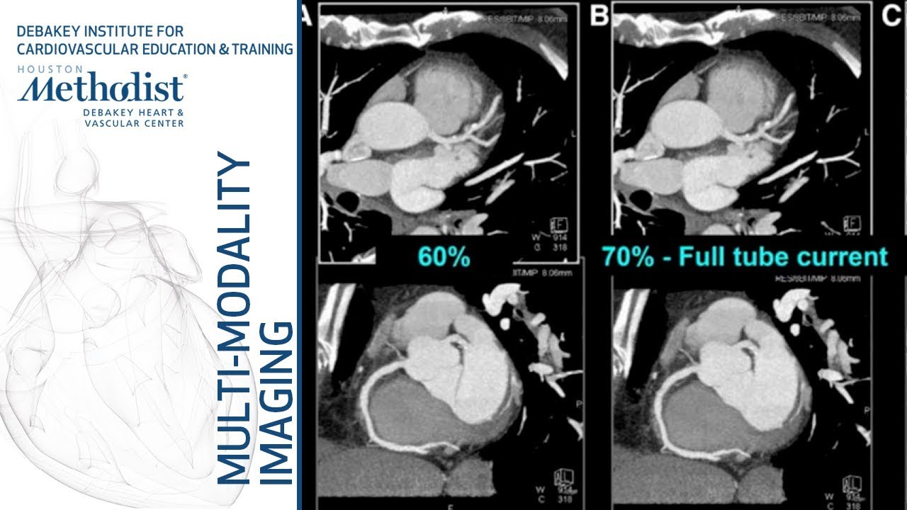
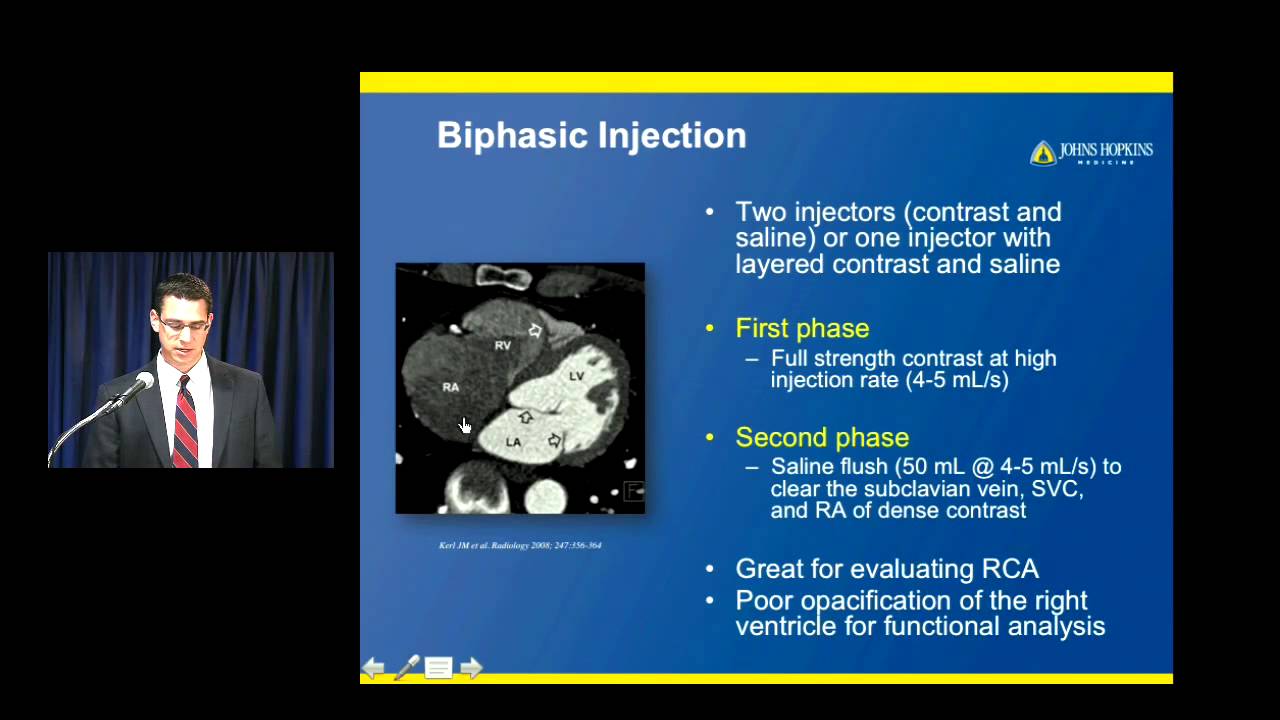
the on the right here we see the white arrows pointing to two areas of stenosis in the coronary arteries two areas that show that dye is kind of cut off is kind of blank for a moment indicating that there's not much blood there's not much fluid passing through the arteries at the point that the arrow is indicating and lastly we have cardiac CT CT is computed tomography and cardiac is obviously computed tomography of the heart which is a more complicated x-ray it's not quite as simple as a normal chest x-ray it's a series of x-rays
that are that are put together by a computer to produce an image and can also be used to produce a three-dimensional image cardiac CT is best to assess the extent of coronary stenosis so we can identify coordinate coronary stenosis best using cardiac catheterization we can we can assess the extent of stenosis we can get a higher resolution picture and see just how bad that artery is being as being clogged up using cardiac CT cardiac CT is also good to look at the thoracic aorta so if we have any kind of a or two problems such
as an aortic dissection cardiac CT can identify that the benefit of cardiac CT is that it's less invasive than catheterization you're not actually putting anything into the body and you can also produce a three-dimensional model which is something that catheterization cannot do and the drawbacks of CT are a pretty intense radiation exposure more so than check stray chest x-rays and more so than MRIs and of course to get some some pretty images you usually have to use contrast that can damage the kidneys so that's all we have for these imaging modalities of the heart I
hope this was helpful and thank you for listening