Researchers at Harvard Medical School have captured extreme closeups of bacterial membranes and their cell wall exoskeleton during cell division, providing never-before-seen views of the processes involved, that may provide new clues for fighting antibiotic resistance. The work focused on the double-layered membranes of E. coli.
Some of the most dangerous bacteria that infect humans have this same double membrane structure, which makes it hard to get drugs into the bacteria and kill them. The researchers used live cell fluorescent imaging to get a full view of the bacteria. This allowed them to see, in real time, how the different cell surface layers changed relative to each other during cell division.
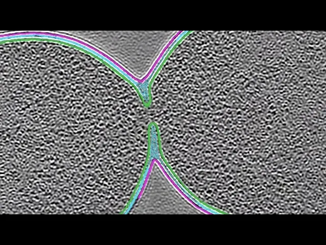
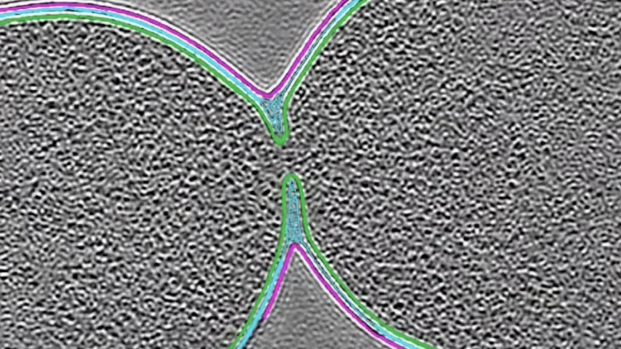
They then used cryo-electron tomography to get the first ultra-detailed, three-dimensional views of what happens within the double membrane structure during division. No other tool could reveal such detail inside cells. Here, a small opening between the membranes of the two daughter cells is captured moments before the cells separate.
The team gained new insights into what controls the push and pull between surface creation that makes a bacterial cell grow longer and surface creation at the division site that allows new cells to form. Studying mutants that alter E. coli’s DNA revealed that this bacterium contains genetic instructions that shape the cell’s division site.
Surprisingly, the mutants divide in several different ways, forming distinct shapes seen throughout the bacterial kingdom. By revealing more about how bacteria divide, the study promises to drive fundamental research and help in combating the worldwide antibiotic-resistance health crisis.