welcome to EKG explained clearly by the end of this course you'll have all the skills needed to confidently interpret EKGs in a systematic way you'll understand the electrical activity represented on EKG paper that allows this process to happen the heart to beat and cycle blood through our lungs and bodies this is the final product whether or not you have experience with EKG interpretation I think you'll find this series of videos very useful if you understand the key foundations of how the heart works it'll be much easier to learn and remember the nuances involved with the
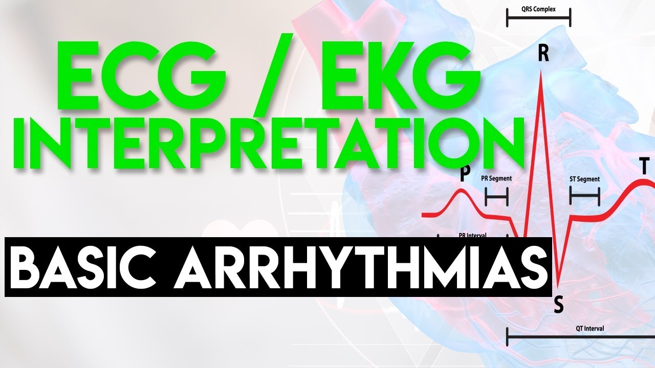
EKG that we'll cover later in this course so we'll start with the anatomy and physiology of the heart depolarization repolarization and then on to leads and specifics about EKG paper and how a tracing is captured next are specifics on EKG tracing the P wave the QRS complexes the QT interval the r2r interval etc we'll cover the impact of our nervous system and neurotransmitters on the heart then on to rate rhythm axis escape rhythms PVC bigeminy tachyarrhythmias ventricular tachycardia and the key differences between ventricular tachycardia and paroxysmal supraventricular tachycardia with aberrancy the QT c and the
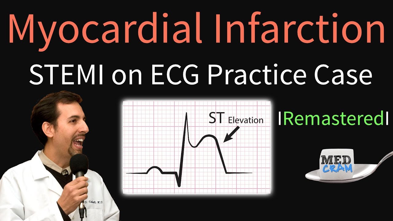
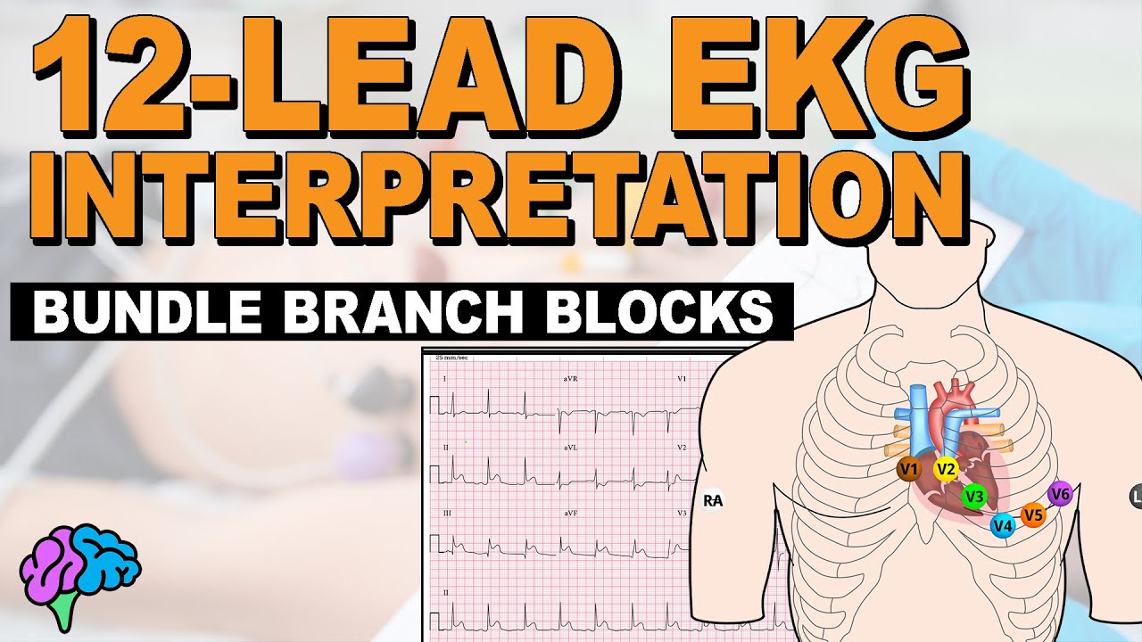
potentially life-threatening Prasad's the points atrial fibrillation and flutter widened QRS complexes and the various types of heart blocks using precordial leads to hone in on certain areas of the heart atrial enlargement and ventricular hypertrophy acute coronary syndromes myocardial infarction and pericarditis we'll talk about T waves ST segment and T wave changes that we see with ischemia bundle branch blocks and vesicular blocks and then at the end we'll put it all together and teach you how to systematically read an EKG we'll go through a normal one first and then on to a variety of abnormal EKGs
liked sinus tachycardia atrial fibrillation multifocal atrial tachycardia first second and third-degree heart blocks h'lo largeman tricular hypertrophy x' hyperkalemia rare anomalies and many many more abnormal EKGs so let's begin with anatomy this is a animation here of the heart beating and there's a number of things that I want you to see so you'll understand this when we talk about the conduction system of the heart and the EKG so the EKG as you know is a way of looking at the electrical activity of the heart and it's really the electrical activity of the heart that is
responsible for what we're seeing here right now just for those that don't know the anatomy of the heart we've got the right ventricle here on this side we've got the left ventricle here on this side and these areas up here are the atria so this is the right atrium here and the left atrium is over here interestingly here what we've got is the tricuspid valve this is a valve that allows blood to flow in from the right atrium into the right ventricle and then gets pumped out through the pulmonic artery and this is the pulmonic
valve here the semilunar valves as you can see and then what you can't really see very well is the aortic valve which is here at the top and this is where the left ventricle is pumping out into there now one of the things that I want you to notice first off is if you look carefully you'll see that the right atrium here beats before the right ventricle right atrium right before the right ventricle and you'll also notice similarly that the left atrium beats right before the left ventricle now this is because of the anatomy of
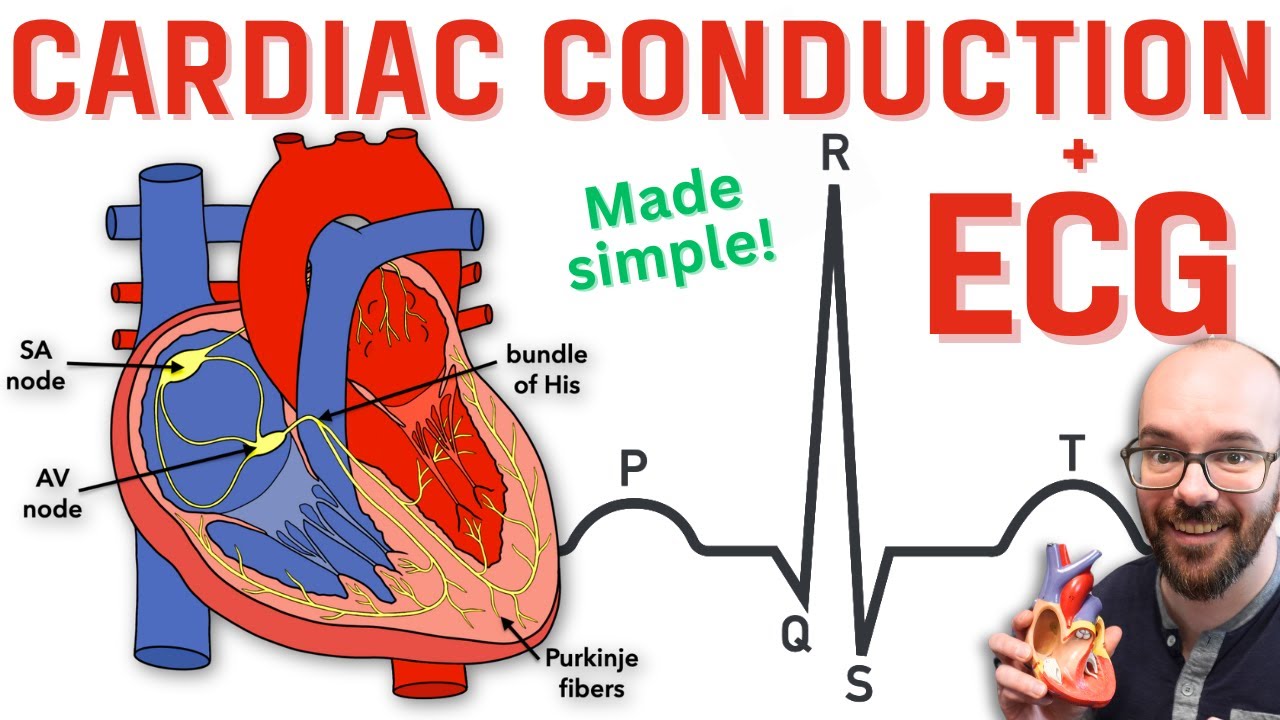
the conduction system there is an electronic conduction system in the heart that is not made up of nerves but instead of modified specialized myocardial sites okay up here is the sinoatrial node which because of its relatively quick activity is the pacemaker for the whole heart it then goes to the atrial ventricular node which is here and then goes down this conduction system here called the hiss Purkinje system and as you can see it goes up and it basically conducts the electric signal to the myocardium now we're gonna talk more about this in detail but I
wanted you to notice that it's this conduction system that basically makes this movement that we're seeing right here like a perfectly well timed Symphony depends on the analogy that you want to use there are several different analogies that you can use the conductor that is conducting a symphony and you want to make sure that the violins and the cellos and the flutes and the viola and the timpani they're all acting all at the same time so you get the maximum effect in other words everything is working in concert the other way of looking at it
is a little bit more destructive you know that if people are trying to demolish a building they will have certain areas of the building that are drilled out and weakened with sticks of dynamite and when they activate that dynamite it has to be activated in just the right order in just the right fashion so that the building comes down right on top of its footprint well this is kind of what's going on here with the heart this electric conduction system the sinoatrial the atrioventricular and the hiss Purkinje system is activated in such a way that
the electrical conduction goes and stimulates the heart in concert so the electrical conduction starts up here and then goes to the ventricle the reason for that is so that the atria contract right before the ventricle the purpose of that is to get the blood from the atria down into the ventricle to increase the size of the ventricle right before the ventricles contract that causes an increase in freeload and allows your cardiac output to be improved that's happening on both sides so you can see that the atria are contracting first and then the signal is placed
down here to the history Kinji system it travels down very quickly down this hiss Purkinje system and then up here into the myocardium so the effect is is that essentially the myocardium is contracting all at the same time and you want that to happen instead of having contraction occurring from the top down you want contraction occurring all at the same time because of this very unique conduction system this is going to look very unusual or look very unique I should say on an EKG what is an EKG it's a way of measuring current you know
that when you take a battery and you place it on a voltmeter if you put the positive end to the positive meter and the negative end to the negative meter you're going to move the dial up so that you see that there is a difference in the voltage when that voltage starts to move that is when you have an electric current and so what an EKG does is it sees how is this depolarization or this movement of positive charges that's going down the hiss Purkinje system how does it look from the electric vision if you
will in other words if I have an ultrasound machine I could see if there is fluid or fluid moving on the ultrasound what an EKG does is it does exactly the same thing except instead of fluid moving and objects that you could see you're seeing electricity moving and so that is what we're going to look at today atria contract first then ventricles contract second but they're contracting in concert and then what happens is everything resets back to normal and you have the same contraction happening again the next series that we're going to talk about is
we're going to break things down to the smallest level and then we're going to build it up so that we can finally see how we get back to this point again okay so zooming in a little bit more now in terms of where we were let's pull away all the muscular activity here and let's look specifically at this conduction system I want to be very clear the thing that you've got to understand that these are not nerves okay these are modified myocardial cells that conduct electricity very quickly and we'll talk about how that happens so
the first one that you've got to know is the sinoatrial node it is at the very top and the reason why it is there and as the pacemaker is because its intrinsic activity is the fastest and since it is the fastest it's going to cause depolarization to occur all the way down and everything else is going to have to be in concert so this is kind of the conductor of the concert now there are different pathways to get to the AV node you can see here that the SA node can to the AV node now
the AV node is right here and it kind of holds things up it kind of delays things we'll talk about that in another lecture so that what you have here is you have the sinoatrial node saying okay it's time to contract and we'll talk about how that happens and the atria contract and then the electrical conduction gets down to here and it holds up what that effectively allows the heart to do is to pump blood from the atria down into the ventricles and allows it the time to pump it member this happens so quickly SA
node to AV node then when the blood is in the ventricle the AV node then conducts this depolarization that's occurring into the Hispano system and it travels down and around so quickly so very quickly that essentially the entire myocardium that this innervates okay or that it penetrates basically depolarizes almost all at the same time so that again is sort of a map of this cardiac conduction system and really it's this cardiac conduction system that allows the depolarization to occur in such a nice and timed way now on an EKG when we're actually looking at the
electrical depolarization are we actually seeing the electrical movement down this conduction system the answer is no this is such a small amount of electricity that it's almost imperceptible on an EKG what is it that we're actually measuring what we're measuring is the depolarization of the muscle the depolarization of the muscle is very large and amplitude and that is what we are picking up on the EKG kind of like if we are demolishing a building what do we see on the video do we actually see the electricity going into the wires to the dynamite no we're
actually seeing the dynamite blow up and the building coming down that's in other words what we're seeing on the EKG even though there is electrical conduction going down this what we're seeing electrically on an EKG is the depolarization of the actual muscle cells so just be aware of that okay so to get a little bit more of the insight about what's going on let's go down to the microscopic level and I want to show you a myocyte here now the thing that you should know about a myocyte is just like any other skeletal or smooth
muscle cell it has a collection in it called the sarcoplasmic reticulum we'll call that SR and the SR is full of calcium and the reason why it's full of calcium is because this calcium can be released into the cell upon depolarization of the cell we'll talk about that and that's gonna cause contraction of the striated muscle so we'll just put a bunch of muscle cells here it's going to contract okay and if you were to look histologically at the areas of the myofibrils that are in cells you'll see how muscle cells work but basically calcium
is what's going to activate them to contract and they're made up of troponin and all of these other things that we won't get into the key here though is the sarcoplasmic reticulum causes calcium to be released and that's what triggers it the question is is what triggers the sarcoplasmic reticulum well what happens is as you may know on the cell surface of these cells you have a sodium potassium pump and the purpose of that is to pump sodium out of the cell and as you know sodium has a plus one charge and at the same
time pump in potassium and potassium has a positive one charge but it pumps out three sodium for every two potassium and so what ends up happening is is there's a very high concentration of sodium okay one plus outside the cell and there's a very high concentration of potassium inside the cell now as it turns out the cell is very important so sodium stays very high outside of the cell the way of thinking about it is because three are going out and two are going in there is a negative charge on the inside of the cell
okay negative charge on the in of the cell and there are positive charges if you will on the outside of the cell we'll ignore those for now because the outside of the cell is very very large and the inside of the cell is very very small so as a result of that there's a far more negative charge that we see here now the other thing that's important to know is that the cell while it is very important it is very permeable to potassium and as a result of that potassium leaks out somewhat now as it
leaks out it's losing a positive charge so that even makes it more negative and as a result you have a very negative resting potential on the inside of the cell now with that basis of history let us go forward here in the next lecture and talk about why this interior negative charge is so important to the electrical conduction that we're going to talk about here in our EKG course thanks for joining us