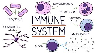
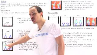
welcome to my new video series known as bio techniques explained in less than five minutes where I explained biological concepts in less than five minutes or so so if you haven't yet subscribe to my channel please subscribe and hit that subscribe button so today's topic is enzyme linked immunosorbent assay or Eliza so this assay is a colorimetric test that uses antibodies and color development reaction to identify as certain substances like antigens or it could be also used for detection of antibodies now Eliza gives a qualitative and quantitative information about the presence of an antigen or
an antibody Eliza is widely used as diagnostic test for many viral diseases including HIV AIDS now in Eliza the reactions are set up in a 96-well plate and that is read in a common metric machines so where which is a colorimetric detector basically which measures the absorbance of specific wavelength and the result is displayed in a computer screen and the result is basically absorbance versus concentration from that we understand how much we get the quantitative and qualitative information about how much quantity of substance is present now there are three variants of Eliza we would discuss
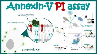
one by one by one so the first variant is indirect Eliza which is used to detect an antibody against a known antigen so when we know our antigen we want to understand whether there is unknown and what antibody binds to it we would do our indirect Eliza in this situation wells of the microtiter plate are coated with known antigen here the antigens are marked in yellow then the antibody to be tested is added into the wells hopefully that if the antibody is specific against that antigen it would bind to that antigen now our enzyme-linked secondary
antibody is given and while substrate is provided the enzyme would give rise to product and that results in color development so if the color is developed that is an indirect proof that the sample that we provided has that specific antibody against the antigen which is coating the surface of these 96 for the plate so that is how we understand whether our antibody is present in a particular biological sample example a patient's blood or a patient serum etc so this kind of technique is used to detect HIV so let's say this human is infected by a
HIV virus and have has AIDS so it's a hiv-positive human being so as for time for diagnostic tests his blood is collected and centrifuge to form the serum so from the from the serum fraction we could get the antibodies generated against the HIV virus proteins or HIV viral antigens so we know if HIV virus effect that person and the person is HIV positive so the antibodies would be produced in his body if we can detect those antibodies then we would have a idea that whether the person is infected or not so definitely in order to
understand that we would do a indirect laser so we would see if the antigen is present that means the person is HIV positive if the antigen if the antibody is absent the present is HIV negative now in a lab what would happen is they would give the patient serum serum into the well so which is previously coated by antigens present on the viral surface so if the serum contains the antibody against the viral antigen then it would definitely buy it in the antigens coated on the surface of the web and thereby when we put the
secondary antibody enzyme-linked it would develop a color yeah upon giving a substrate but on the other hand side if the patient serum doesn't have that antibody produced that doesn't have the antibody produce should that should be produced in a viral response then there would be no color reactions because the secondary antibodies doesn't bind to a primary antibody hand unable to give a color reaction so that would be HIV negative there are other variants known as sanducci lyza which is used to detect antigen of interest for example we wanted to detect an antigen with our hand
each end is present in a patient's blood sample or not so what we can do we know that antigen binds to specific monoclonal antibody so we can coat the well of the microtiter plate with monoclonal antibody after that we can give the patient's serum or patient blood or anything like bodily fluid like that and then we can give another monoclonal antibody coupled with enzyme and then we put the substrate now if their second if the monoclonal antibody coupled with enzyme is binding to that particular antigen then in a color would develop that would tell indeed
that antigen is present in the patient sample or patients blood so that is how we understand whether antigen is present in the sample or not whether indirect eliezer tells us about whether antibody is present or not now once we know and the gene is present another question is that how to reject how much antigen is present and competitively lanza give us an understanding about that so you're going to be competitively Liza you know you you are you are asking a question that how much antibody is produced in a patient sample or huh so definitely how
much antigen is produced in a patient sample so what you would do you previously incubate some antibodies which you know it is produced against these yellow antigens and then what you would do would coat a surface of the well DCL Oh antigens now if the antigens are present in ample then all the antibodies we provide against it would bind and there would be very less amount of free antibodies available to bind to these antigen coated surface so once we give it so what would happen is most of the antibody would be enough floating condition because
it cannot bind to the antibody Co and the antigen quote of the quills so ultimately faint color would be developed so there are two possibilities one is there is a dark-colored reaction happening that means there is very I mean if the dark color is developing the antigen concentration is very low because as the antigen is low when we incubate with antibody few antibodies bind to the antigen but most of the antibodies are now free which can in turn burn bind to the antigens coated onto the surface of the well and when we provide secondary antibody
it can give rise to a color reaction but if there is too much of antigen present then once we provide our antibody against that antigen almost all the antibodies are occupying that antigen and now when we provide the whole solution into the Eliza will if there is no more free antibody which can bind to the surface coated antigens that's why once we provide the secondary antibody after you faint color or no color is developed so that is how we understand that how much antigen is present so if it's more antigen then so if a lot
of antigen is present then a very faint color would be developed in a competitive in East Eliza but if there is very less amount of antigen then the dark color would be developing in the competitive Eliza so that is all about Eliza so I hope you enjoyed this video if you liked my video give it a big thumbs up don't forget to Like share and subscribe thank you
![ELISA [Full Training Video]](https://img.youtube.com/vi/R416jtA6gCE/mqdefault.jpg)