the liver is the largest internal organ in the body and it weighs about 1.5 kgam it's surrounded by a capsule of fibrous connective tissue called gissen capsule if we look at the liver from an inferior view which is A View From the Bottom of the liver we can see that it is divided into a large left lobe and right lobe as well as two smaller loes called the quadrate and cadate loes the liver parena or functional tissue of the liver is organized into thousands of hepatic lobules which have a dual blood supply that comes from
terminal branches of the hepatic portal vein and hepatic artery the blood then flows through sinusoids surrounded by hepatocytes before draining into the lobules central vein hepatocytes are the main functional cells of the liver that perform a large variety of functions including the production of bile a number of plasma proteins and non-essential amino acids the metabolism of fat carbohydrate and protein the storage of glucose vitamins and iron and the breakdown or detoxification of metabolic waste products drugs and toxins the hexagonal shape of the H hepatic lobules can be identified by their slightly darker edges and the
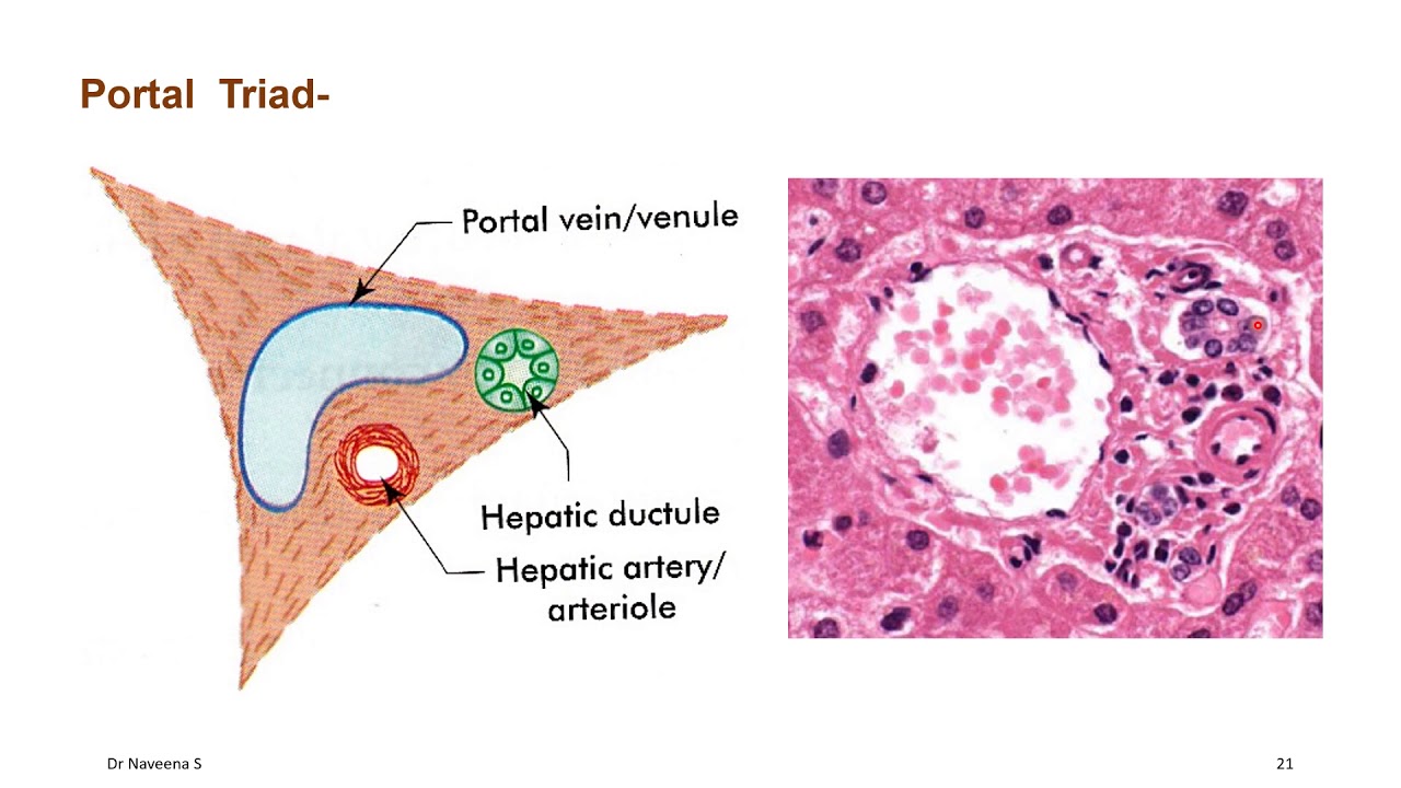
prominent Central veins in the center of each lobule the portal Triad consists of a bile ductule portal venal and arterial after identifying the lobule it can be easier to locate portal Triads in an image since they're typically located at the corners of the lobules if we take a closer look at just one portal Triad we can more easily identify the portal venel by its large diameter and thin walls compared to the arterial which has a much smaller diameter and thicker walls similar to this image the portal tract can sometimes have more than one bile duct
the bile ducts can be identified by their prominent simple cuboidal epithelium also in this image are a couple small lymphatic vessels which have even thinner walls than the venule now let's take a closer look at the hepatocytes which are large polygonal epithelial cells that form branching plates that are only one cell thick separated by sinusoids and radiate outward from the central vein the sinusoids blood from the hepatic arterial and portal venil to the central vein while the bile canaliculi or capillaries carry the bile produced by hepatocytes in the opposite direction in order to drain into
the bod ductules the hocy cytoplasm is very eosinophilic or pink because they contain a lot of mitochondria many of the cells in this image also have fine Brown granules within the hepatocytes called lipop fussin these granules are considered a sign of where and tear and it's normal to see the amount of lipop fussin gradually increase with age the hpy plates have a supportive tissue or stroma of reticuline fibers these fibers are difficult to see with an h& stain but a reticuline specific stain can be used in order to visualize the fibers and hocy plates better
a reticulin stain is actually used to help diagnose hepatocellular carcinoma since a liver with plates that are more than three cells thick is diagnostic which we can see in this image all right as a quick recap the liver is organized into thousands of hepatic lobules at lower magnification the lobules can be identified by their prominent Central vein as well as a slightly pale central portion of the lobule compared to the edges of the lobules the portal Triads are found at the corners of the lobules each portal Triad contains a portal venil hepatic arterial and bile
duct the venil can be identified by a large Lumin and thin wall the arterial will have a thicker wall but a much smaller diameter and the bod duct can be identified by its simple cuboidal epithelium the hepatocytes are large polygonal cells with very eosinophilic cytoplasm and basophilic nuclei the hepatocytes are organized into branching hocy plates of cells that radiate out from the central vein and lipop fussin is a sign of wear and tear seen within hepatocytes as fine Brown granules helping current and future clinicians Focus learn retain and Thrive learn more