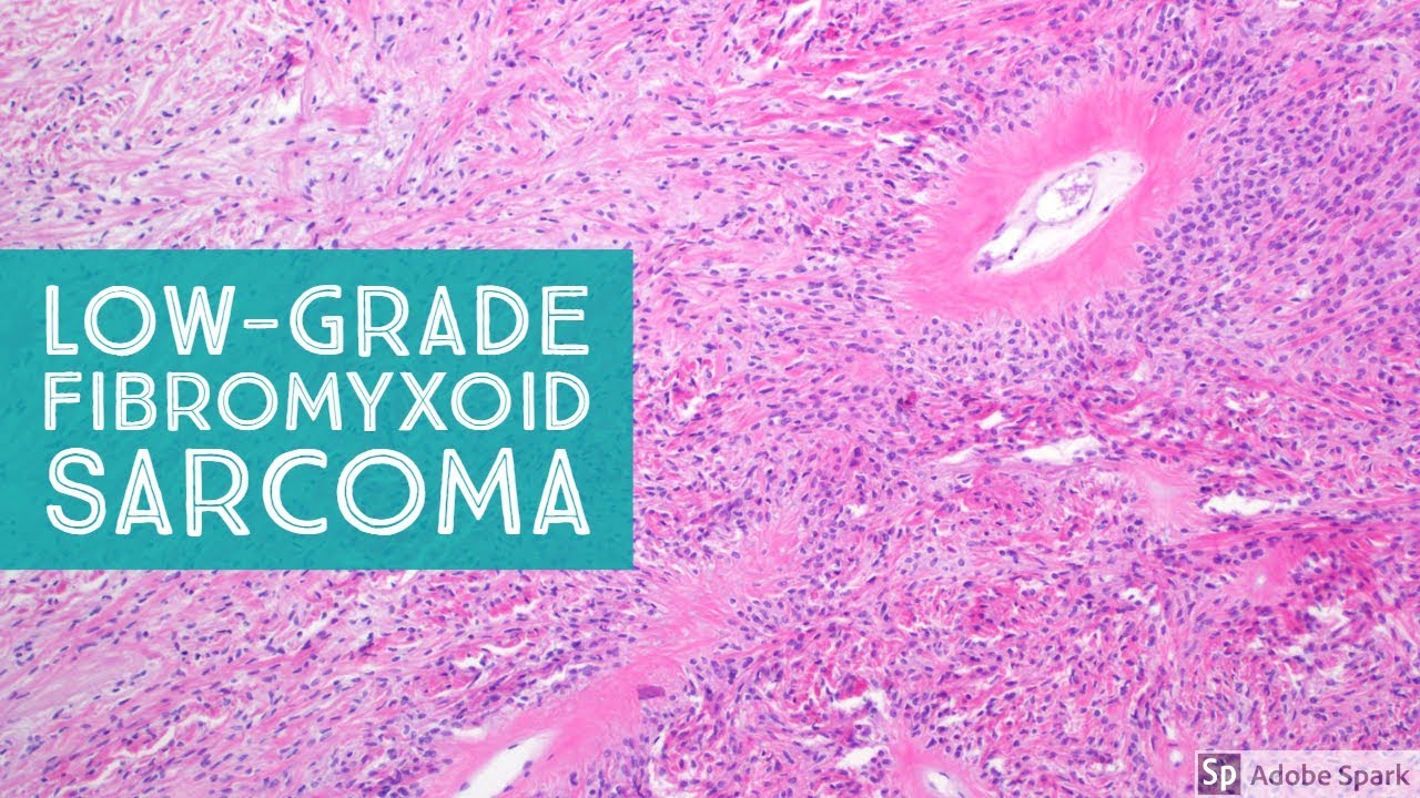
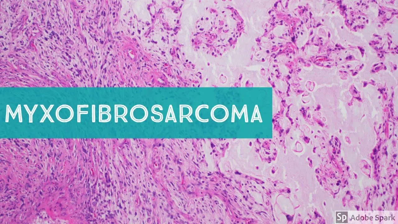
hi I'm Jared gardener and today we are going to be discussing Mick so fibrosarcoma so this is one of those tumors that's confusing because there's another soft-tissue entity that sounds very similar and that's low-grade fibro Mick's oeid sarcoma so it's really important to not confuse those two the name sounds similar but they're totally unrelated and very very different tumors they're actually quite easy to tell apart and I'll have another short video to quickly discuss the ways to distinguish mix of fibrosarcoma what we're talking about here from low-grade fibro mix oeid sarcoma mix of fibrous sarcomas
are pleomorphic sarcomas they're run a range from low-grade to intermediate grade all the way up to high grade at the high grade end they have areas that resemble undifferentiated pleomorphic sarcoma and they probably represent at least the high grade ones I think probably represent a mix of undifferentiated pleomorphic sarcoma and I'm gonna tell you the features here these are these are you know all sarcomas are rare compared to other types of of usual cancers like carcinomas but among sarcomas mix up fiber sarcoma is one of the more common ones at least in my practice I
see these on a regular basis usually a couple times a month at least in a routine sarcoma service work so a few things they usually occur in elderly patients they're in older adults very very rare and young adults and I've personally never seen a case in a child there are some reports in the literature although I still remain skeptical a little bit about those I just have a hard time understanding how it occurs in a young patient so it usually is older adults the lower extremity is the most common sight but also you can see
it on the upper extremities or other places but usually the lower extremities of elderly patients they are oftentimes in deep soft tissue but another you unusual thing about these tumors compared to other sarcomas is that they tend to involve the suprafacial these the superficial soft tissue above the fashion layer so the sub cutest in the dermis about half of cases go above the fascia up into the sub cutest or the dermis so that's important to know and the other thing is many sarcomas and in deep soft tissue at least are very round and circumscribed they
grow quickly they push other tissue out of the way but as you can see here this tumor is very very infiltrated look it's playing apart these skeletal muscle bundles and infiltrating into the soft tissue beyond and because of that they tend to extend much farther than it appears clinically they tend to extend out if it looks like a you know 3 centimeter mass on the skin of the thigh once the surgeon takes it out they may think they'll have wide margins around it but the tumors often going to extend out to the margins even when
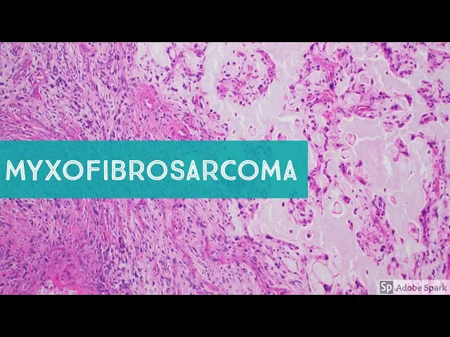
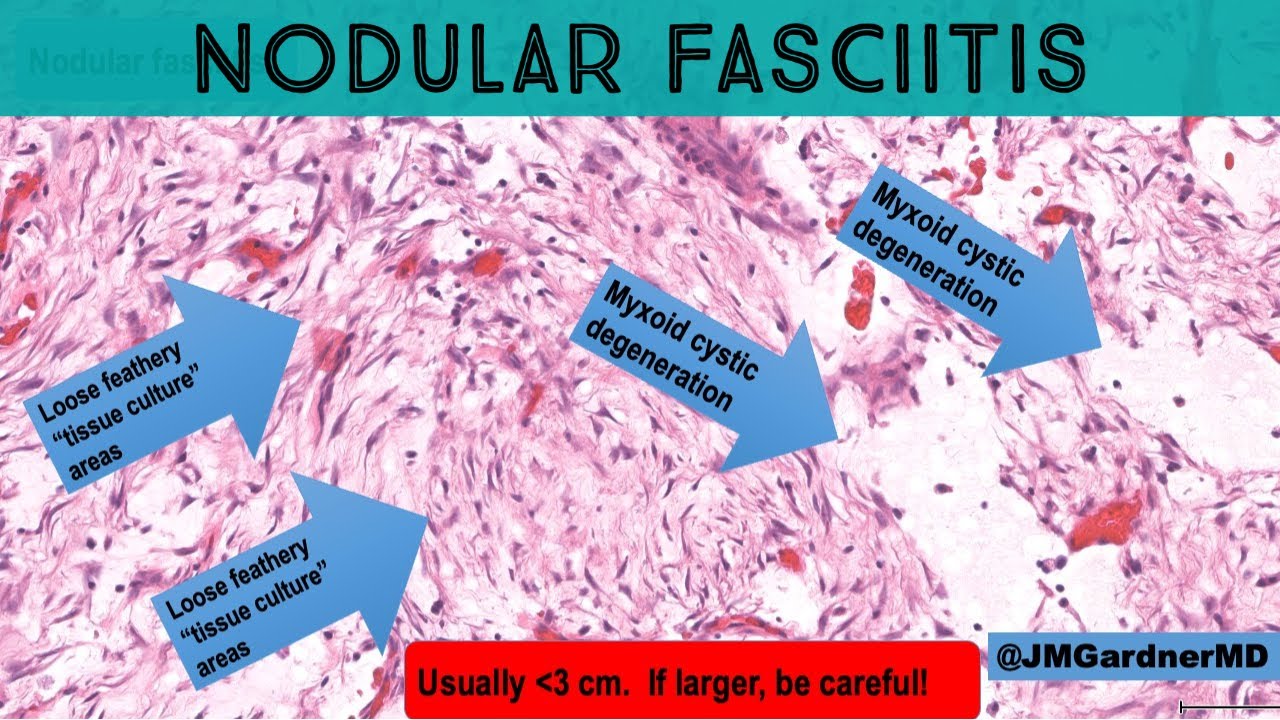
the surgeon thinks that they've gotten around it and so that's a problematic thing and if you don't clear these tumors because of their infiltrative growth they have a high risk for local recurrence ok so let's talk about the histologic features we've covered some of the clinical information that's really important and important to know first of all these tumors as the name suggests they are mix alike they have a blueish background and areas that are hypo cellular like this ok the low-grade forms are almost entirely hypo cellular with bluemix lloyd background at the high grade end
you might only have small pockets of mix oeid stuff and most of the rest of the tumor will be sheets of high-grade pleomorphic sarcoma so the mix toyed change is abundant and I think that's one helpful way to remember mix so fibrosarcoma the word mix though comes before fibro they usually look more blue than pink as opposed to low grade fiber mix so in sarcoma which the fibro word comes before mix oh they look usually more fibrous and pink and less mix Oy in general there are exceptions to the rule of course and one thing
you can begin to appreciate we're on Forex objective here already you can see big hyper chromatic pleomorphic cells scattered in the background that is basically the the hallmark feature of this tumor to make a diagnosis of mix of fiber sarcoma you essentially always have to have hyper chromatic pleomorphic atypical cells at least some scattered even in the grade one low-grade form there should be pleum or fizzing nuclear atypia and the pleum or fizz 'm is is a sign of what's going on molecularly these tumors like almost all other pleomorphic sarcomas they have random chromosomal gains
and losses they don't have any recurrent translocation translocation sarcomas usually have uniform monotonous cells and aneuploid tumors that have random gains and losses usually have plea amorphous of some of the daughter cells are small some are big so you'll see a typical mitosis and pleomorphic cells that's true for most pleomorphic adults are comas so here you can see these big ugly nasty hyper chromatic pleomorphic cells no one even a medical student a look at this and say I think that cells a typical right and it's because it's so ugly and so much bigger and nastier
looking at its neighbors but even the background cells that are smaller you can still appreciate really have a lot of hyper croatie very atypical nuclei and the tumor cells are floating around in this background of blue ground substance myxoma Tyrael hyaluronic acid and other other glycosaminoglycans in the background here so that's a very characteristic thing ugly tumor cells floating in a mix or background okay the other feature that you often see in mix of fibrosarcoma are these blood vessels look at how prominent the vascular network is and the vessels tend to be long and they
kind of gently undulator kind of curvy linear they're long and kind of slightly wavy you can you can imagine this vessel probably goes a lot longer than we're seeing but see how it goes up and down it's this kind of curvy oh I don't have it quite on the screen here there we go this one you can see it's going up and down up and down this kind of curvy linear elongated vessels so that's a very characteristic feature of mix of fibrosarcoma as well and like I mentioned look at the tumor infiltrating out here it's
clearly splaying apart the muscle you can easily tell that's tumor not just reactive mix oil change in the soft tissue that's tumor that's still tumor now it's a infiltrated beyond the muscle and out into the fat and we see normal blood vessels in the background still see tumor cells in there and the tumor will go all the way out and it's probably positive out at the margin somewhere so again this is a very characteristic finding on imaging at least if imaging studies are done the deeper soft tissue masses when mix of fibrosarcoma is a large
deep mass if they've done an MRI or CT you often can see these strands of tumor extending out from the main tumor mass and in between the fashio in between the muscle along the the septa I'm sorry along the subcutaneous septa and along the fashion layer that divides the sub cutis from the skeletal muscle underneath so there's a nice example again of those very prominent elongated vessels now sometimes you can see a little bit of delicate vessels that are a little branching don't confuse this with a mix oil i po sarcoma no way is this
going to be a mix or liposarcoma the reason is mix with liposarcoma x' they do have mixed Lloyd change they are hypo cellular they do have delicate branching vessels but they rarely ever have pleomorphic again because they're translocation associated sarcomas so when you see big ugly cells in a mixed Lloyd tumor you are almost almost certainly not dealing with a mixed so I'd like the sarcoma even the high-grade round cell form of mix Lloyd liposarcoma still has relatively uniform cells they're round and they all look like each other because they have the same molecular abnormality
this tumor again has random gains and losses here's another important point that is often seen in mix of fiber sarcoma look at this cell right here let's see if we can get them back on the screen now this cells very plain or 'fuck but it has bubbly cytoplasm and you might be tempted to think that it could be a pleomorphic lipo blast now plea a morphic lipo blasts are not seen in mix oyd liposarcoma but they are seen in pleomorphic liposarcoma and occasionally in d differentiated liposarcoma we'll talk about that in another video but this
is not a lipo blast this is a pseudo lipo blast and it's a common finding in miksa fibrosarcoma and you can also see it in other types of sarcomas that have prominent mix oi backgrounds what's happening here is the tumor cell is sucking up the background mix or material it's eating it up and it's making vesicles filled with that same bluish mix oide material see how this is blue and mix oeid the inside contents of each of these vacuoles is blue and mix oi just like the background that's the easiest way to tell this apart
lypa blast should have totally clear white looking back heels because they're empty they were filled with lipid during processing the lipid washes away and dissolves during tissue processing and what's left behind is an artifact xual space a round circle that often indents the nucleus sudo Lipa blast on the other hand the vacuole is usually are not perfectly round because they weren't lipid lipid has to be a spherical shape in aqueous solution our bodies are made of water so lipid droplets make bubbles look at look at adipocytes look at sebaceous cells look at true light but
bless they all have perfectly round little bubbles that are white and washed out and have nothing inside them so a true lipid droplet should do that whereas mix soy Tyrael it's been sucked up and eaten by a tumour cell will make these kind of loose irregular floppy looking vat of vesicles or vacuoles in the cytoplasm it'll of a bluish color it may sometimes indent the nucleus but it won't be that sharp scalloping that you usually see in a lipo blast most of the time see there's maybe a little exception down here there's a little indentation
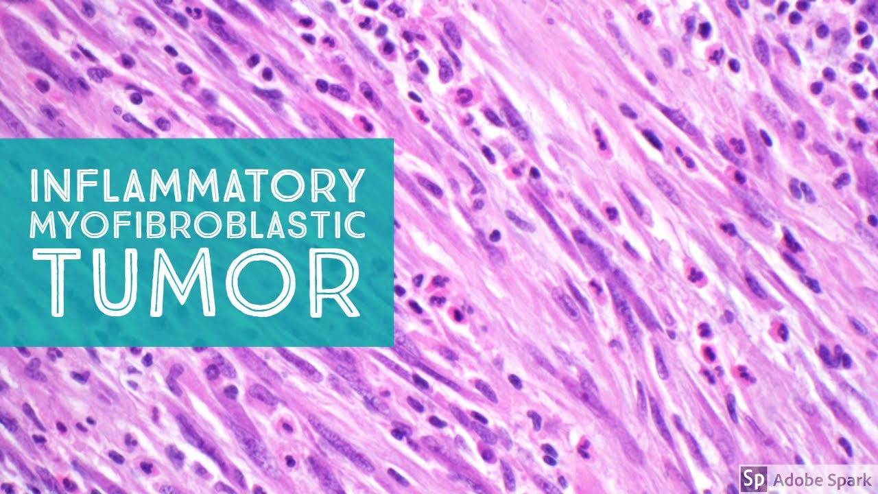
of a nucleus here but so see the the main thing to me that helps is just recognizing that there's blue stuff inside the vesicle that's not a lipo blastn that's a pseudo lipo blast and it should make you think of miksa fibrosarcoma also mix so inflammatory fibroblastic sarcoma a much more rare and unusual tumor we'll have to talk about in a different video look here's another one to other ones this one is you can see the tumor nucleus over here maybe this is a next-door-neighbor nucleus lots of bubbles here all of them are blue here's
a tumor oh that looks maybe like it's clear and indenting the nucleus but look right next to it blue stuff so again these are pseudo lipo blasts this is a very common feature in miksa fibrosarcoma let's look at a few more cases now here's an example in the skin and like I said these I also do dramatic pathology in addition to sarcoma pathology and I think this is a really important tumor for dermatologist and dermat of pathologists to know about because they will often when they get into the skin they can be kind of small
only a few centimeters they may look like a cyst or a lipoma which is a common thing many sarcomas and other rare malignancies like malignant sweat gland tumors things like that that occur in the skin they're deep to the epidermis and so they often get misdiagnosed clinically as a lipoma or just a cyst because they look like a skin colored nodule like a cyst would look like an cysts are much more common than sarcoma so it's understandable how this happens but it's important to remember that they can sometimes get be thought to be benign clinically
and then once the the dermatologist or the surgeon removes the lesion it becomes clear that it's not a cyst and then it gets sent in for consultation and I end up seeing them so this is one here you can see it's involving the dermis and this is a very hypo cellular case so a case like this I would probably call based on this area here I would call this a grade one and I you could also say it's a low grade miksa fibrosarcoma but I really hate doing that even though it's technically correct because it
sounds so much like low-grade fibro mixed soil sarcoma which is incredibly confusing for everyone else except for soft tissue pathologists so what I like to do is say mix of fibrosarcoma comma grade one and that helps to distinguish that by the way the name is a written out to help people distinguish that this is not low grade fabric mixed lights are coming this is mix of fibrous sarcoma that is low grade and again the key is hypo cellular you could even think it might be a myxoma but even from this power you can see big
hyper chromatic cells floating around in here myxomas benign myxoma should never have hyper chromatic ugly cells floating around at least not in my experience so again you can see here the backgrounds of mix Lloyd stuff sometimes we get artefactual little bubbles in the mix Lloyd material I don't always see this just sometimes happens on processing but you can clearly see the tumor cells are hyper chromatic and pleomorphic very atypical mitosis if you look around mitosis are usually present they're often typical but in a hypo cellular grade one lesion like this the mitosis may be infrequent
or hard to find but you've got ugly pleomorphic cells and they're floating in a mix light backgrounds if you look around we'll probably find vessels you can see some elongated vessels in here and the vessels these are delicate but a lot of times they're thicker and bigger than the vessels of say a mix oil I post sarcoma but again you don't have to worry about that because you've already got the plea morphism mixer liposarcoma is almost never gonna have plea morphism look at those beautiful pseudo lipo blasts there sorry I know beautiful is it a
great term to apply to sarcoma but those are a really great example of tumor cells that have sucked up and eaten a lot of that mix soy bluish stuff and even though they look bubbly if you look closely at least on my scope you can see they have a bluish color they're not true lipo blasts alright and then let's go back to lower power here and look we got one nodule here and it's infiltrating up into the dermis but look what it's doing in the sub cutest remember the sub cutest is made of mature adipose
tissue arranged in lobules the lobules are divided by thin fibrous septa and we often in normal subcu tusen skin we don't often see those septa very well but in pathologic processes whether reactive or inflammatory or tumor all we often can see the subcutaneous septa highlighted more clearly all right and look what's happening here you can see the SEP that is very widened here's a here's one fat lobule here's another one here's another one and look here is fibrous tissue expanding that lobule but look expanding that septa excuse me septum and look closer though ugly hyper
chromatic tumor cells are in the middle there they're infiltrating between the fat and look where they're going down here you can still see tumor going along here there's still more tumor we're way away from the main tumor mass and we're coming all the way out to the Inked margin here and we still have still have tumor cells so grossly it would sometimes be hard to see these subtle areas infiltration certainly it's going to be hard even for experienced sarcoma surgeons to detect these I work with some really awesome start a surgeon's and there's still times
that they're surprised when the margins are positive because they really thought they were rounded in the operating room and again this is because the tumor is sneaky and infiltrative not because the surgeon doesn't have the skill to recognize it it's just these are hard to murrs to see the edges of and you have to plan accordingly so I usually add comments to my report when I diagnose this tumor that they're often very infiltrative they're often hard to clear surgically you have to really get around them with a wide margin and you should follow these patients
because they do have a higher risk of local recurrence okay so this is a great example again here's the the main kind of tumor mass over here and it's infiltrating all the way out here along the fascia and the in between the subcutaneous lobules on the the septa okay here's another example in the skin again look at that infiltrate of growth I always worry about what will happen if I ever get a superficial biopsy of one of these so if I see on the extremity of an older person if I see anything mix oeid and
with atypia in the dermis I like to usually add a comment that you know make sure that to the to the dermatologist to make sure that this is not part of a bigger deeper mass because it could be as superficially sampled mix of fiber sarcoma in that setting and again you can see the the pleomorphic cells the elongated there's the cells lots of them here's the elongated vessels the mixed backgrounds and down here though it becomes much much more cellular and you have stuff that starts to resemble what you'd see in an undifferentiated pleomorphic sarcoma
here we just have cellular sheets of pleomorphic very ugly tumor cells so in an area like this I would just call this an undifferentiated pleomorphic sarcoma if this was all I had and I had done stains and they were they were negative for the usual markers but out here you see it transition in the areas that are becoming less cellular more mixed soil long elongated curvilinear vessels look here's an atypical mitosis I told you usually we'll see those if I can get it back on screen there it is atypical mitotic figure so I would call
this a mix of fibrosarcoma high grade or grade three and occasionally you'll have some that are kind of in between and you call them grade two and that the exact grading of mix of fibrosarcoma can be a little bit tricky and subjective and so not everyone does it quite the same way to me the most important thing is recognizing the ones that are totally hypo cellular and don't have any cellular areas if I start to see any areas where the cells are really clumping together like this then to me it's at least the grade -
if it's large areas like this then it'd be a grade three that's the way I do it and both grade two and grade three in my opinion should be regarded as high grade sarcomas with metastatic potential the risk of metastasis is actually a little bit less in some studies than for say other pleomorphic sarcoma is like a like an undifferentiated pleomorphic sarcoma but they still have full-blown metastatic potential and again the local recurrence is a much more common issue here than with a lot of other sarcomas the low grade grade one mix of fibrous sarcomas
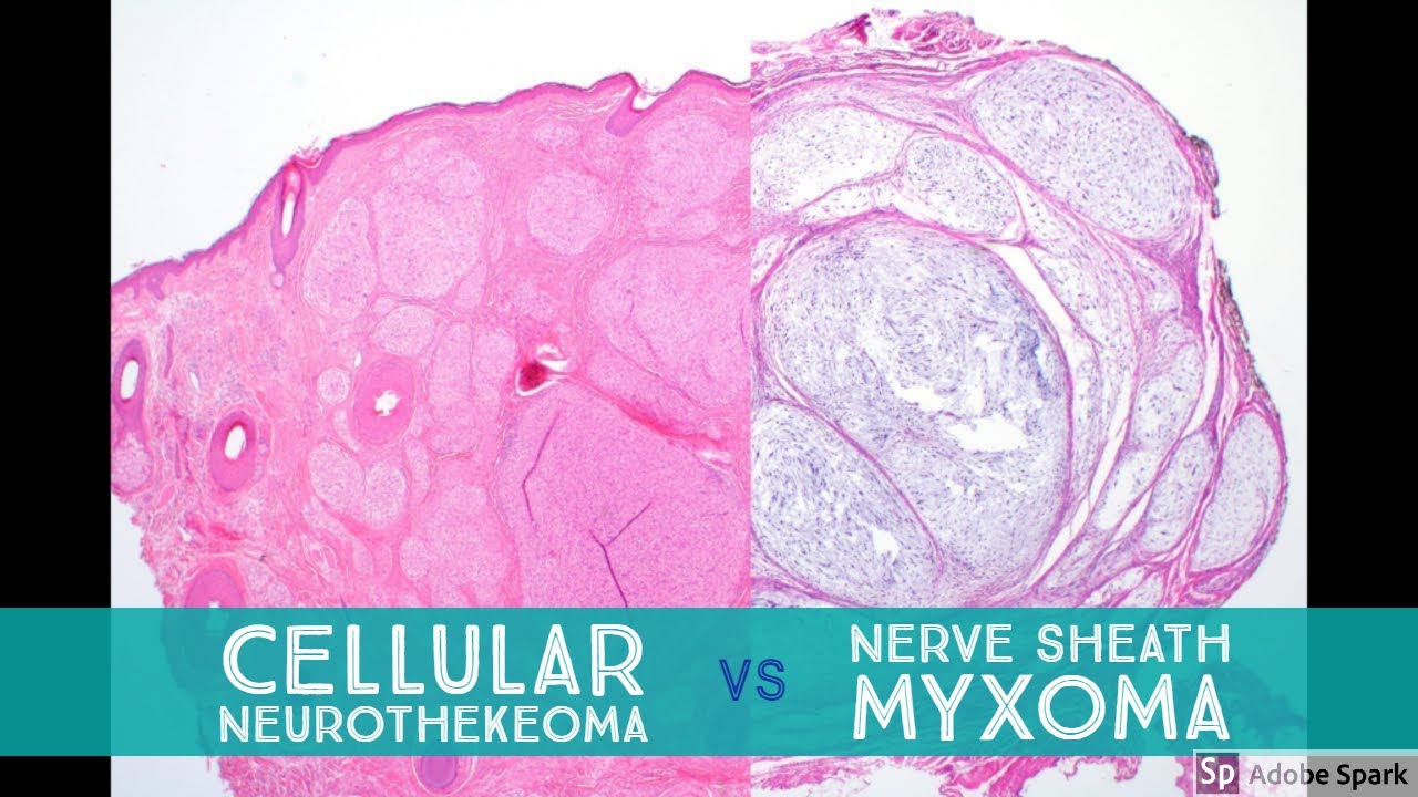
when they are confined to the sub cutis and skin and they're just grade one they almost never metastasize so I think it's important to distinguish those very hypo cellular grade one examples like the here's an example here here's a nodule in the sub cutest it's small it looks hypo cellular and mix oi you could think certainly from low power you could wonder about is this a myxoma but once we get closer even though it's not very cellular it is very plain or 'fuck very hyper chromatic the nuclei of myxoma are small kind of bean shaped
nuclei they usually a pale chromatin these guys are ugly sometimes you'll get multinucleated hyper chromatic cells sometimes it's hard to get the nuclear detail to show up on the video but you can see how hyper chromatic and ugly these cells look so even though they're scattered if I see something that looks like a myxoma and it's got scattered ugly pleomorphic cells then it's probably a mix of fibrosarcoma one important pitfall to avoid is that particularly for intramuscular myxomas they can entrap skeletal muscle that's a common finding and when skeletal muscle fibers get atrophic the nuclei
clump on top of each other and can mimic plee amorphous I'm so you want to make sure that you're not making that mistake and over calling and trapped atrophic skeletal muscle as pleum or fizzle in a intramuscular myxoma but in this case we have a mix or lesion that's hypo cellular but as scattered plea morphism in its in the sub cutest I'd call this mix of fibrosarcoma comma grade one now for a lesion like this that's grade one and confined to the sub cutest or the dermis the chance of metastasis again is is close to
zero probably the problem is though that local recurrence is still a big issue even for the grade one examples and sometimes when these tumors recur they tend to recur as a higher grade so I've seen cases that started out as a low grade and when they recurred they had more and more cellular and more and more atypical areas and eventually became full-blown high grade mix of fibrous sarcomas so that's an important thing to remember when when you're looking at these cases here's another example here and this is one that you might I might think about
putting in like a grade two if I just had areas like this much of it looks like the grade one hypo cellular long vessels ugly cells and the ugly pleomorphic cells have this tendency to kind of clump and cluster around the edges of vessels see how they're kind of making these clusters around the vessels here and then you can begin to see areas where those those clusters expand so you might call something like this a grade two or an intermediate grade and especially when I just have a fragmented partial specimen if I see one of
these and I think it's got cellular areas but it's not quite enough to call grade three I'll usually say miksa fibrosarcoma at least grade two and then put a comment that you know obviously this is a fragment of specimen this was probably taken out assuming the lesion was benign that's what usually happens in this kind of a case and then I'll make the comment that there's probably a lot more tumor left in the patient they need to do a wide local excision and then examine that wide local excision specimen to see if there any other
higher grade areas present before making a final grading assessment on this tumor here's one more case again showing the variability running from very hypo cellular areas like this to more and more and more cellular areas and eventually sheet-like areas that look like other high-grade pleomorphic sarcomas but even still even when you have a high grade form there's usually going to be little pockets of mix oyd material and that's gonna be one clue that you're probably dealing with a mix of fibrosarcoma so if I see sheets of high-grade pleomorphic spindle cells and I see pockets of
mix oi stuff like this if I see some prominent curvilinear vessels those things make me suspect I'm dealing like if I have that right there to me this if I had even only a little areas like this I would still call it a high grade or a grade 3 mix of fibrosarcoma to me even an area like that is enough there's not a good standard in the literature again it's kind of a subjective but if I start seeing even pockets like this it's in my experience these usually end up on full excision being a mix
of fibrous sarcoma and again the reason that's important is to make sure that the surgeons aware of the infiltrate of nature and the potential for local recurrence and again in this case you can see it infiltrates out into the skeletal muscle and beyond and I think the heterogeneity this is true of many Mick's Lloyd tumors they tend to be heterogeneous they can have more cellular and less cellular areas and this is particularly true of mix of fiber sarcoma and the way I kind of think of it I don't think this is biologically what's happening but
but there's all this mix oy gooey stuff in there and the tumor cells are floating around in it and sometimes they float around and they clump up at one area like if you have a bunch of you know driftwood or debris floating on top of a lake sometimes at all the wind blows it all to one side and so you'll get an area with with very little debris and then an area over here where it's like a log jam and everything's all packed together at one end of the lake so in my mind I kind
of used that as a memory device and I think it's the same same kind of concept - when I think about the infiltrative potential I feel like the mix Lloyd stuff loses out of the main tumor and then the tumor cells kind of come along with it and that's part of why it has this very infiltrative kind of a leading edge and even in intramuscular myxomas which again are unrelated but they have a bunch of mix oy background you can see that same thing where you can have a lot of myxoma Tyrael losing out and
in between the adjacent skeletal muscle so I feel like Knicks oi tumors have something about the mix like background gives the tumour cells this ability to spread out into the adjacent tissue and here look at this look at this curvilinear vessel here's a really prominent one that runs from here all the way up it's like the Grand Canyon it just continues to go and go and go and branching off in different directions so these very prominent elongated Kerbal your vessels are a classic feature of Mick so fibrous or Cola just a couple more cases and
then we'll finish here I think we've covered the details again and again but sometimes repetition is a good thing here's one again look at out blue I mean from low power you could even almost wonder if this is a chondrosarcoma or something a cartilaginous tumor because it's so blue Mick's oeid material and chondroitin can look quite similar but here we've got tumor cells very ugly some that are multinucleated they're floating around in the mix Lloyd background and again look at other cytoplasm many of them have a phagocytose they're sucked up and eaten the bluish mix
oyd material from the background so those are all pseudo Lipa blasts some of them look more bubbly than others and if you do a stain for the mix oi background material like an ocean blue or something you can usually really highlight this stuff but you don't need to because it's so obvious on H and E but it is pretty so if you want to do something for educational purposes that's a kind of a fun thing to do and then look over here very very wild nasty atypia very ugly sells more pseudo Lipa blasts with the
mix would gooey bubbles inside their cytoplasm another mixer fibrosarcoma so I think that pretty much covers it keep this entity in mind it's a it's a really important disease to know about you will probably encounter these in your practice at least at some point and there are other tumors that are sarcomas that can have a mix light background and it's important to not confuse mix of fibrous sarcoma with those because it's kind of a unique tumor that has some very important points that need to be considered both clinically and histologically I hope this videos helped
you understand it and again if you haven't watched my low-grade fiber mix oil sarcoma versus mix of fibrous come a video I encourage you to do that I'll put a link in the video description down below I think's for watching click like if you enjoyed the video and please subscribe to my channel to get updates about more term path and sarcoma videos in the future