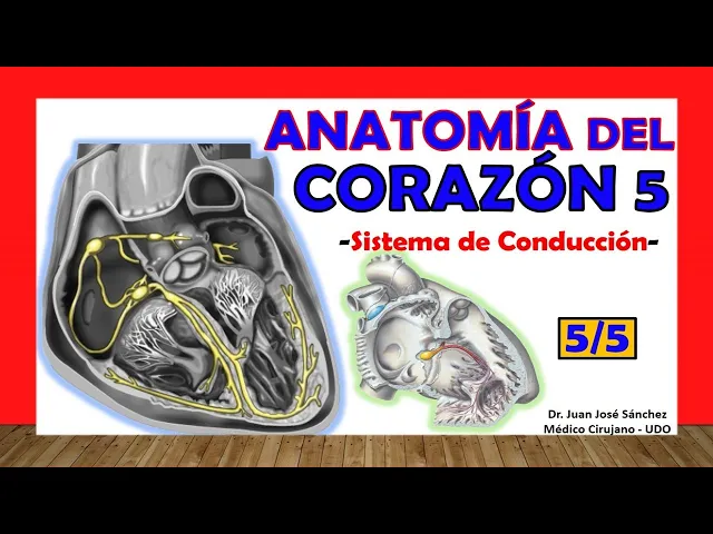
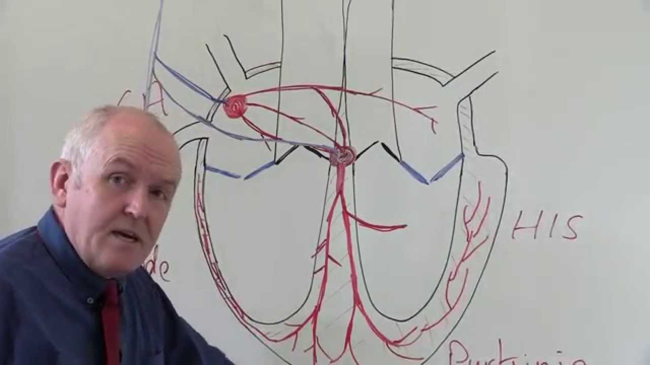
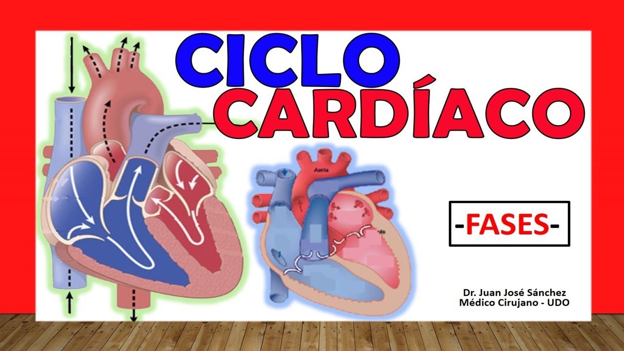
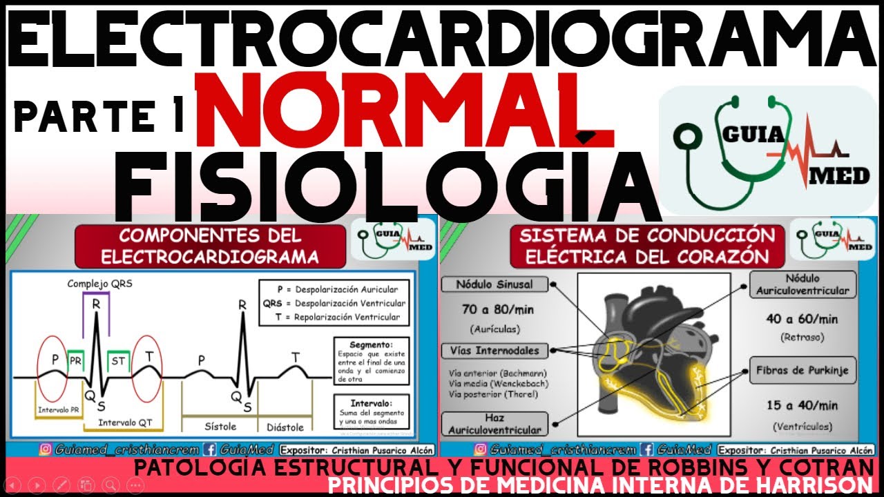
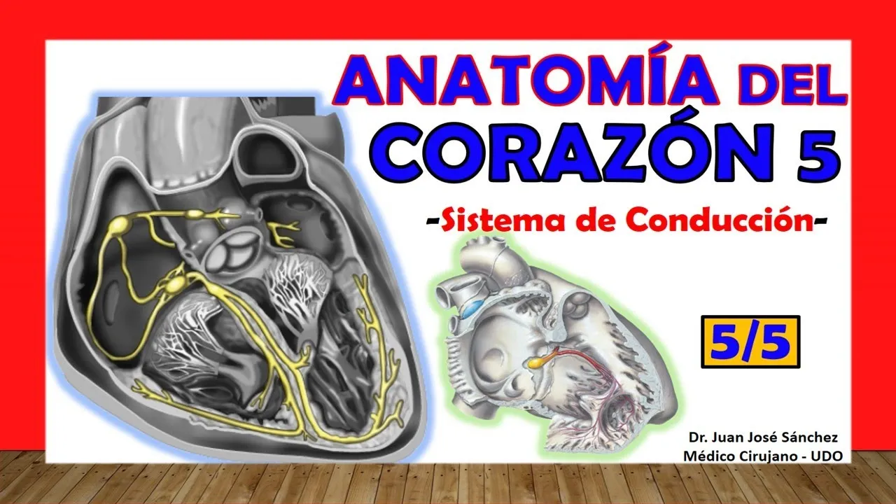
Greetings my dear friends, now if we enter the last heart video, the fifth installment, today we are going to talk about the cardiac electrical system, the conduction system, how that heart does to generate its own impulses and how that heart does to contract, this is basic in the study of the electromechanics of the heart; If we understand anatomically why it contracts, what structures make it contract, we will be able to understand how the cardiac cycle occurs and how pathologies can affect the heart [Music] I invite you to subscribe here in the lower right corner, click [Music] ] and don't forget to like the video. So the cardio-reading system of the heart is actually quite simple, there is nothing complex about it because the structures are very well located and very well studied, one thing that you should know is that these are a set of nervous tissue structures, that is, This is not a nerve, it is not a nerve fascicle that you are going to see when dissecting the heart; For us to find these structures of the cardiac system, we would have to do a special dissection of the heart because these structures that I am going to name: what are the nodes, what are the bundles, are structures of specialized cardiac muscle, that is, they are fibers. very similar to that of the myocardium itself but that specialize in electrical conduction, they play the role of a nerve but it is not that it is a nervous fascicle that we are going to see like any other nerve in the body, it is very important that you know from the beginning that They are nothing more than the same myocardial figures specialized in electrical conduction.
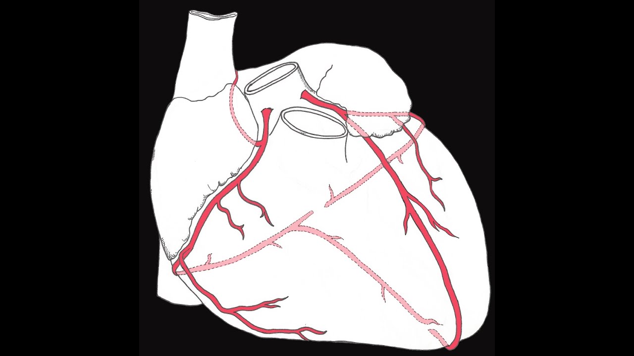
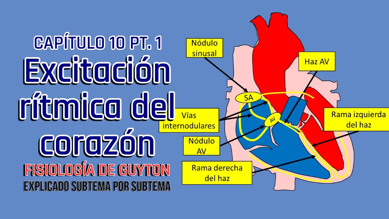
So it all starts with the pacemaker, remember that the pacemaker is a device that many do not know externally as it starts the electrical current of the heart and makes it contract, anyone can have an external electrical pacemaker of those artificial ones but the heart has its own pacemaker , that is, it has a structure that governs all the others, it is the one that first initiates the cardiac impulse, this structure has several names: we can find it with the name of node or nodule in both ways, it is also sinoatrial, also We can get it with the name of the sinoatrial node or node, we can also get it simply with the name of sinus node or node which is the old name of Keith and Flack, remember that that is an eponym and that should not be used in anatomy. This is a structure that is found, the first thing you need to know, below the epicardium, that is, it is a fairly superficial structure of the heart, as I reminded you in the previous video, it is supplied by the right coronary artery in most cases. , rarely supplied by the left coronary artery, measures approximately 7 millimeters in length, never exceeds one millimeter in width, does not exceed one millimeter in width, is located just posterolateral to the mouth of the superior vena cava in the right atrium, remember that When we talk about the internal anatomy of the ventricle of the atria, on the right there was a structure called the terminal crest, in the upper part of the terminal crest is where the sinoatrial node is located, so it is the pacemaker, it is the one that initiates the cardiac impulse.
The second nodal structure, we will find it far from it, that is, we will find it at the level of the interatrial septum, this structure is the famous atrioventricular or atrioventricular node or node , often only abbreviated with the name Av node or nodule. , it is not a there is an A and a small v assuming that A and v would mean atrioventricular, how is this node different from the previous one? The previous one told you that it was below the epicardium, this is below the endocardium, that is, it is very deep in its location in the heart, it is located at the base of the interatrial septum far in front of what is the mouth of the venous sinus and far in front of what is the insertion of the fibrous annulus of the tricuspid valve.
Now this atrioventricular node has to have some communication with the sinus node or else how would the impulse get from one place to another, well, What happens is not reflected in this image but there are some beams, beams is the plural of beam beam with z, which are called internodal beams which are the ones that connect these two nodes and are the ones that make the electrical impulse travel the sinus node to the atrioventricular node, these bundles are three weeks is internodal we would have an anterior internodal bundle, we would have a middle internodal bundle whose eponym is Wenckebach bundle and then we would have a posterior internodal bundle which eponym is Thoral bundle. Now it is important to know that this anterior bundle, this anterior interatrial bundle, gives a branch that is the inter auricular branch or bundle, this bundle that originates from the internal part of the anterior is called the Bachmann bundle or bundle and is the one that carries the impulse then from the right atrium to the left atrium because someone has to contract the left atrium, we know that there are three internodal bundles that cause the right atrium to contract and that travel and transport the impulse from the sinus node to the atrioventricular but someone has to contract the left atrium, that will create the Bachmann bundle. Some authors say that the anterior internodal is called the Bachmann bundle, but you really start looking and most of the literature talks about the Bachmann bundle being a separate bundle from the anterior one and that it is simply the same fascicle.
interatrial that derives from the previous one; After the impulse being in that atrioventricular node it now passes to a fascicle or a bundle that is the atrioventricular fascicle, very generally known by the name of fascicle or bundle of His, this Bundle of His is going to go from the Av node and is going to pierce the right fibrous trigone, you must have seen the previous videos to understand this and once it pierces the right fibrous trigone it passes to the part of the interventricular septum that is membranous, remember that first is the membrane, they pierce that septum membranous, that is, it is in the thickness of its walls and then divides into two branches that would be: the right branch of the bundle of His and the left branch of the bundle of His. What is important about this? This branch is placed outside the myocardium of the so-called muscular part of the interventricular septum, the right branch continues to the septum marginal trabecula which is the arcuate band and then continues to the anterior papillary muscle and then it divides into branches that go to the septal papillary muscle which would be this and the posterior papillary muscle which would be this one seen back here derived then from the right branch of the Bundle of His, the left branch we would have to see a left image, see that it goes above the myocardium or the myocardial part of the interventricular septum and there it would divide into two branches: an anterior division that would be the anterior division of the left branch of the Bundle of His and a posterior division that would be the posterior division of the left branch of the Bundle of His; These branches will then go to the anterior papillary muscle and the posterior papillary muscle respectively and will divide at the end into a fiber called the purkinje subendocardial plexus, that is where the cardioreading system ends, which is the one that goes through all of them.
myocardial walls, if we see again I have to show you this image, the same thing happens with the right branch of the Bundle of His which ends in a purkinje subendocardial plexus which carries the final endings of this fabulous cardiolection system or cardiac conduction system.