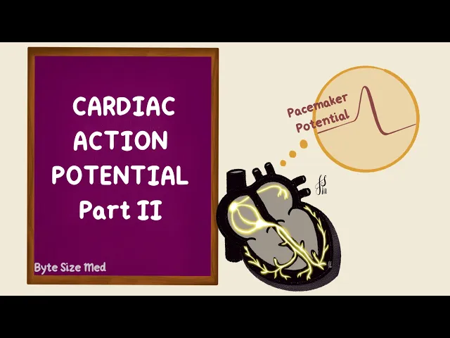
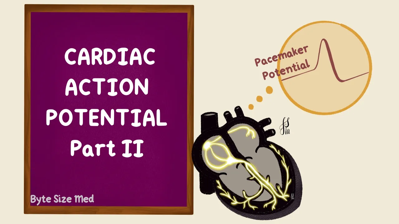
Hello. Welcome to Byte Size Med. This video is on cardiac action potentials.
And in part two, we're going to look at the action potential in the sinoatrial node. In skeletal muscles, what initiates contraction is an action potential. That's generated from a neurotransmitter released from a nerve that supplies it.
Cardiac muscle contraction is not initiated by nerves. The autonomic nerves that supply the heart can modulate contraction, but they don't initiate it. The heart has specialised muscle cells which are capable of self-excitation.
They can spontaneously generate action potentials without nerves telling them to do so. These cells form the cardiac conduction pathway. This includes the sinoatrial node, the atrioventricular node, the atrioventricular bundle and the purkinje fibers.
Self-excitation is because their resting potentials naturally drift up to threshold and an action potential fires. In part one, I talked about the ventricular action potential, which had five phases. Zero to four.
Phase zero is the upstroke, that's depolarization. Phase one is initial repolarization phase, two is the plateau phase, three is the final repolarization and phase four is resting membrane potential. There are three important ions that are mostly involved.
Sodium, calcium and potassium. The pink is the inside of the cell and the blue is the outside. The resting membrane potential of cardiac muscle is negative at around -90 millivolts.
Sodium uses the fast sodium channels to enter the cell, causing depolarization. A small amount of this is also from calcium entering the cell. The sodium channels get inactivated and the potassium leaving the cell results in the initial repolarization.
Calcium influx balancing the potassium efflux creates the plateau at the end of the plateau. The calcium channels close and the potassium efflux results in repolarization back to the resting membrane potential. That's contractile cells.
In the conducting cells of the sinoatrial node, it looks a little different. There's phase 0, phase 3 and phase 4. But there are no phases one and two.
So there's no notch and no plateau. The ventricular muscles have a resting membrane potential of around -85 to -90 millivolts. These cells in the sinoatrial node, they have a less negative resting membrane potential.
Around -55 to -60 millivolts. Since that's higher, any time the membrane potential becomes less negative than -55 millivolts, the sodium channels get inactivated. So they can't contribute much to the depolarization in the action potential.
It's actually the calcium channels that make the up stroke happen. But the calcium channels are slower than sodium. These are L-type calcium channels, slow to open, slow to close.
So the upstroke is slower. It's not as steep as the ventricular action potential either. At rest, the membrane potential is negative.
Calcium entering the cell makes the inside less negative and more positive. That's depolarization. Once the calcium channels close and potassium leaves the cell, the potential starts becoming more negative again reaching the resting membrane potential.
But it doesn't stay there. The sinoatrial membrane is leaky and it allows sodium to enter the cell. The movement of ions creates currents and this is called a funny current.
If sodium keeps leaking into the cell, the membrane potential is obviously going to become less negative and drift upwards to the threshold of around -40 millivolts, and that fires off another action potential. This is phase 4 of the action potential, which is spontaneous depolarization. This is also called the pacemaker potential or the pre-potential.
It's mostly because of the sodium entry creating the funny current, but towards the later part of the pre-potential calcium also contributes. This is through the T-type calcium channels. The T stands for transient.
The up stroke is by the L-type calcium channels. So phase zero is depolarization, phase three is repolarization and phase four is spontaneous depolarization. Those are the phases of an action potential.
That gets generated in the Sinoatrial Node. This phase 4 depolarization is important. The structure which has the fastest rate of phase 4 depolarization will be the one to generate the impulse, that then gets transmitted through the rest of the conduction system.
The fastest rate of discharge happens mostly in the sinoatrial node, making that the pacemaker of the heart. Because it sets the rhythm. The other structures, though capable of self-excitation, get suppressed because the sinoatrial node discharges faster than them.
So before they can reach threshold, the sinoatrial node has already sent an impulse. Now what would happen if the rate of phase 4 depolarization were to increase? The potential reaches threshold faster.
So there are going to be more action potentials in the same amount of time. That would increase the heart rate and this is what happens with sympathetic stimulation. Remember that the heart is supplied by the sympathetic and the parasympathetic nerves.
They don't initiate contractions. But they can change the rate and this is one way that they do that. Parasympathetic stimulation would do the opposite.
It slows down the rate of phase four depolarization. So there are lesser action potentials and a lower heart rate. Now this action potential, it starts the whole path.
It gets conducted through the atrial pathways to reach the contractile cells of the atria. It travels to the atrioventricular node through the atrioventricular bundle and the purkinje fibres to reach the ventricular muscles, generating action potentials there, such that the atria and the ventricles can contract one after the other. And that's the action potential in the sinoatrial node.
I hope this video was helpful. If it was, you can give it a like and subscribe to my channel. Thanks for watching and I'll see you in the next one!