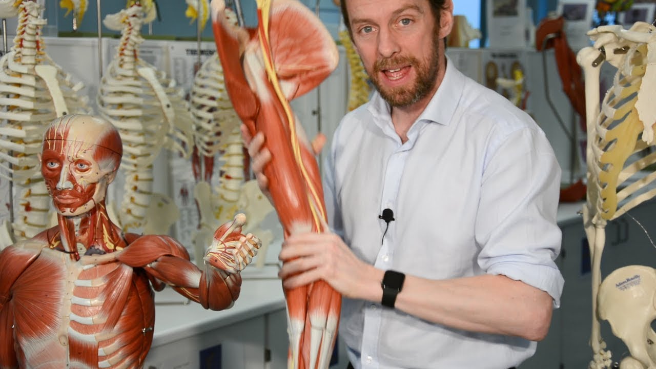
[Music] in this lesson we will review the key structures in the posterior arm including the muscles and neurovascular structures let's start with a simple line drawing of the posterior part of the arm and note the orientation here this is the medial side and this is the lateral side over here and we're looking at the posterior side of the scapula and the posterior side of the humerus there's one muscle in the arm which is the triceps it has three heads as the name suggests tri meaning three and SEPs meaning heads the lateral head of the triceps
is seen here situated on the lateral side of the post year humerus the second head of the triceps is known as the medial head of the triceps and it is situated at the name suggests on the medial side of the posterior humerus and is seen here the medial head of the triceps is mostly covered by the third head which is known as the long head of the triceps and the longer the triceps in fact is the only head of the triceps that also has an attachment onto the scapula note that the medial head is mostly
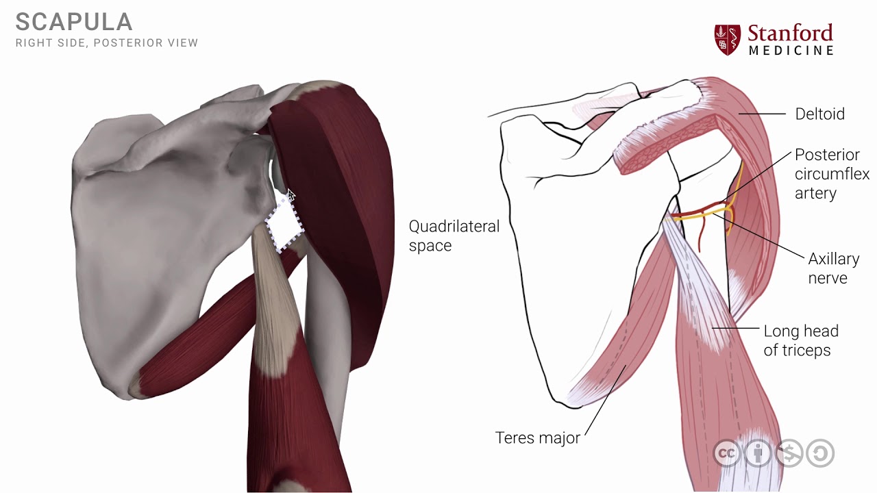
covered now so I'm going to remove a segment of that long head of the triceps to continue to keep the medial head exposed and to add some neurovascular details in order to understand some of these structures the neurovascular structures we need one other muscle as a landmark muscle which is the teres major muscle the teres major is a muscle that attaches on to the scapula the inferior posterior part of the scapula and the fibers then run laterally and superiorly to attach on to the anterior part of the humerus as it does this it has a
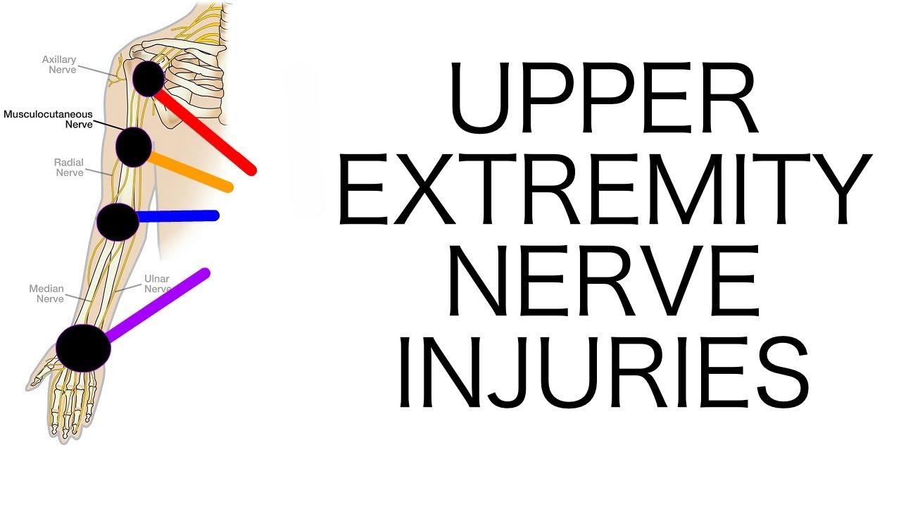
very crucial relationship to the long head of the triceps in that the fibers of Cheri's major are on the anterior side of the longer the triceps note that the axillary artery is in its location in the axillary as it crosses the teres major muscle it becomes the brachial artery there is an important structure that is situated in this area which is a branch of the axillary artery known as the posterior circumflex artery or the posterior circumflex humeral artery which is seen here this artery is accompanied by a nerve known as the axillary nerve and both
of these structures wrap around the surgical neck of the humerus this is an important area where injuries or fractures might occur and this type of an injury can damage the axillary nerve and the posterior circumflex artery the brachial artery then continues from the axillary artery and gives off another branch known as the deep brachial artery this branch of the brachial artery the deep brachial artery exits through the triangular space and wraps around the middle third of the humerus the position of this artery around the humerus has a groove which is a very shallow groove and
is called as the spiral groove of the humerus this deep brachial artery is accompanied by a nerve which is a branch of the brachial plexus which is known as the radial nerve the radial nerve and the deep brachial artery traveled together in the spiral roof and are very closely related to the mid shaft of the humerus this is another very important relationship to keep in mind when there are injuries in the mid shaft of the humerus such as a fracture of the humerus these structures the radial nerve and its accompanying artery can be damaged the
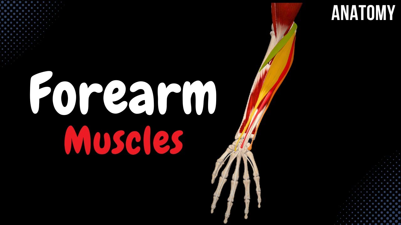
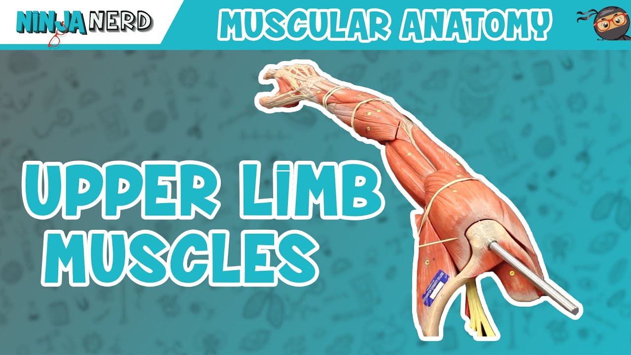
three heads of the triceps muscle attach more distally and become one tendon that then crosses the elbow joint and attaches on to the ulna specifically the olecranon process of the ulna since this muscle attaches onto the ulna and crosses the elbow posteriorly it has a function of extension at the elbow joint all three heads of the triceps muscle are supplied by the radial nerve let's look at the posterior arm in a photograph where the dissection has been completed and in this photograph we note that the deltoid muscle is seen here that has been cut on
its surface in order to expose some of the deeper structures the post year fibers of the deltoid have been removed from this area the first structure that we can see is the long head of the triceps which is seen here and remember this is the only head of the triceps that has a scapular attachment the other part of the triceps which is visible here is the lateral head of the triceps which is seen here these two halves unite and they become tenderness as they go into the distal part of the posterior arm note that in
this view there is nothing of the medial head that is visible in order to understand and in order to see the medial head we need to make a cut as shown here and the result of that would be as seen in the photograph on the right side if we make the cut and separate these two heads we can now see the deeper structures the lateral head has been separated from the long head of the triceps the medial head of the triceps is now seen in the depths remember the medial head is also sometimes called rightly
so the deep head note the neurovascular structures in the spiral groove the radial nerve is seen here and accompanying the radial nerve is the deep brachial artery which is seen here these two structures wrap around the mid shaft of the humerus in the spiral groove and have the propensity to get injured in fractures around this region [Music] you