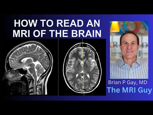
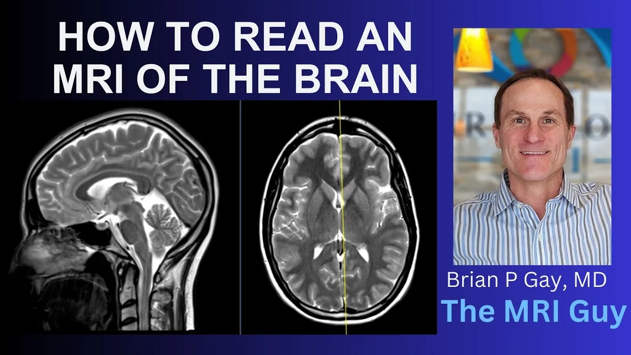
hello this is dr. gay from first second Mariah and I'd like to show you how to read an MRI of the brain so this is a patient has a normal in might of the brain and I just wanted to go over some Anatomy and show you what we see and what we look for so I'm gonna start with this view is called a sagittal image if we scoot the patient over we can see that the nose is off on the left hand side over here and we can see the tongue is down over here the
sinuses are right here in the middle and the forehead is right over here this area of the frontal bone which is the forehead if we roll towards the right hand side we can see the right eye there's a round area here you can see the lens in the front and we see the left eye over here now right in the central part of the brain at the midline we see a little round ball here this is the pituitary gland we look carefully at that to make sure the pituitary gland is nice and normal in size
and it sits in a little cup here called the sella and the sellers normal inside is not expanded and again the pituitary tissues look normal and we can see a pituitary stalk that comes down into the pituitary gland now if we zoom back out here we can see the cerebrum the cerebrum is up top here starts over here in the front it goes over the top and all the way to the back over here is the frontal region this is the occipital region and back we have the parietal region over here if we go off
to the edges we have these areas called the temporal lobes of the brain and so again this is all the cerebrum and the cerebrum has a right side a right cerebral hemisphere over here here's the right eye and then it has the left side and the right and left sides are joined by this broadband it's like a wiring harness right here it's called the corpus callosum so we look carefully at the corpus callosum sometimes they can be developmental abnormalities as that or sometimes people with multiple sclerosis will have little lesions to go right up to
the edge of that so we look at that it looks good in this patient now down below we have something in back here this is called the cerebellum the cerebellum looks like cauliflower here on this and also we have some structures here in the middle the middle structures are the midbrain and the brainstem so the midbrain is in this region over here and then the brainstem is this area to here this is the pons which is his ovoid area and the medulla is more of a straight area and then if we go down here we
can see the spinal cord that goes down through the neck comes off the bottom the brainstem this is the cervical spine down here now if we change the orientations this is that same sequence where we see fluid is really white and then brain parenchyma is more dark and gray and we're gonna go down from the top down down down now we see the front is over here the back is over here this is the right side left side and this is what we call an axial image or slicing going down down we see these longitudinal
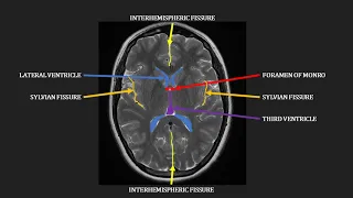
slits these are the ventricles and they're filled with fluid and we look at the ventricles and see are they normal in size are they shifted from right to left when they're enlarged we call that hydrocephalus but the ventricle is a nice normal in size there's no hydrocephalus and we come down here we see the right eye left I look at the lenses we see make sure the eyes are normal in contour make sure they're not poking out which we call proptosis and we see these linear bands coming off the back of the eye this is
the optic nerve so we look carefully at those to make sure that the optic nerves are normal in appearance and they are and we look at all these fat which is in the orbit here right on the left side the fat looks preserved and nice you may notice these little areas of darkness these are blood vessels normal flowing blood you have two things in the front that are like each other this is the right and left internal carotid arteries or the cavernous credit arteries there's another one back here this is the basilar artery and we
call this the interior circulation post your circulation if we follow that circulation up he goes up up the internal carotid arteries will end and give rise to this and this this is the left middle cerebral artery right middle cerebral artery and if we keep going up they give rise to these this is the intership arteries and so we look there and say do we have nice flowing blood which is just jet black and yes we do and both of the internal carotid artery the basilar artery here looks good it gives rise to some posterior broadly
so I'll just kind of glance here make sure that there is normal darkness which is flowing blood in there in there very very back there's also a dural venous sinuses a venous channel where the blood return through here and we look here is that a nice dark area or is your abnormal brightness but it looks like nice dark flowing blood within that dural venous sinuses that looks very very good again the eyes look good now would go down a little bit further and we look at these two things these are called the internal auditory canal
is their right side left side we see these little circles here these are the semicircular canals to give balance and also we have other little things here called the cochlea in this little area here a little layer over here and it's going to glance at these make sure that there is no tumor within this little canal here the internal auditory canal and make sure that these structures are morphologically normal as we continue down we go down to the bottom of the cerebellar hemispheres is the back cerebellum or at the bottom of the cerebellum and over
here we see an area of Darkness is called the mastoid so patients can have fluid in these they can get inflamed called mastitis or I have other findings sometimes tumors can be in here or inflammatory conditions but these are nice and dark filled with air you can see air outside of the patient is dark and air inside here is dark as well the other thing that can be dark is compact bone so sometimes you'll see some dark band around the back like this if you have a dense skull bone here this would be dark and
if you have air in here it'll be dark there's also bone in here too but the air and the bone are both just black so the whole area just looks dark same within the sinuses they are very dark cuz there's lots of air in the sinuses this is another orientation where we're looking straight at the patient if we go very far forward we'll see the eyes there's a right eye left eye we can see the lenses there in the very front this is the nose area sinuses and with the eye we can see the optic
nerve coming off the back on the right here's the optic nerve there and we see this bright stuff around his orbital fat and we see these little dark bands these are the extra ocular muscles these are the muscles that make the eyeballs move you can see how this one contracted were to pull the eye upwards on the left here so we look at all these structures make sure these are symmetric in size and make sure the orbital fat is normal and so everything looks really good with the eyes if we continue on back here we
eventually get to the internal auditory canals again take another look at those we see them coming across here the seventh and eighth nerves go through there we can see these similar circular canals going up and also we can see the cochlea on the left hand side here and we have thin images I don't have them on this patient but we get really super thin images where we can see all the details very nicely and see the individual nerves going through here and let me go to another Sigma so now we go to a sequence called
an axial image and this is a sensitive sequence for any abnormalities within the brain parenchyma so around the periphery we have what we call gray matter with the leaves religionists bright signal around the edge and then centrally we have more dark uniform signal here the central stuff is called the white matter and the peripheral brighter stuff is called the gray matter and we look very carefully if patients have migraine headaches if they have small vessel problems called chronic small besos Kimia abnormalities there they can have strokes or bleeds in the brain and we'll see in
geographic regions of abnormal brightness or darkness within the brain and we also can sometimes see the brain pushed to the right or left and so this is called a flare sequence again it's very very sensitive for any abnormalities especially in the white matter so we look really carefully patients with multiple sclerosis will have little white matter lesions this is just another view that really shows the difference between the gray matter again the gray matter on the outside and then that white matter which is the dark uniform stuff centrally and again we look really really carefully
and all the white matter make sure there's no evidence of multiple sclerosis there's no evidence of chronic small vessel ischemia there's no small tumor within the white matter or within the gray matter and you go up and down and we also look at the ventricles again so and then this is a nice normal patient you can see these blood vessels here left middle shrimp artery right middle super artery and the eyeballs again here on this sequence as well the fat is really dark so you can see fat around the edges dark and also fat within
the orbits is very dark as well so if there were an inflammatory condition involved in the eyes that would stand out and we can see these optic nerves here again coming off the back so that is how I read an MRI and thank you very much