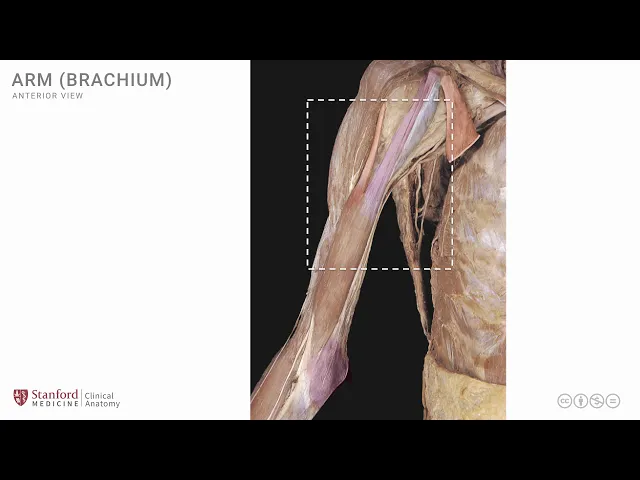
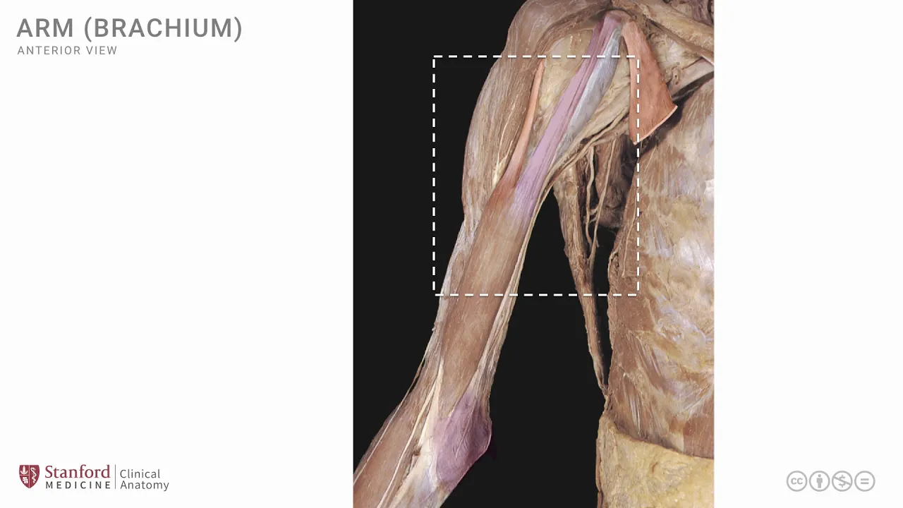
[Music] in this lesson we will review the structures on the anterior side of the arm we will look at the muscles and we will look at some of the neurovascular structures that are in this region let's start by looking at a photograph of a cadaver dissection that shows the proximal part of the shoulder area and the anterior part of the arm in order to get ourselves oriented let's see where the coracoid processes which is over here and the clavicle which is seen over here these two structures can orient ourselves and we can now identify our
first muscle which is v which has been cut and the cut portion is seen here the pectoralis minor the pectoralis minor is attached to the coracoid process and then extends up to the chest wall the distal part of this muscle the inferior part of this muscle has been cut away we do see some neurovascular structures in this region that are traveling from the root of the neck area through the axilla and into the arm the first one of these structures is the axillary artery which is seen here and it's highlighted in red just end here
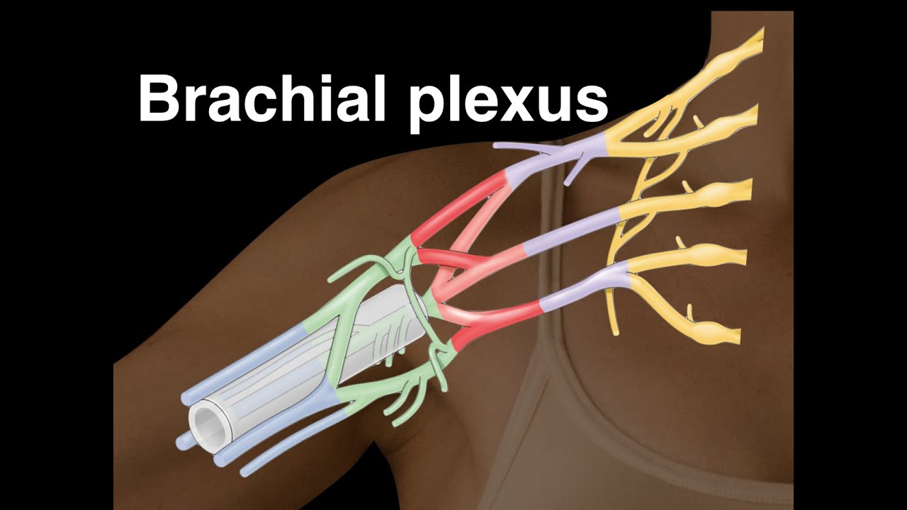
to it we see another structure which is the axillary vein which is seen here and highlighted in blue you can see clearly there are many nerve like structures in fact they are part of the brachial plexus it is difficult to identify exactly which part of the brachial plexus these elements are from and in order to undo that one would have to separate out these structures and follow them to their destination but for our purpose we'll just call them brachial plexus collectively if we now focus a little bit on the lateral side of this photograph we
can see that the deltoid muscle has been cut away in order to expose some of the deeper structures the deltoid muscle is highlighted in red here and because the fibers the anterior fibers of the deltoid has been removed we can see the humerus this is the head of the humerus as well as its bony tubercles the lesser tubercle that is covered here with some capsular fibrous tissue but note very clearly seen is this long head of the biceps or the biceps brachii which travels through the intertubercular groove and it's going distally to join a short
head of the biceps which is seen here the short head of the biceps also attaches onto the coracoid process and it continues on its way into the arm the long head of the biceps and the short head of the biceps unite to form the biceps brachii muscle and this is the muscle belly down here note that the short head of the biceps has been cut in the middle in order to expose an area that is seen here which we will talk about in some more detail in a subsequent slide but it does expose one muscle
which is known as the coracobrachialis muscle which is seen here and I'm going to shade this in a slightly bluish grayish color the coracobrachialis muscle is a relatively small muscle it extends from the coracoid process onto the mid shaft of the humerus as the name would suggest coracobrachialis khorgo comes from the coracoid process and brachialis is the humerus the arm this is a small muscle and we'll look at its detailed attachment in the subsequent slide here we have a simple line drawing that shows the scapula and the humerus and we will look at the anterior
muscles on the of the arm the first thing in order to orient ourselves is to identify the coracoid process and we'll put a label to it here there is a muscle known as the coracobrachialis that we just mentioned and it has attachment more approximately on to the coracoid process and the fibers then go laterally and inferiorly to attach onto the mid shaft of the humerus on the medial side this corker brachialis muscle is a minor muscle in terms of function but it's a very important muscle as a landmark muscle in order to identify structures in
this area when we are performing surgery in this particular anatomical region the musculocutaneous nerve which is a branch of the brachial plexus travels in this region and in fact pierces the coracobrachialis muscle and supplies this muscle and continues on its way to the arm to supply other muscles and is very clearly seen here the second muscle in this region is the biceps and the long head of the biceps brachii as well as the short head unite to form the biceps belly we see the Bryce EPS long head here traveling through the intertubercular groove and the
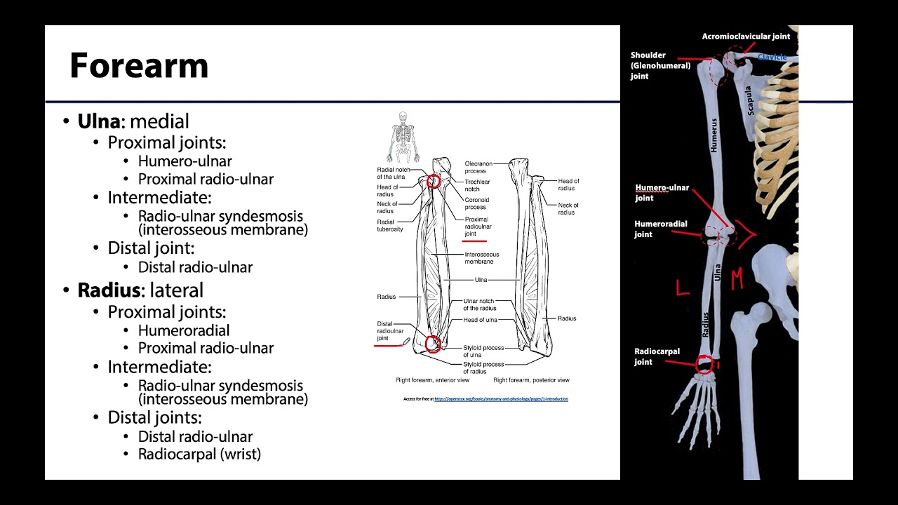
short head of the biceps brachii is seen here arising from the coracoid process along with the coracobrachialis muscle it covers some of the coracobrachialis muscle as shown here and then the biceps belly ends as the biceps tendon near the elbow joint as shown here the biceps tendon muscle crosses the elbow joint on its anterior side and it attaches on to the radius bone which is one of the two forearm bones and because this tendon crosses the elbow joint and on the anterior side it is a flexor it flexes the elbow joint the biceps also has
an extension known as the bicipital aponeurosis which is an extension from the distal part of the biceps that extends medially and goes and attaches on to the deep fascia as well as on to the ulna on the medial side the ulna is the second forearm bone on the medial side and the bicycle aponeurosis provides some sort of a stabilizing action in terms of the flexion movement that it produces at the elbow joint if we look at a superficial dissection of the anterior arm in its full length we are presented with the full biceps muscle as
seen here in order to orient ourselves let's again put the coracoid process in place which is seen here and one can note the pectoralis minor which is seen here as we just saw the coracobrachialis has its attachment on to the coracoid process and it's seen here and is partly hidden by the biceps muscle the short head of the biceps which is seen here which is also attaching on to the coracoid process this muscle is joined by the long head of the biceps which is seen here to form the biceps muscle itself or sometimes described as
the belly of the biceps muscle one can see the bicipital aponeurosis around the elbow joint area and it runs onto the medial side near the ulna and some of the deep fascia in this region we don't see very much of the biceps tendon itself here in order to do that one would have to do a slightly more deeper dissection let's look at the short head of the biceps and have a close-up view of this region if we make a transverse cut in the short head of the biceps as shown here and reflect the distal fragment
laterally we arrive at this particular deeper dissection photograph and we can now take a close-up view of this and see the deeper dissection and the relationships that are exposed so the short head of the biceps is seen here and the shorter the biceps also is seen here this is the short head of the biceps which has been cut and then reflected laterally so that you see the short head of the biceps at two different locations we also see the long head of the biceps which has now been lifted a little bit because of that retraction
and it is going on its course distally and it joining the shorter the biceps to form the biceps itself the biceps muscle which is seen here note that this is the deep surface of the biceps muscle not the superficial surface because we have reflected the shorter of the biceps laterally the coracobrachialis is seen here and the musculocutaneous nerve enters the coracobrachialis muscle piercing it and it then continues on its course to supply the biceps muscle from its deep surface and in fact continues even further to supply a muscle that we can still see in the
depths this is a third muscle in this area known as the brachialis muscle and is highlighted here in red in order to understand the deep muscle of the anterior arm let's look at a simple line drawing here and the deep muscle is known as the brachialis is situated in the distal half of the anterior arm as seen here it crosses the elbow joint on its anterior side and is attached on to the ulna and as one would imagine the movement that this muscle produces is flexion this muscle is supplied by the musculocutaneous nerve the musculocutaneous
nerve lies on its superficial surface and it continues on into the forearm as a cutaneous nerve known as the lateral cutaneous nerve of the forearm note that the musculocutaneous nerve supplies all muscles in the anterior arm the coracobrachialis the biceps muscle as well as the brachialis let's now look at a photograph of a deeper dissection of the anterior arm in this photograph the biceps muscle has been removed in order to expose the brachialis muscle which is seen here note that it is situated in the distal half of the arm the nerve that one can see
that is situated on a superficial surface is the musculocutaneous nerve it is piercing the coracobrachialis muscle in its more proximal location and then continues on the superficial surface of the brachialis muscle it then continues as the lateral cutaneous nerve of the forearm if one notices structures a little bit more medial to the musculocutaneous nerve one can see two different structures there's an artery which is the main artery of the arm the brachial artery which is seen here and just medial to this brachial artery is a nerve an important nerve known as the median nerve and
it is situated here running down the medial side of the arm and then entering into the forearm note that the median nerve does not supply any muscle in the arm it does have supply to muscles in the forearm and the hand [Music] you