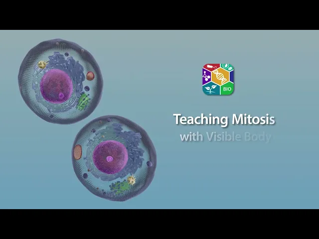
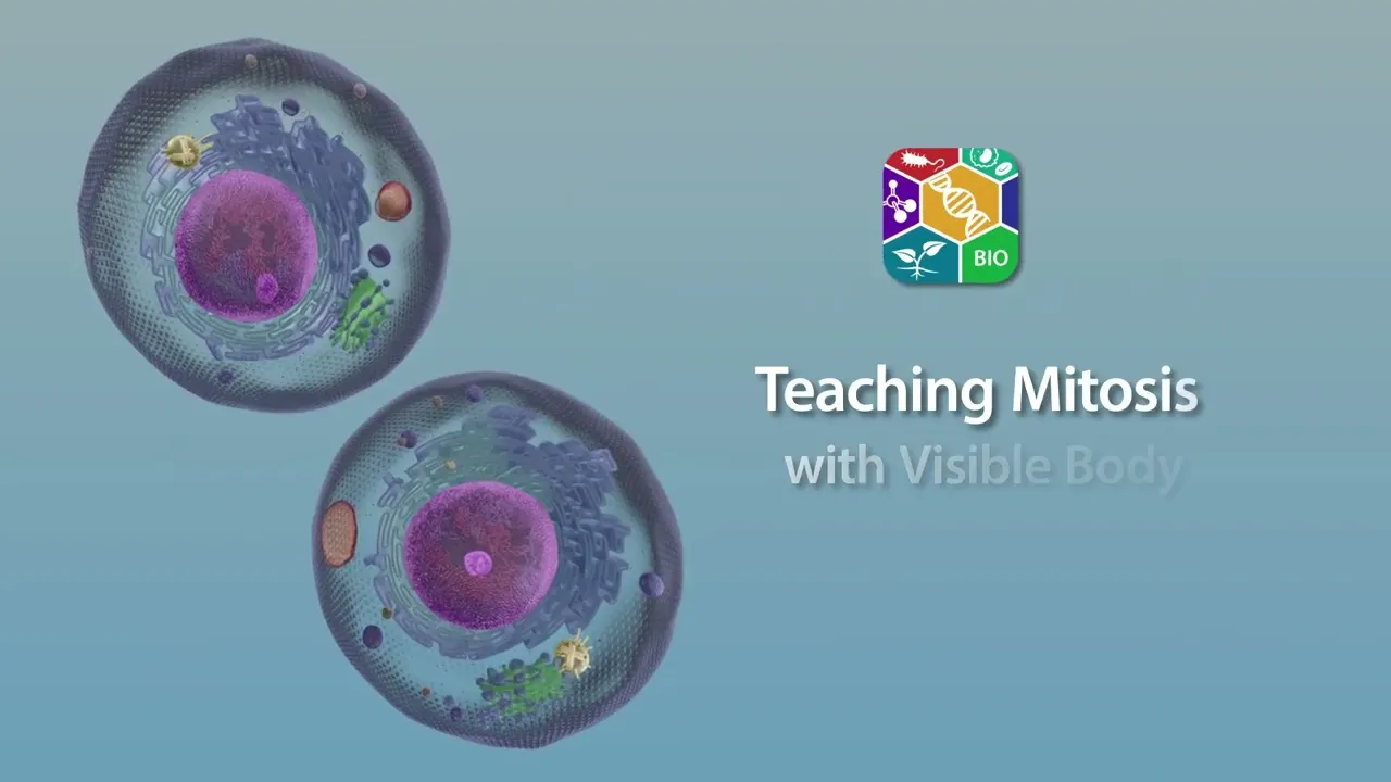
With the mitosis moving model in Visible Biology, students can visualize the steps of this important biological process in interactive 3D! Mitosis is how body cells - or somatic cells - replicate themselves. It starts with one diploid cell, and ends with two genetically identical diploid daughter cells.
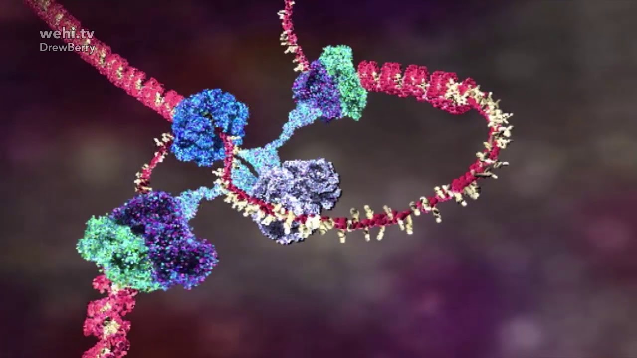
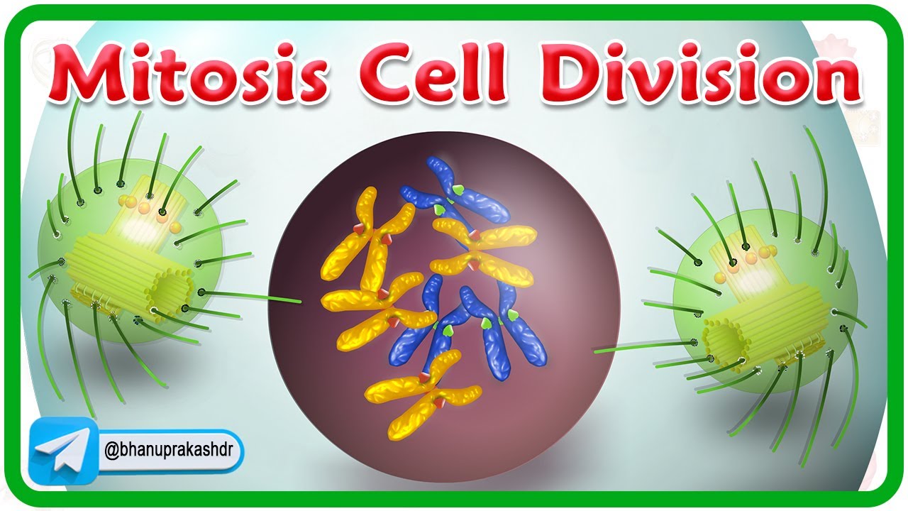
Before mitosis, the genetic material - DNA - in the cell's nucleus duplicates. In a normal diploid cell, half the genetic material comes from an organism's mother, and the other half comes from its father. In Visible Biology, we represent this using red for the mother's genetic material and blue for the father's genetic material.
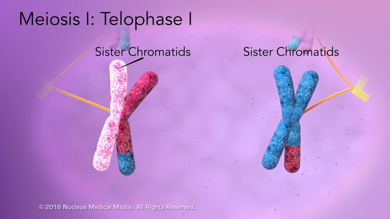
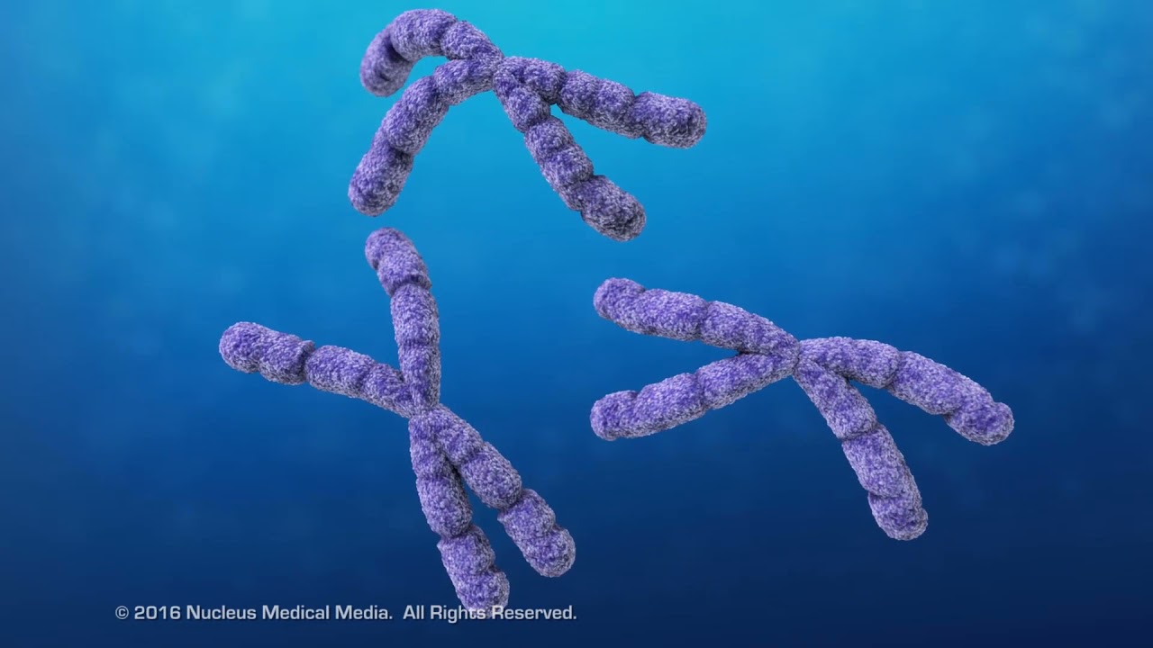
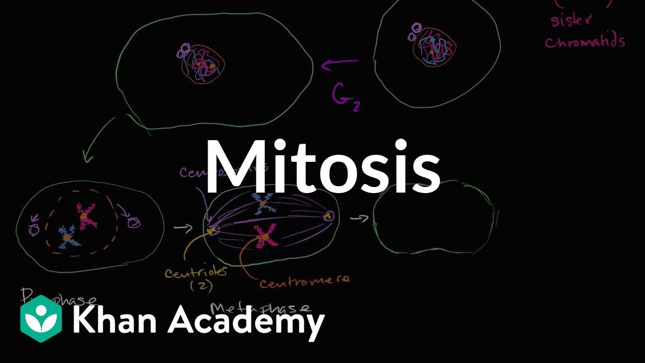
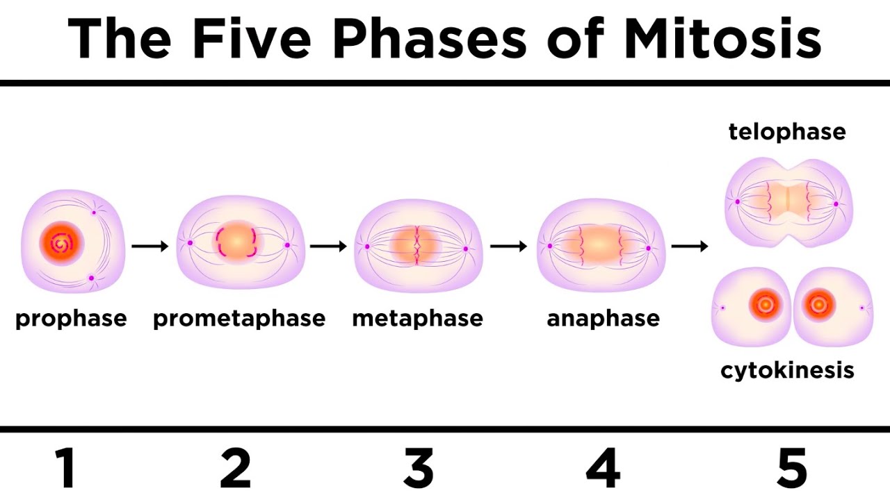
Now let's see how the different steps of mitosis are illustrated in Visible Biology's moving model. During prophase, the duplicated DNA condenses into duplicated chromosomes called sister chromatids. The centrosomes move towards opposite ends of the cell.
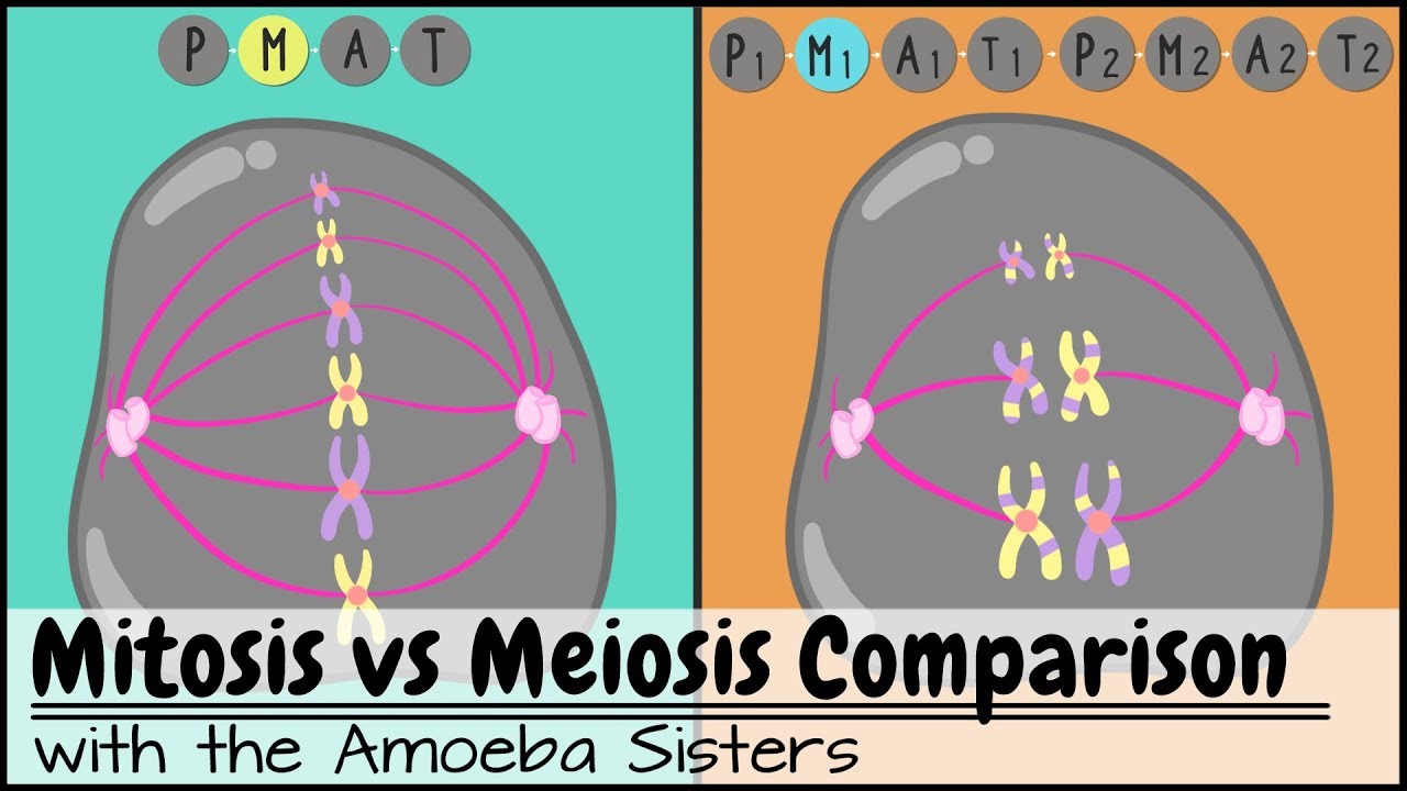
During prometaphase, which Visible Biology shows as its own step, the nuclear envelope dissolves and the spindle fibers extend and attach to the chromosomes. During metaphase, spindle fibers line up the chromosomes in the middle of the cell. During anaphase, the spindle fibers pull the sister chromatids apart.
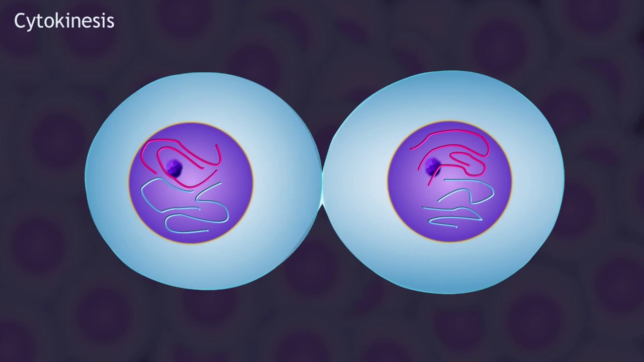
During telophase, a new nuclear envelope forms around each set of chromosomes. The mitotic spindle fibers get pulled back into the centrosomes, and the cell forms a cleavage furrow in preparation for splitting. Then, the original cell pinches off along the cleavage furrow, forming two new, genetically identical cells.
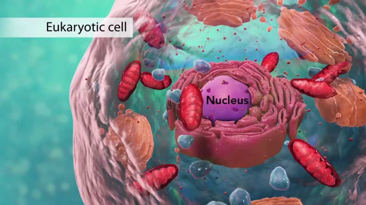
You can see that each of these cells has a full set of chromosomes as well as all the organelles it needs. To learn more about how to use models like this one in your classroom, and how to use them to create assignments for your students, visit visiblebiology. com!