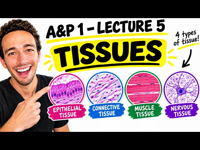
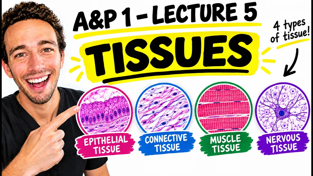
Okay, everybody. So, by the time you get to this lecture, you usually need some of these because you will be crying a lot. And no, I'm not talking about these tissues. I'm talking about these tissues because these can be a little complicated. So, in this video for lecture five of 1, [music] we're going to make them simple. Hey everybody, Organized Biology here. We make difficult biology concepts simple. And Real quick, I wanted to mention if you email
[email protected], put in the subjectline tissues and send that email, you will get a free PowerPoint breakdown of the
tissues because I know a lot of these tissues are observed under microscopes. I give you pictures of the microscopes, locations, functions, those types of things. But in this video, we're going to be drawing a lot. We're going to be talking about the levels of Organization, again, where tissues fall. We're going to talk about the four tissue types and then the specific types of tissue so that you can hopefully ace your test and also see why this is incredibly important to know now so the later parts of this course it'll make a lot more sense when
we're talking about some of these tissue types. So first off what the heck are tissues in terms of&p well tissues simply are clusters of similarly functioning cells. So clusters Of similarly [snorts] functioning [clears throat and cough] cells. Now, we know the drill in this class. When we see cells plural, we know that we're not just talking about the cellular organization. Now, we're talking about the level that's going to be considered tissues. So, tissues are basically clusters of these cells and they're similarly functioning. As you can probably guess from core concept number One, similarly functioning also
likely means that they're similar in structure as well. So these cells in each tissue type are going to be similar in structure, therefore similar in function. Now to give you an image of this real quick before we get into the four tissue types, I want to actually draw the next level of organization which most people know pretty well and that's going to be the organ level. Right? So you guys know organs like the Lungs, the liver, the kidneys, the heart. All organs are are just two or more tissue types put together for a bigger function.
Okay. So, if we were to draw a heart, for example, I'm going to use several different colors. I've got I'll just tell you so that you can be prepared. I've got a blue, green, orange, and red. I am not colorblind today. And we're going to draw the heart structure and see what different colors we can bring out in it. So, check this Out. [clears throat] First part, we're going to draw the outside part of the heart. outside part of the heart. By the way, the heart kind of looks like this. Almost like a teardrop shape.
I don't know how you want to call that. So, the apex of the heart, the bottom part points down towards your hip is right here. And then the uh base of the heart is actually on the top. It's kind of strange, but it's shaped like this. Now, we know the outer part of the heart is Something called the paricardium. Pericardium literally just translating to outside or around the heart just like the perimeter of a field or something is the outermost part of the field. Okay. Now if we look inside of the heart we also have
some chambers of the heart that will contain the blood as it's pumping. And these chambers are called the atria and the ventricles. And they're going to have some structures that are lining the inside of the heart Itself. And that's going to be called the endocardium. Okay. endocardium. See, we can see there's kind of a perimeter outside and then endo within the inside. So, both of these are kind of lining the heart, whether it's on the outside of the heart or on the inside of the heart. But not only that, we also have some structures called
the myiocardium. So, in the myioardium, it's going to be this muscular layer on both sides. In reality, the left side of the heart be Really thick. I kind of drew it that way. So, this layer we call the myioardium. We've learned that words matter as we're seeing myo meaning muscle and cardio meaning heart. So this is the heart muscle tissue itself. Yes. But then also we've got some connections between all of these structures. And and fun fact by the way we also have some myioardium up in the atria. It's a lot smaller. So here's some
myioardium here as well. But We also have to kind of connect everything together. So we have some valves that will connect atria to ventricles. Atria ventricles. We're going to have the septum of the heart kind of keeping the heart anchored both ways together. We're also going to have some other connective tissue or some other connective things around the heart maybe up here holding things together etc. And then lastly, we're also going to have fascinatingly Some tissue. One being the SA node and one being the AV node. Once again, this is the SA node of the
heart. You are like what the heck is a node? So SA node, AV node, [clears throat] these are basically the signaling tissues of the heart that tell the heart when to contract. So fun fact, if you were to take the heart out of the body, uh until it runs out of ATP, it will continue to tell itself when to contract, which is kind of fascinating. So why did I draw these in different colors? And why did I use the words connect or line or signal? Well, because this is a heart. This is an organ level
of organization. But the organ is made of in this case four tissue types that build itself in the paricardium the endocardium. These were all tissues called epithelial tissue. epithelial tissue. We also had the myioardium which is typically the muscular tissue. It is the muscular tissue not just typically It's the muscular tissue specifically the cardiac muscle. Then we also have these nodes. These will be considered nervous tissue. And then lastly, all of the connective tissue that binds things together all over the place is connective. So we've got connective tissue, we've got nervous tissue, epithelial tissue, and
muscular tissue. Those are our four tissue types. So if we look at any organ, you will find these tissue types embedded within Itself. Now, we need to understand the main goals of these tissue types so that when we see them for the remainder of the semester, we'll know what we're looking at. So, we're going to start first with just talking generally about what these tissue types do. So, I'm just going to write E for epithelial here. Epithelial. When you look at that word epi whoops, if I can spell it right, epithelial tissue, epithelia Refers to
upon and then sheetlike. Sheet like upon. So these are clusters of cells that are sheetlike and they cover tissues. That makes sense, right? Because this was lining the outside of the part heart and also lining the inside of the heart. So typically when we look at organ the outermost part and the innermost part will have this epithelial tissue. I like to think of this if you take your finger and you just stick it wherever you want. So Stick it anywhere in your body. Okay? So you can take your finger and you can touch the outside
part of your skin. You can touch your eye. You can the inside of your mouth. You can stick it up your butt. Wherever the heck you want to touch, you are actually touching epithelial tissue because you're touching tissue that is literally lining the structures of your body. All right? So that's epithelial tissue. Goals of epithelial tissue. You could probably Predict if we're lining structures like on the outside of the heart, we're likely going to be protecting structures, right? So we're going to be doing some protection. We're also going to have some fluid we may be
secretreting to help lubricate the heart. So a lot of the times some epithelial tissue will do some secretion as well producing some sort of product typically a fluid based product or maybe even a hormone some excuse me a hormone Sometimes will also have some specialized epithelial tissue that will absorb right so the inner lining of your digestive tract lining your digestive tract this these tissues are going to help absorb some nutrients into the bloodstream okay and then you could just say kind of lining and then support for other organs like inside lining of the heart
allows that blood to flow nicely. So there's other functions but these are the main ones. So you can think of it as Epithelial tissue always has a good public service announcement PSA. All right. So that's epithelial tissue. Let's get into the generalities of the others. So easy one is going to be muscular tissue. I'm going to go to muscular tissue next. Draw it in red. And as you can guess, muscular tissue starts with the letter M. So I want you to remember muscular equals movement. These are tissue types that can contract to generate force. So
think of if Anything in your body is moving. Like right now, my mouth likes to move a lot. I'm using muscular tissue to help move obviously my jaw, my vocal cords to help make sound. Uh you can also argue hey my intestines are moving moving through the tract. It's also muscular tissue. Sometimes if you turn the lights on and off your pupils will dilate and constrict. That's also muscular tissue. So anything moving in your body is moved by muscular tissue. Now I want to go Through the three types. These are pretty easy of muscular tissue
and those are skeletal, cardiac and smooth. Skeletal, cardiac and smooth. Now what do the names tell you? Well, skeletal muscle obviously is going to be attached to your skeleton. So any motion of your bones will be conducted by skeletal muscle tissue. And you need to know that skeletal muscle tissue as you are demonstrating it is voluntary. Okay? So you have a portion of your brain that you can consciously control called the primary motor cortex. And you can actually control when those neurons fire and consciously decide when to move your muscles. All right. Now, you can
also, interestingly enough, have reflexes where like you tap a certain part and your muscle contracts involuntarily. But since you can control skeletal muscle voluntarily, it's considered a voluntary muscle. Now, also you'll notice that Skeletal muscle tissue under a microscope looks something like this. You've got all these lines. They kind of look like bands or accordions. We call skeletal muscle striated. So it's got some stripes to it. And the reason for that is structure fits function, right? All these striations in a way are going to come closer together and further apart to contract or relax the
muscle. It's very similar to like an accordion if you've ever seen those weird Instruments before. Now I want to mention that cardiac muscle is also striated. So both skeletal and cardiac muscle are striated. So if you look at cardiac muscle, it kind of looks more like this. They're a little more rod shaped and go like this. But they also have those striations. So in a way the contraction mechanism of cardiac and skeletal muscle will be the same. They kind of shorten to generate force like this. But the difference is couple Things. You can't control cardiac
muscle. It is involuntary. And thank God for that. Um remember the heart basically contracts on its own. You don't have to think about oh I need to contract my heart now. Oh and now. Oh and now. Oh, and I would probably forget and probably die. So, thankfully cardiac muscle basically can control itself both within the heart itself as well as signals from the brain stem. We'll talk about that later on. And also, cardiac Muscle isn't only involuntary, but obviously it's only going to contract the heart itself. That's what cardiac refers to. So, it's only going
to move that heart so that blood can be pumped throughout the body. Now, smooth muscle is different. [snorts] It's named smooth because it's kind of organized like this where you've got a lot of cells that kind of contract similar to how a python eats food. Um how basically it expands All together and then contracts all together kind of like this. And so you see that the cells themselves do not have those striations. So we say smooth muscle is not striated. It contracts in a different way than these other two striated muscle tissues. But it is
also involuntary because this is in your intestines. It's also in your arteries. So arteries can adjust their diameter to control basically pressure of blood flow. Um Your iris in your eye like I was talking about the pupil dilating and um dilating and constricting. That's conducted by iris smooth muscles. So basically smooth muscle is a variety of different places. It's involuntary. Um so think about this. If a muscle is involuntary and it's not the heart, right? It's not cardiac, it's going to be smooth muscle. All right? So, that's what you need to know about smooth muscle.
Now, we'll get to this later on in the class, but I Want to mention it now. Since these both are involuntary, cardiac and smooth, it will be regulated not by your conscious thoughts, but it's going to be controlled by something called the autonomic nervous system called the sympathetic and parasympathetic organizing their contraction as well as hormones, which would be the endocrine system. So, these cell types are going to be controlled by these two things mainly. All right, awesome. So, that's The muscular tissue. Pretty straightforward and simple. Now we're going to move on to nervous tissue
and give the generalities of nervous tissue. So as I mentioned earlier, the nervous tissue is going to tell the heart when to contract. And in the same way, nervous tissue just exists to send signals. So signal sending. [laughter] Sorry, I've got a cold recently, so I actually needed those Kleenexes. I don't know where they went. I chucked them over here. Oh, they're by my feet. Sweet. So I'm going to use a tissue in a tissue video. Thank you very much. Uh you should comment below. You have terrible dad jokes. Uh Mr. Jay has terrible dad
jokes. That is true. Okay. So signal sending. We're sending signals. So like when we were talking about the heart contracting, this essay node is going to send a bunch of signals called action potentials throughout the Heart muscle to basically tell the heart muscle when to contract. We call those again signals are going to be called action potentials. We'll talk about them later in class. It's basically when cells get very positive. And remember in the uh two videos or one video ago I talked about how cells are typically a negative charge. Once they become positively charged
they either send signals or they contract or they do something cool. That's the job of Nervous tissue is to send those signals. Now nervous tissue will typically consist of a couple different cell types. The first one will be these very large cells that typically have a couple branches to it and they kind of look something like this. These are simply called neurons. These are the signaling cells themselves. So signaling cells. So they'll have different parts. We'll talk about in the nervous tissue. They'll have denderrites to receive Signals from other things. They've got a soma, a
cell body, and then an axon to send that action potentially that signal to something else, a different tissue, different neuron. So these are the main ones, but then you'll also see a variety of different cells perhaps like holding on to them like these guys that'll kind of look more like this. You'll also see other cells potentially wrapping their membranes around the neurons. And these are all just going to be called GAL Cells. GIA means glue. And so they're literally they were named that because it looks like they're glued to the neurons. And their job is
to basically just support the neurons functioning. Pretty straightforward and simple. We will find nervous tissue neurons in the brain, the spinal cord, and nerves. So, that's all I got for nervous tissue. Now, connective tissue, I'm not going to break it down yet. I'm actually going to break down both um epithelial tissue and Connective tissue here in a second, but I just want to give you an overview for connective tissue. What I imagine connective tissue as is basically a bunch of anatomists were looking at the different tissues of the body and they're like, "Oh, nervous tissue.
That looks like a neuron. There's some glue cells. Cool. Neurons and ga. Oh, the muscular tissue. It just contracts and moves stuff. Cool. Easy. the epithelial is lining the body and then they found This category where it's just like there's a bunch of weird looking cells and stuff around. So they put it in a big bucket they called connective tissue because it's all connecting everything else together. So I want to briefly just say that connective tissue will be made of three main components always. The first one will obviously be the cellular components. So there's going
to be cells in connective tissue. But then even more importantly, there will also be gels and Fibers. Cells, gels, and fibers. So connective tissue always has these three components. Now the cells will basically produce these gels and fibers. So I want you to write cells produce it. So as an example, fibroblasts will be fiberb cells that will produce fibers outside of themselves and allow some fluid to surround it. So therefore, the cell is the unit obviously the functional unit of life and they're building these two Things. What we call these two things is the matrix
or the extracellular matrix. Sometimes you hear both. Extracellular matrix literally translates to the stuff outside of the cell. So you'll usually see connective tissue is the following. You'll usually see some sort of weird looking cell, maybe like this, like a little fibroblast. Then outside of it, you may see some thick or thin lines like this. These are going to be those fibers. Okay? So these are the fibers Outside. Then you've got the cell and all the space in between that you don't really like notice or see is just going to be the gel. Now the
gel may be soft, may be fluid based, it may be hard like in bone tissue. But again, you can determine that something's connective tissue because you see cells, fibers, and gels within itself. Now, you can probably guess several functions of the connective tissue based on what it looks like. So, connective tissue is likely Going to connect or bind, right? Sometimes you hear wrapping or binding, like I said. And what are they connecting wrapping and binding? well, different tissues to each other. So in this case, the connective tissue is kind of holding the epithelial tissue together,
helping anchor the muscular tissue together, helping hold everything just tightly, properly, right? But it can also help to transport things because in the blood case, blood is a Type of connective tissue. We're transporting nutrients, oxygens, fluid all the way throughout the body. It can also serve as kind of a structural support. Okay, structural support. We'll talk about bone tissue later on. Obviously that holds our bodies up. Uh you also have verticular tissue where it provides support for white blood cells to kind of survive and live inside of itself. Um as well as just strength in
general, strength in binding Specifically. We'll talk about tendons and ligaments, how they bind bones, muscles, and muscle uh bones to bones together. Um so all in all, connective tissue does a variety of different things because it has a variety of different cells, gels, and fibers within it. So now what I'm going to do is I'm actually going to erase the board and we're going to go into some details specifically about epithelial tissue, how it's kind of organized and Structured and then we're going to hop into connective tissue and all the subdivisions of that. So stick
around. You guys are doing great. All right, so let's get into some epithelial discussion. So epithelial tissue again is the lining of the body. Now we organize epithelial tissue in two different ways. The first way we're going to do is based on the cell's shape. So I want you to write cell shape. And then the second one we're going to talk about is how many layers there are of those cells. So basically we determine the epithelial tissue type based on the cell's shape as well as the layers of how many cells there are. All right.
So first one, let's talk about cell shape. There can be a few different cell shapes in terms of epithelial tissue. Cells can look like this. Cells can look like this, kind of like a cube. Cells can look like This, where they're kind of tall. And then sometimes they can be even funkier and kind of look like this and they've got little hair looking things on there. Okay, so let's talk about these different cell shapes that can be in epithelial tissue. Well, the first one looks squashed, does it not? So this is a type of cell
called the squamus cells. So flat thin cells, not much to them. Then obviously this one looks like a cube. So we rightly name it cuboidal. Sometimes anatomists are nice to us and say things that are very obvious. So that's a cubuidal cell. Then this one, same thing. We call this a columnar cell because it's tall like a column. And then this one is going to be of a weird tissue type. We actually called pseudoratified columnar. Pseudoratified. Oh man, I'm losing space. pseudoratified columnar columnar [clears throat] okay and sometimes the columnar by Themselves can have little
projections as well but we're not going to get into that quite yet uh because these projections are different from those projections so we'll get to that in a second now that being said I want you to remember that core concept number one structure fits function right the structure of the cell will determine what the function of the cell is now remember we talked how these cells can be mainly for secretion. Secretion I think I said protection first as well as absorption PSA. Yes. So if you think about it, which ones do you think would maybe
secrete something? Okay. Produce a product? I always ask this question in my class and usually students can't come up with the answer. Well, [clears throat] if you need to produce a product, say you have a small business, you make quilts for a living or something. Uh, wouldn't it be nice to have a large space for you to produce Your quilt, to have the knitting machines, to have kind of the editing machines, to have the whatever? I don't know how you make a quilt. Why did I choose that? I'm not sure. But you need space for
that, right? You need space to produce your product, right? Of these cells, which one has the least amount of space that would be the least uh uh beneficial to produce a product? Well, the squamas cells, right? They would they wouldn't have like any space To produce anything because they're just flat and thin. It'd almost be like those galley kitchens in New York where like as you're cooking on the stove, your butt is in like the dishwasher because you have no space, right? That's a squam cell. It doesn't have anything inside of itself really. So, it
can't typically secrete much. Whereas the cubuidal [clears throat] cells, the columnar cells in the pseudoratified cells, they've got a lot of space to work with Inside of themselves, right? So maybe this guy has a little vesicle with a product inside and he can spew it out and do some exocytosis. Right? So these three cells are typically more secrettory in nature. They produce a secretion. Whereas protection wise, [clears throat] okay, what we do here is we actually take the squamas cells that are very thin and flat and we can layer them. So let's talk about the
layering aspect of this. We can have either Simple or stratified epithelial tissue. What does that mean? Well, simple [clears throat] just means one layer, one layer of cells. One layer of cells. Whereas stratified means two or more layers of cells. So what does that mean? Well, [clears throat] if I were to have cells, these squamas cells just in a line like this, we would categorize those as simple, right? one layer squamus is a Simple squamus epithelial tissue. But what if I had right a bunch of flat thin cells on top of each other like this?
Well, now I no longer have simple squamas. I have what? I have stratified squamas. Yeah. And think about it. [clears throat] Do you think this guy versus this guy will have very different functions? Yeah. Structure fits function. This guy would likely be better for protection because now we've got these large layers of thin flat Cells that are usually heavily embedded with proteins that can protect us. So, interestingly enough, this is the outermost layer of your skin. Outermost skin layer. So, what you see on my skin right now is actually stratified squamus epithelial tissue. Lots of
flat thin cells stacked on top of each other to protect from the outside environment. Makes sense? Whereas if we had simple squamus like this, right? What if it was like this in The lungs and it is this is the alvoli of the lung. a lung alvoli and that is embedded or not embedded it's buted right up against the bloodstream in your pulmonary circuit and that allows for oxygen to diffuse across the cells easily into the bloodstream and for CO2 to diffuse out into the lungs to be breathed out. So since we have such thin flat
cells diffusion can happen really easily. Now I kind of group that in with absorption. We're kind of bringing something into the blood which is technically absorption but you could also say that we have some diffusion function inside epithelial tissue as well because the thin flat lining of simple squamus allows that diffusion to happen whereas this outermost skin layer called the epidermis has the stratified epidermis I can't spell epidermis okay of the skin which means out on the outside of the dermis the top part Excuse me of the skin I'm struggling This cold has been hanging
on for a couple weeks, y'all. Okay, [snorts] so that's kind of the differences between cell shapes and layering. Let's talk a little bit more about where we'd find these more secrettory cells. Well, these cubuidal cells, we like to see them a lot of the times in glands. So, we see a lot of glands using cubuidal cells. Uh, one such gland would be your sweat glands. They actually use stratified Cubuidal tissue. lots of cube- shaped cells that are producing sweat, right? And those cells actually die and spew their uh uh their products out. It's pretty fascinating
as uh actually that's in um a I think it's apocrine glands. I haven't looked that up for a while, but just know that those cubuidal cells are typically in glands. We'll also see them interestingly enough in the endocrine system. The endocrine system usually uses cuboidal cells to produce hormones. So we'll secrete these things out and the hormones travel in the bloodstream and act as little signals that can go everywhere. So we'll see these cubuidal cells in the endocrine system as well which is typically why we call them endocrine glands. Make sense? Now with those glands
I just mentioned two words. We can have both exocrine glands or we can also have endocrine glands. So exocrine and endocrine. What does that mean? Well endocrine means into the Body, right? So indocrine is all the hormones being secreted into the body into the bloodstream. Whereas exocrine means outside secretion. So this is anytime we produce a product and goes outside of the body. Now what's interesting about exocrine though is that what about your stomach? So let's let me draw the stomach right here. You have some cells inside your stomach called parietal cells and they actually
produce a product called hydrochloric Acid and they throw that into the opening of your stomach here. This is called the lumen of your stomach, the inside lining of your stomach and these cells are pumping out hydrochloric acid into the lining of the stomach. Is that an exacrine or endocrine function? This would be exacrine. Why? Because technically the inside lining of your gut tube is outside your body. Like we learned about in the first lecture, remember location, location, location. If we talk about the blood, we're inside the body. If we're in a opening like a lumen
opening of a tube, we're actually outside of the body. So technically that is an exocrine function. All right. All right. So moving on, let's get into the columnar cells a little bit. So the [clears throat] columnar cells are often lining the intestinal wall. So I'm going to write intestinal wall. And those are typically simple columnar. So simple columnar line the intestinal wall and the projections on those columnar cells are actually called micro villi. Microvilli translating to tiny finger like your fingers are villi right and they're tiny finger-like projections of the cell membrane. So essentially the
cell has extension of its membrane that's doing this. It almost looks like glove world when we're talking about Spongebob. They go to glove world. They have all those Projections on their gloves. I'm a Spongebob geek. Make fun of me in the comments. I don't care. I love Spongebob. But those projections increase the surface area for absorption because think about it. If we have nutrients coming through here, look at how many ways the nutrients could get in through the cell. They can go in these invaginations and pop into the cell which then gets transported into the
bloodstream etc. So we're increasing That surface area for absorption which is pretty fascinating. Okay. There's other uh there's stratified columnar. It's very rare along the urethra uh specifically in the male u reproductive tract and urinary tract, but they're not as common. So simple columnar in the intestinal wall. Pseudoratifi is a little more interesting. You see how we have projections here, but these projections are typically a lot longer. Okay? And they're usually uh formed by a Variety of different proteins that are helping uh move stuff outside. So we're actually going to see these projections kind of
move back and forth. These are called psyia. Okay, this pseudoratified columnar tissue is located specifically in the respiratory tract. Respiratory tract. [clears throat] Okay, so where's the respiratory tract? Well, through your nose down through your trachea and specifically more in the brona and the bronchioles of the Lungs. Well, what's the point of this? Well, the psyia are going to help move mucus. So, I like to draw this out a little better. Hopefully, I can erase this. You guys have already emailed me here. you've gotten this PowerPoint to help you out. But the pseudoratified colometer, think
about them lining your tract. So think this is like the trachea that goes down to your lungs. I'm going to draw a kind of big picture and we're going to zoom in basically on the edges Of the trachea as it goes down. What you'll notice is there's a bunch of these columnar pseudoratified columnar cells. They look a little different than that. Be more like this. I can't really draw very well on the spot, [clears throat] but you'll have them all lining here. Perfect. And the psyia will be on this side. It's called the apical side
facing the hollow tube, the lumen where all the air is traveling down. So now that air Obviously is going down there to the lungs. Well, what if [clears throat] we had maybe some bacteria or some big dust particles that were coming down? Well, what will happen is there will be other cells lining this tract called goblet cells. They literally look like a goblet full of mucus and they will be secretreting a pretty decent size amount of mucus here. What will happen is this mucus will get all of this dust particles and stuff get trapped in
it And then the pilia rather than like moving it all the way down, they actually move it upwards and upwards and upwards. [clears throat] So the psyia are moving that mucus upwards and I'm demonstrating it right now. And once it gets high enough, it tickles your throat a little bit. So then then you cough. Okay? So that's all the way throughout your respiratory tract. So they'll be moving mucus, moving mucus. And then once it gets high enough, you Can cough it out so that you prevent the alvoli of your lungs, really tender tissue from getting
embedded with any dust particles, bacteria, other crud. So structure fits function here. The secretions of this epithelial tissue and the psyia that's moving it help prevent lower respiratory infections which is very very important. Okay. Now go back to the word pseudoratified. It's kind of an interesting name. Pseudo means false stratified. The reason they call it that And you'll see it in the PowerPoint that I email to you. It almost looks like the columns are stacked upon each other because sometimes the next one right here is only like this tall on the apical side and then
really thin down here. And the nucleus might be here and this nucleus might be like over here. So it almost looks like the column cells are stacked. So that's why they called it pseudo like false stratified columnar. All right. Now there's one Other epithelial tissue type you may get asked in your class and that's going to be something called transitional epithelial tissue. Now transitional epithelial tissue is primarily in the bladder and the urer. So in the um urinary tract urittors and that is a type of tissue similar in in structure to stratified squamus but rather
than just being really thick and in and for protection it's able to stretch and then kind of contract back Not really contract because it's not muscular tissue but it's able to kind of stretch and bend. All right to allow urine to essentially flow through it. So that's transitional epithelial tissue um found in the bladder and the urers. looks a little different. So, I'm not going to draw that one out, but that is also likely on some of your tests. All right, so that's the overview of epithelial tissue. Hopefully, that was helpful. But the last thing
with Epithelial tissue is that it is exposed to the environment, right? It is exposed to the outside environment, the heat, the the UV radiation of the sun, uh g acid that your stomach is producing here. There's a lot going on where these cells are getting hit by kind of dangerous and things. So, one thing I want you to know about epithelial tissue, it is highly mitoic. What in the world does that mean, Mr. J? It means these cells divide a lot. Cells divide a Lot. So, think about it. [clears throat] Your skin, right? You've got
all these cells in the top layer of your skin. They are constantly getting hit and pushed and all that stuff, right? You lose millions of skin cells every day. Well, you can't just lose them and not get them back. The cells on the bottom layer of them way down here called stem cells, I'm going to label these guys. These stem cells are going to be constantly Dividing and basically producing new cells that will then push upward and upward and upward and they'll keep dividing and keep dividing and keep dividing to keep your epithelial tissue uh
present. Okay. Now the problem with these stem cells dividing and dividing and dividing a lot and getting bombarded by the environment is there's an increased risk for DNA mutations. DNA mutations. So as you learned way back in cell biology when cells divide they have To duplicate their DNA, their instruction manual on what to do. And when you do that when you duplicate like 3.2 two billion letters of your DNA. Sometimes you can have errors, right? And sometimes if you get hit by the UV rays of the sun or are exposed to harsh chemicals, sometimes those
mutations become more common. So therefore, if these stem cells accumulate some mutations, they have an increased risk of cancer. Increased risk of cancer. That is why the majority of cancer types are cancer types of epithelial tissue because they're getting exposed to the environment. So, they're getting damaged. They increase in mutations and they're already constantly dividing. And cancer quite literally is rapid unregulated cell growth and division which causes a tumor which could metastasize and move and cause damage to other organs. So once again, there's an Increased cancer risk with all epithelial tissue, which is why it's
important to put on sunscreen, right? Take care of your skin. It's all epithelial tissue here. That sunscreen helps protect from the UV damage from the radiation from the sun so that you have a decreased risk of skin cancer. All right, so all in all, that is epithelial tissue. Let's roll into that wonderful fruit basket upset of connective tissue. All right, the moment You guys have all been waiting for is connective tissue, right? Because when you go to look under the microscope and look at these things, you're just like, "What the heck are these different types?"
So, let's just overview connective tissue a little bit. We're going to make like a little schematic where basically we're going to have connective tissue subdivide and then branch off into different things. So, make this with me. I think it'll be Helpful. So, remember connective tissue, what three things is connective tissue comprised of? As a reminder, it's going to be cells, gels, and fibers. Okay? Cells, gels, and fibers. Now with that, depending on which cell type we're talking about and which uh fiber type specifically we're talking about, we'll have a different type of connective tissue. So
think of this as like the big picture bucket connected tissue like a schema, right? It's a big picture box. We will have two subdivisions out of connective tissue. One will be called proper and then one will be called not proper or improper. Sometimes they don't even give it a name. Okay, proper versus non-proper. Now, what does that mean? Well, [clears throat] in proper connective tissue, the cells, gels, and fibers are very clear. Okay, it's very clear, defined, and organized. Like you look at these tissue types, and it's like, yeah, that looks like connective Tissue. Whereas
not proper, sometimes those cells, gels, and fibers are less clear, less clear and obvious, like, oh, that looks like a bunch of stuff. I don't really see the gel aspect. I don't really see the fiber aspect. So, it's just a little harder to uh distinguish if that makes sense. So, as you can probably guess, let's start with the ones that are clear, then work our way to the ones that are not as clear. So, we're going to have great vision and Then it's eventually going to get very blurry, but we'll try our best to make
it easy. All right. So, with proper connective tissue, since we love subdivisions, there are two subdivisions of proper connective tissue. And it'll make sense why. One will be considered dense, okay? and one bucket will be considered loose. And I think you could guess why. Dense tissue is going to have very dense fibers. [snorts] So dense tissue is Likely going to have dense fibers called collagen. So the collagen fibers are very dense fibers. In fact, if you look under a microscope, the collagen fibers typically look like very dark red or dark pink lines. So collagen fibers
are very strong. uh think of them as like ropes essentially. So think of collagen as kind of like a rope. Very strong uh fibers. They don't rip easily. They hold things together well. So think of that As rope. Sometimes you hear the analogy of like steel rods because they can also provide support uh in certain directions. So either way, they're just really strong fibers. All right? And you'll see them in dense connective tissue. The first one I want to talk about is going to be dense regular. So I'll write dense regular connective tissue. And then
the next one we could probably guess is going to be dense irregular. Okay, so [clears throat] we Define that dense means collagen, right? So dense fibers are going to be those collagen fibers. Now if it's regular versus irregular, we're going to have these collagen fibers, but they're going to be organized in a different manner. So, what we see with dense regular is we're going to see all these collagen fibers kind of in one direction and they'll kind of be lined up in the slight wave that looks like this. And you're like, "Well, Mr. Jackson, I
Thought connected tissue cells, gels, and fibers." You're exactly right. Embedded inside will be a variety of different fibrolast cells. So we have fibro blast cells and fibrolast means fiber building cells. Blast always mean building cells. So you'll see blasts later on. They just build something. And what do these guys build? Obviously the fibers, the matrix outside. And the majority of dense Regular connective tissue is just these cells, but mostly 99% collagen in one direction. Now, if you're thinking about this, [clears throat] you've got a lot of strong, thick ropes in one direction. You're going to
have a very, very strong, tight connection. So, therefore, structure fits function. These are found primarily in tendons and ligaments. Tendons and ligaments. [clears throat] Now, what are those? If you never heard Of them before, uh maybe you've heard of an ACL before in your knee, right? That is a ligament that's going to help hold bones to bones. So, bonetobone connection. We're anchoring them together. We don't want these guys to stretch. We just want them to anchor those suckers together. Same thing with tendons, but we're going to connect muscle to bones. So, muscle to bones in
this case. Once again, you've [clears throat] probably heard of Achilles ruptures, right? The Achilles tendon holds that calf muscle to the calccanous bone, right? So, we're keeping a very strong, sturdy connection between those two. Now, these are very, very strong in one direction. If you put enough force on them, they can rupture, but it takes a lot a lot a lot of force. Fun fact, your Achilles tendon can take on about 2 to 3,000 lbs of force per square inch. So, essentially, if you had a square inch of your Achilles tendon, You could hang 2,000
lbs on it and it wouldn't rupture. That's pretty miraculous. It's pretty miraculous. Now that being said, we [clears throat] have dense regular but dense irregular. What the heck is that? Well, in this case, you will see a lot of irregular looking collagen fibers. Maybe something like this, [clears throat] maybe something like this. And you'll see some fiberblast kind of embedded inside, but you'll see a little more space around It. And those will be just the gels, the fluid basically embedded within it. So, why do we call it dense irregular? Because the collagen fibers are very
irregular in nature. Now, they're not all in one direction. So, they're not going to be super duper strong, but they will be allowed to stretch. So, I want you to write for dense, irregular, strong yet stretch. This is pretty common uh connective tissue right underneath your skin, your Epidermis layer. So, basically the epidermal layer is the outermost layer of the skin. The dermis is dense, irregular. So, I'll write dermis of skin. Why is that? Why do we have the dermis, right, the lower layer of the skin dense irregular? Let me draw it for you. Remember,
we have stratified squamus on top. Yes, that's for protection. So, here's your stratified squamus epithelial tissue. But then right Underneath is going to be this dense irregular connective tissue in the dermis of the skin. Why? Well, look at your skin. Does it get hit often? Does it potentially get stretched and pulled? Is it anchored together, though? Yes. Because that dense irregular, it's strong. It's going to keep things together, but it'll allow a little bit of stretching and flexibility so that when you move your body and your skin has to stretch a Little bit, right, it
doesn't rip. Okay, it doesn't rip. So, that's the function of dense irregular connective tissue. And you'll see this in a variety of other places in other organs. Um, it's going to be in most most other organs, just in different layers of them, just keeping it together, allowing it to stretch a little bit, but anchoring things strongly together. Awesome. >> [clears throat] >> Now, uh, with that, we're going to move Now into a little bit more of loose. I know that there's a subdivision in here, but I don't want to mention it until a little later
on. So, let's get into loose proper connective tissue. We will have three subcategories here. One will be atapost tissue, one will be areolar connective tissue, and then one will be reticular connective tissue. Reticular connective tissue. >> [clears throat] >> Now with these three we call it loose. Why? Because the connective tissue itself looks very loose in shape. All right? So if you look at atapost tissue, this is basically just fat tissue. And the fat tissue is going to be comprised of a bunch of atapost sites where you have this large looking cell that has just
a lot of storage of fat inside of it. So they'll look like this. So we've got the adiposytes literally translating to fat cells And their job essentially is to just store fat in this sort of matrix so that it gives padding so that it gives um basically uh structural compression and cushion maybe uh you could argue insulation as well. So underneath interestingly your dermis this dense irregular portion you also have a lot of atapost tissue right underneath it to pad basically the skin from all the underlying organs which is very important then we have areolola
I just Want you right with areola it's very airy in look you have a variety of different uh fiber types in this one so you'll have your fibroblasts here okay but then you'll also have potentially some collagen fibers not much Okay, maybe a couple. You also might have a few maybe reticular tissue uh fibers that look more like this. They're a little darker, thinner. You might even have some elastic fibers, but all in all, it looks just kind of airy. There's Space in between. There's a lot of gel in between. And I just want you
to write with aerial or connective tissue, it is very widpread. Widpread. If you've ever done dissections before, aerial connective tissue looks kind of wispy. like you kind of peel the skin back and you'll see some wispy almost like cotton balls coming off. That's aerial connective tissue. It's also surrounding in the mezzent connecting The internal organs. So, it's very airy. Just keeps things pretty tightly together, but it isn't as strong as dense irregular connective tissue. Then we've got reticular tissue. So, reticular connective tissue is based upon the presence of reticular fibers. And these are a little
more durable but not as strong as collagen. So, kind of in between. And what it'll look like is you'll see just a lot of dark reticular fibers. And the reason it's called Reticulum, that literally means like mesh. And so this looks kind of like a net like mesh. Okay? And also the reticular fibers are very dark. Like you'll see dark lines through them. But inside the reticular fibers, you know it's reticular tissue because you see a variety of cells that don't typically look like fibroblasts. They actually look more like white blood cells because they are
white blood cells like to hang out in reticular connective Tissue because it's almost like scaffolding. If you ever seen that picture of the New Yorker sitting on the scaffolding on like the 100th floor of a building, it's basically like these white blood cells are just sitting there and whenever they're called to action, they can get out easily because there's space in between. [clears throat] So that's the white blood cells. So therefore, you can guess that all of these are in lymphatic tissue. So the Reticular connected tissue is in lymphatic tissue. So things like your lymph
nodes, things like your spleen, uh bone marrow [clears throat] also has some reticular connected tissue. It's all the spaces where white blood cells like to hang out so that if you have an infection, they can get out easily and go into the blood um and do all their stuff. Which brings me to a very key point. Connective tissue is quite vascular. Vascular. We've got a lot of Typically [clears throat] blood flowing through it, but it's not all the same. As an example, reticular fibers, you've got great blood supply here. Like there will be blood supply
lining there. They can hop into the blood anytime. In aerial or connective tissue, you typically have a decent blood supply, too. Dense irregular, you also have a decent blood supply right underneath it. Uh, so obviously it's like feeding all of this Tissue, feeding that kind of the lower cells of the epidermis. But then we get over to here in dense regular mostly dense regular. I'm going to say that dense regular kind of lacks a blood supply. Lacks a good blood supply. Lacks a good blood supply. Why is that? Well, look at it. What is packed
in there? It's just a bunch of fibers. Like there's really not a lot of space to pack other things in there. And so therefore, uh basically it's like Squeaky wheel gets the grease. What's the squeaky wheel? We need to anchor our bones and muscles together, but we're going to sacrifice blood supply, right? So, the goal is anchor these things together, but we won't have much space for blood. So, if this lacks a good blood supply, these tissues typically heal slowly because blood is the river of life. Blood is the river of healing. If you don't
get blood to a place, it's not Going to heal as well. So that's why with like tendonopathies, like uh diseases of the tendons, diseases of ligaments, uh any of that, it's really difficult to heal quickly because the blood just doesn't get there too much. Okay? Because it's mostly just fibers. Remember blood, it feeds cells and there's not many cells. It's mostly just fibers. So interesting [clears throat] topic there. So that's mainly the uh dense and loose. There's also one kind Of middle uh connective tissue that can kind of fall into a variety of different buckets
and that's going to be elastic tissue. Now elastic connect connective tissue is not as clear because it's not just like elastic and then not elastic. What do I mean by that? Well, [clears throat] elastic tissue contains elastic fibers. But did you know that there are some elastic fibers in dense irregular specifically in the arteries? Remember Arteries, they've got to be these blood uh vessels that basically take on a lot of blood constantly. So they have to stretch, right? So have to be stretchy and strong. Well, they have to be stretchy and strong. So we're going
to use dense irregular for a lot of that. So they can stretch, but then they snap back. But they also be able need to be able to snap back, right? They need to be able to stretch and then snap. Stretch and then snap. So certain Arteries have dense irregular, but they're also going to have elastic fibers embedded inside. So we call that dense elastic connective tissue. So elastic fibers are kind of weird. They're in certain places exclusively like um the earlobes. Is it elastic connective tissue? Um but it's also going to be elastic cartilage. So
I'm actually just going to say elastic fibers exist in a variety of different places. We'll talk about where in some Of these not proper, but the only place that they're located in proper connective tissue is specifically in arteries. So, let's move on into the not proper connective tissue. And hopefully it'll give you a little more evidence as to why they are not proper. All right, moving on. Okay, so improper or not proper connective tissue, I want you to remember BBC. So, BB C, what is that going to stand for? It's going to stand for bone.
It's going to Stand for blood and it's going to stand for cartilage. Blood, bone, and cartilage. All right, so those are kind of the buckets of improper connective tissue. Now, bone is very distinct. Obviously, you know what your bones are. And you'll find that usually under a microscope, we're looking at the compact part of the bone. And so, you'll see this very dark circle in the center, central canal, and you'll see some branches off of it. It kind of looks Like a little pin wheel. And then you'll see a variety of dark dots inside of
them. Maybe looking like this. And you might see some lines like this. Maybe some more dots like this. Connect. Uh bone tissue is very distinct. Like you look at it um under a microscope and it's very distinct. So it kind of looks like this. What's going [clears throat] on here? Well, we have these cells inside called osteocytes. Translates to bone cell and they are Creating these kind of concentric circles of matrix. So the connective tissue gels and fibers and that matrix is going to be called the lamele. Lamela or lamelea that typically holds a calcium
phosphorus combination called hydroxyappatite. Hydroxy appatite. Think of that as like the concrete of the bone. Okay? Kind of the hard hard part of it. But also your bones are under stress. And so embedded inside of the hydroxyapatite, we're also Going to have collagen fibers. but you won't actually see them. So the collagen fibers are not seen. So remember collagen strong in one direction. It's like a steel rod going through the bone. And then we're also going to layer it with some concrete so that it not only can come under stress but it also will not
shatter. It won't break right easily. So that is the matrix. It almost be like the gel [clears throat] and the fiber. But we don't really see it and That's why it's improper. But we do see those osteoccytes. you do see a good blood supply with the bone. So, we'll talk a little bit more about bone later on in this class, but it's the compact part of the bone. You'll also have some um tacula, which is actually the spongy part of the bone, but it's also hard. We'll talk about it later on. Now, blood is also
very distinct. Typically, [clears throat] when you look at blood, you will see a few different things. First, you'll see a lot of usually red looking, but sometimes pink looking small cells. A lot of them. You'll just see a crap ton of these guys. And this is usually on a 40x magnification under a microscope. And then occasionally you'll see dyed blue cells that are a little bigger. So maybe like this. Okay? And maybe some that are a little smaller like this. Well, [clears throat] what do we see here? Well, we see some White blood cells. I'll
just put WBC's. Then you also see some red blood cells, which will be the small red ones. You have a lot of them, about 6 million per drop of blood. And then you'll also see just space in between. So all this space in between is something called plasma. Okay, plasma, which is the watery portion of your blood. And this contains obviously water, proteins, and a variety of other nutrients, etc. So what's the gel and fiber here? Well, we've got the Cells and we've got the gels and the fibers, but again, we don't really see those
necessarily. So once again, that's why we call it improper connective tissue. It's kind of hard to distinguish. Now, cartilage. Cartilage is interesting. I've said that about every single one I'm noticing. Cartilage is interesting because it has several subdivisions as well, very similar to the proper connective tissue. So, what I want you to write down is cartilage has Three main types. The main one will be hyelin. Hyelin is going to be pretty easy. Okay. The second one is going to be fibroartilage. Fibroartilage. And then the last one's going to be called elastic cartilage. And that's a
little more rare. We don't really talk about a whole lot. So elastic cartilage, highland cartilage, fibroartilage. What do these mean? Well, I want you to remember highland cartilage, H stands For hard plastic. It doesn't actually stand for hard plastic, but I think of highland cartilage as a hard plastic. We've got bone tissue, right? That's really hard, structurally sound, but then cartilage is kind of like the in between, right? It's kind of hard, but it's also a little bit more flexible. So, we have highin cartilage. It's hard plastic in our ribs anchoring the rib bones to
the sternum. So, you'll see some cartilage there. You'll also see Highland cartilage in the trachea lining the trachea. It's kind of bendable, right? But it's strong. It's not going to break down very easily, right? So, it's kind of a blend. And you'll typically look at highland cartilage and it'll be this very smooth picture. So, I'm going to like zoom in on this picture. It won't look like this, but you'll see a smooth picture and it almost look like little blackeyed pe structure all the way throughout like This. What all of these are are condroytes. I'm
going to draw these. These are all condroytes. Condro meaning cartilage. Condroytes. And they have made this matrix and it's really glossy. It's like smooth all the way throughout. And it's just this kind of hard plastic gel kind of a blend between I would say almost blood and bone right in between again where it's kind of bendy kind of strong at the same time. So that's what the highland Cartilage is. Hard plastic keeps things kind of anchored and pretty strong. Whereas fibroartilage will have more soft fibers inside but it's also going to be cartilage. Now this
is going to be I remember like a tempropedic mattress. Temper I don't know how to spell tempropedic. Is it you? Tempropedic mattress where they always argue the tempropedic mattresses are like they're firm but they're also very supportive and soft, Right? They're firm but supportive and soft. Well, this is all for padding of your body. Okay, so padding places like your meniscus in your knee. It's basically taking on a bunch of force from your whole body and there's a pad inside your knee called the meniscus to help keep that kind of supported. You also have it
in your intervertebral discs between your vertebrae. Your vertebrae are constantly being compressed like if you're jumping up and Down. So there's a little pad inside of there to keep it supported. And it's also in your pubic symphysis. Your pubic bones down here, they come together really close and anytime you basically step or jump, they kind of push together. there's a little layer of fibroartilage to help pad that. So that's the importance of fibroartilage. Now, same thing here. It's cartilage. So we're going to see the condroytes, the cartilage cells. But in this [clears throat] case, you'll
see those teardrops, but then you'll see a lot of kind of wavy looking lines like this. Okay? It almost looks like a nice soft pillow inside of it. So that's how you can determine that is a fibroartilage. All right? Last one. Elastic cartilage. Remember E elastic because it's located in the epiglatus and the ears. Epiglatus and the ears and a little bit in your nose just [clears throat] on the edges here. Okay. So epiglatus. What the heck is that? Well, that is that flap that opens and closes your trachea. So, it's open right now if
you're breathing or talking. But I just demonstrated I swallowed it closed up so that I can push that fluid to the back to the esophagus to get into the stomach. So the the epiglatus, you see, kind of has to be a little flappy, right? It's got to be elastic. Kind of go back to shape and then bend. Go back to shape and bend. So therefore, it's Elastic cartilage because it's pretty firm still. So it's flappy but firm. That's what elastic cartilage is. And same thing with your ears. Once again, can they bend? Well, absolutely. But
do they immediately jump back to their original shape? Yes. That's the role of elastic fibers. So with elastic fibers, usually they're going to be a little darker fibers. I usually get um elastic and fibroartilage mixed up, but I always know the darker those fibers are and I Still see those condondraites, kind of those teardrop looking cells, I know that it is elastic cartilage rather than fibroartilage. Okay, so once again, why [clears throat] were these all called improper connective tissue? Well, it's because the cartilage kind of has a matrix that sometimes is hard to distinguish the
fibers. Sometimes it's easier. And then same thing with these guys. So all in all, tissue types, why do we need to know them? Well, they Build organs, so we'll see them later on. And they all have different structures and functions. So if we know kind of what we're looking at when we're looking at organs, tissues, right? Because tissues build the organs, the better off we will be. So thank you for watching this video. Be sure to check out the next video in this series.