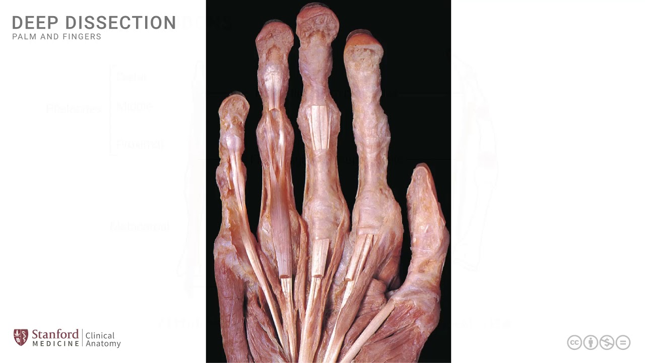
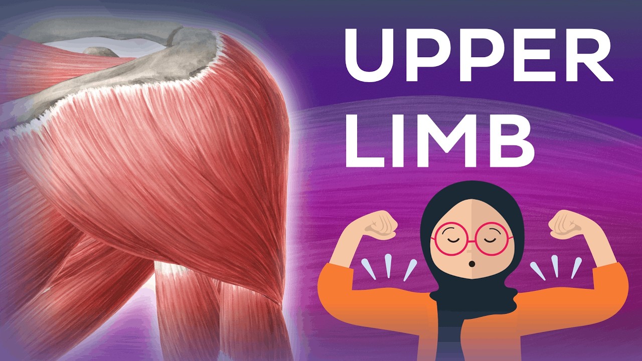
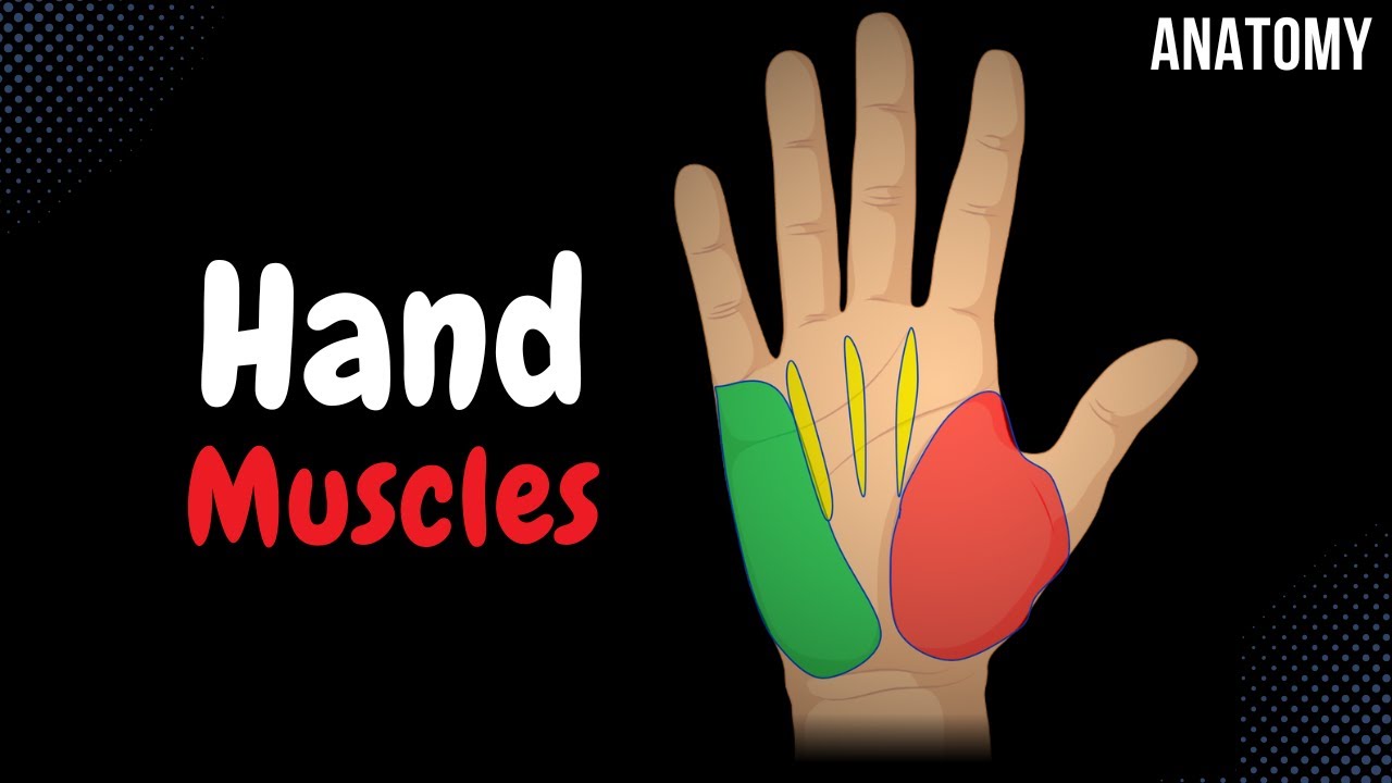
[Music] in this lesson we will review the muscles that participate in the formation of the thenar group of muscles so let's begin by looking at a super visual dissection photograph of the palm of the hand this is the hypothenar eminence down here at the base of the little finger and the thena group of muscles are what formed the thenar eminence over here and in order to understand the thenar group of muscles we will look at it in a diagrammatic representation and these muscles are also arranged in two layers like in many other anatomical locations so
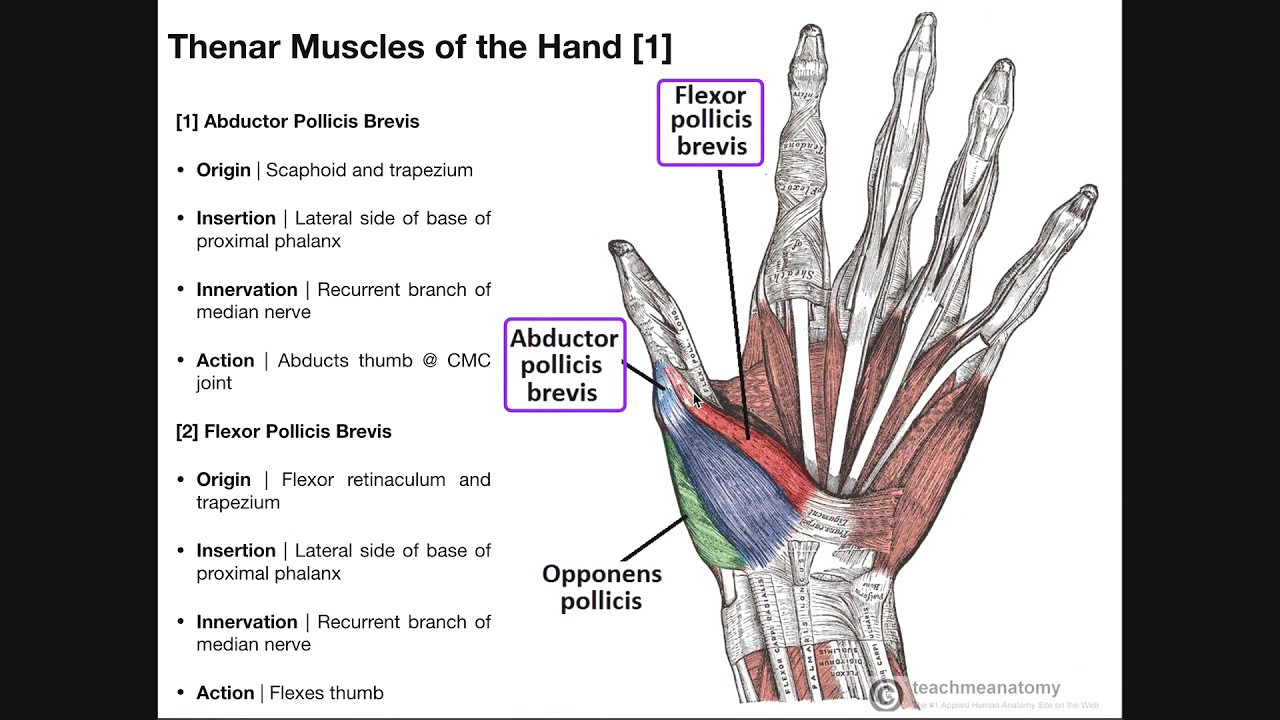
let's begin by looking at this line drawing and this is really the base of the right thumb and the area that is magnified is this particular area the area of the thumb and the adjacent digits in the first image which is on the left side we can put in the flexor retinaculum over here so this is the flexor retinaculum and attaching on to this flexor retinaculum and the adjacent bone is the first muscle of the thenar group known as the abductor pollicis brevis or APB sometimes in abbreviation this muscle attaches distally at the base of
the proximal phalanx as shown in the diagram and is innervated by the median nerve the second muscle of the thenar group is known as the flexor pollicis brevis sometimes abbreviated as FPB it's a much smaller muscle but has a very similar location and attachment extending from the flexor retinaculum onto the base of that proximal feelings and so this is the flexor pollicis brevis these two muscles form the superficial part of the thenar group and if this muscle the flexor pollicis is also innervated by the median nerve if we remove these muscles and we are we
are able to see the deeper muscles and I'll show that in the diagram on the right hand side so let's again put our flexor retinaculum in place which is over here and note that these superficial muscles that I just described have been removed and we can now see the deeper muscle which is known as opponens pollicis and it's a muscle that will have its attachment from the flexor retinaculum onto the metacarpal so it does not cross the metacarpal phalangeal joint and so this is the opponens pollicis and this is also innervated by the median nerve
the opponens pollicis is not visible in a superficial dissection and has to be exposed by either removing the superficial muscles of the thenar group or by cutting them away there's one other muscle that it's worth mentioning here that technically does not belong to the thenar group but if you are reviewing the anatomy in this area you will certainly come across this muscle and it is known as the adductor pollicis it's a much larger muscle than the opponents it is also in the deeper area and it extends from the metacarpal of the middle finger as shown
in the diagram and goes on to the base of the proximal phalanx as shown here it's not part of the thenar group but it does have an action at the thumb and therefore it's worth mentioning here and we can confirm we know that it's not part of the thenar group because its innervation is also different it is in fact innervated by the ulnar nerve and it's an important innervation of the ulnar nerve and sometimes in clinical testing for all nerve injury we actually test for the action of the adductor pollicis similarly when we suspect a
median nerve injury we will often examine for the abductor pollicis brevis and look for its action and examined for any atrophy of this muscle at the base of the thumb as part of that thinner group of muscles so these are some of the muscles that are at the base of the thumb the three that participate in the formation of thenar group and the thenar eminence and then the fourth the adductor pollicis we in the depths of the palm of the hand let's look at these in dissection photograph the left side photograph here is a superficial
dissection it's a close-up view and it is seen so showing the base of the thumb and we see the first muscle here which is the flexor pollicis brevis this is the flexor pollicis brevis right here and it's a much smaller muscle in comparison to the other muscle which is in the superficial teener group the abductor pollicis brevis which is seen here these two muscles are clearly visible as part of that superficial arrangement note also very clearly seen in this close-up view is a branch from the median nerve that is known as the recurrent branch the
recurrent branch of the median nerve supplies these muscles and is an important innovation that needs to be identified in anatomical dissections if we look at the right sided photograph these muscles and nerves have been removed in order to expose the deeper muscles there's a little remnant of the opponens pollicis but not much of it is visible what we really see is the abductor pollicis and so all of this muscle is the adductor pollicis which is seen here one can see it's more transverse fibers as well as it's oblique fibers and the adductor pollicis also goes
on to its way to attach onto the base of that proximal phalanx we see a little bit of the opponens pollicis in this area down here but not very much as it is visible it has been removed in this deeper dissection one other point of note are the cut tendons the cut tendons of the long flexors which that are seen over here note that there are two tendons going into the index finger in this case and we'll look at these in detail in a subsequent lesson [Music] you