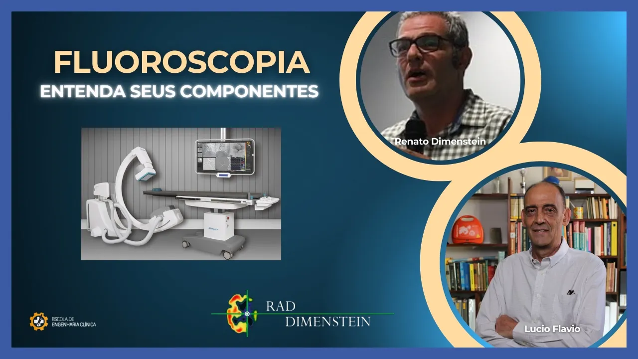
Hey guys who like clinical engineering today another video made with the support of Radi mensin through Medical Physicist Renato Dimenstein who will talk about the fluoroscopy devices in this first part he will talk about all the components in a well way detailed to guide you who enjoy Clinical engineering and understands that it is I need to improve our communication no only with the doctors the technicians who operate the imaging equipment but mainly with physicists who are responsible for ensuring quality and the safety of the operation of this equipment then in this first part If you
have any questions, comment and send your thoughts that Renato's team has willing to help us clarify and make you more secure when dealing with this type of equipment So let's watch the video hello welcome to the engineering channel clinic today we talk about equo de will bloom copy divided into two parts first describe the components and then talk about the operating controls my name It's Renato dein I'm a physicist I'm R specialist shadow physics diagnosis medical the first step is the description of the components we will talk about the generator the tube the filters collimator
radiation detector grid receivers electronic images associated with the resources technological and finally talk about the minimum requirements ah as the beginning of our presentation this are the settings here we can see a remote control on top is the tube below is the detector eh image intensifier on the right is a similar configuration except the tube It's at the bottom, the receiver is at the top it is possible within the settings to have a portable equipment a C-arm in some cases this C-Arch has a smaller size in other cases it has a digital electronics a flat
panel and here on the left configuration of then angiography below the tube above image receiver that may also have as a two-pipe and two-pipe configuration detectors is called biplane here are configure most advanced for Neuro Arches here one conib equipment C These are the main settings like the point of describe the X-ray generator is a generator of power that transforms rectifies your Visa and makes the signal have little variation and this variation is called rip we have here the different generator possibilities power generators with lower power are used for Orthopedic studies and larger for studies
of Cardiology in this illustration a generator configuration Monoblock coupled to the tube and I have here this is the power voltage of operation continuous mode and mode pulsed current and for this type of configuration we have orthopedics for example or studies of urology for cardiac or cardiac studies Interventional radiology needs a greater power to dissipate a larger current to be able to do the patient movement studies here a study of brain and limbs inferior what is important engineering the engineering people Clinic understand This is the relationship as a third point to describe the x-ray tube
this is an illustration of the tube where a difference of potential between a negative pole and a positive pole then produces X-rays and it fine focus emitting filament for details focus Thick thin and thick and here are some intervention equipment has three filaments As the equipment it is described by the manufacturer when you look at the label and your X-ray tube here is the exit where the filters then it is a ceramic tube rotary its diameter fine focus focus Thick as it is refrigerated oh the next tubes they will have this configuration ah m is
a flat emitter that will give better image resolution with conventional and the new flat tubes and here are the oh the new rotating targets it produces a greater cooling capacity and then you can use a better bigger one RB power x an important point of the equipment is the filtration that is placed at the outlet of the beam it removes the low photons energy if an example without filtration kv ma the dose This is the image quality when you filter the kv decreases but the dose compensates it's fine minor some examples of filtration This is
a filtration of a cardiac exam that enters a blade of aluminum and then it compensates for the brightness of the image This is a second example of filtration here without filtration in the cardiac examination with here is with the filtration we can see that this is the compensation filter that does the best adjustment in the automatic system improves the image quality in these dudos cardiovascular another very good example simple but important here is a equipment remote controlled has three and aluminum plates mimicking This is an obese patient This is a detector of radiation then in
most of the procedures the operator leaves black in automatic mode without filtration This is the rate of dose if you had selected here you have a dose of almost 60% less than the staff at Clinical engineering must understand about the filtration is this a fifth component is the restriction of the fe called collimator to left of the image intensifier to the right is a collimator for flat panel flat intensifier panel you can see that there is a distortion image intensifiers the importance of the collimators is the larger the radiated volume greater is the risk of
patient that we can see here a greater scattering of secondary radiation also increases the risk of for collaborators So when you reduces the radiation field you decrease this risk you are better the contrast this is important mainly for you decrease the subposition of the Fields of radiation when you need to do multiple incidences some examples here a simulation with Phantom of skull a detector with collimation adjusted s with open collimation there are times more radiation doses secondary another example Ah this is the Field size This is the dose when decrease the size you decrease the
dose and the one more one another important question is the virtual collimation you get manipulate the collimators on the last image then you don't need to radiate plus the patient is the collimators Ah what is the function of the meter is placed at the output called k kinetic energy right? I mean on the panel You can have time of fluoroscopy which is this one here problem of using fluoroscopy time as a dose descriptor that he does not consider rate for C subtraction may also appear accumulated cumulative cumulative dose and in the study the product of
the dose by area that measures the dap means this part area and this room is really the best de writer is one of the peak of maximum this here is one of the Manufacturers that produce a 2D and 3D mode for dose assessment basically to prevent this and the new equipment must have an alert I mean this is a warning and every half gre has to have a notification a next point is the grid a grid must be removable for studies of Pediatrics which is an example with Phantom here the grid here without the grid
he wants say the grid basically it improves the contrast of image that increases the dose of radiation an important one are the receptors of images, right? In this case, it is intensifiers are the sizes of the centimeter intensifiers inch and basically it magnifies it intensifies the image that's why it is important to put the patient closer possible from the receiver of images the intensifier This is a larger size at the input than at the output to improve spatial resolution here is an illustration of the receiver here at the exit here the electronic system associated and
this is a brief description of how it focuses means you have the input phosphorus that has output phosphorus and you have these lenses here focuser to increase and better resolution spatial at the system output has a char couple camera device this here is an image with image intensifier with a TV of 1 24 has limited brightness in image in the case of flat feathers does not have all that electronic apparatus the signal It is already digital and can be arranged with photodiodes oh and this is the semiconductor and in the end we have an image
using better quality table look than that here a right as he expresses himself resolution of images in line pairs in image intensifiers case you Magnific the image better the resolution and increase the dose in case of flat panel no you can keep the resolution in line pairs independent of magnification some manufacturers used detector pic size smaller and has better image resolution others have proposed a basis of detector putting the semiconductor on top and then you improve the image resolution and can see better resolutions present cardiovascular study you see the stent that fractured for Clinical Engineering
it is important knowing and comparing the detectors here is a example of a flat panel that this is the Pixel size this is the number of pixels and you can magnify the images has Different fields of view is important know that the smaller the field of larger view is the number of line pairs and the higher the resolution of the image to show an example this one is a line pair here with a certain field of view and it is possible to check this number of pairs of line but if I get a bigger detector
with greater number of pixels has greater spatial resolution this is what clinical Eng. it should knowing the comparison is not only in terms of resolution but also in terms of efficiency quantum detection efficiency of sensitivity and then you have the intensifiers with lower efficiency than that the flat panels the flat panels they can be amorphous silicon or they can be of a semiconductor metal is silicon it has a larger detector size and the semiconductor it has the smallest Pixel consequently less noise is more fast but more expensive than silicon love how manufacturers present the results
as a function of quantum dot and of the spine pairs and then the silicon amorphous has more efficiency than the cicor oxide this is how it works and these are the relationships important to understand configuration of equipment fluoroscopy intervention that has to have something called memory of the last image or l do not want say there is radiation in the last image armen is kept on the monitor and then you saves radiation in the dose in patient a second available resource is the roadmap here is the image of a renal angiography and you see there
introduction of the catheter you can do as catheter a third available resource is the subtraction dsa means here a image of contrast graph the image after administration and image subtraction when you see a better resolution in background that he makes a mask subtracts from whole image and from here you have images by subtraction as the last topic of this first part are the requirements Nema's regulatory framework and this Nema is a Standard of the National Association of Electrical manufacturers eh that demands everyone equipment from 2023 T dose indicators rate indicator kerma filtration additional may have
a retention of last image virtual collimation has to store at least 300 frames in mode fluor pulse rate variable a dose report structured There must be limits to what you want to control indication of an anti-diffuser grid especially for studies in pediatrics has that is removable Anvisa's regulatory resources are more limited to mandatory protection the display and to minimize the effect of cataract Ah a petticoat placed below the table to reduce secondary radiation and minimize the risk of epilation the Radiology rooms have to a shielding calculation must be made Ah, there have to be coaches
from operators and doctors must have radiometric survey and testing leakage radiation verification of equipment of epis and finally biannual tests of quality control These are the regulatory requirements Therefore what we saw up to this point in part one were the equipment settings with respect to the detector collimator tube filtration to question of image receptors dali flat panel intensifiers compare the detectors in terms of line pair resolution and efficiency quantum detection and we have now seen the some requirements like subtraction eh keep the latest image requirements regulations of Nema and Anvisa as people who have questions
or doubts about this there is a space on the channel Clinical Engineering and I look forward to seeing you at next week to talk about a part two on controls of equipment operators floria my my name is Renato de manstein and I appreciate the attention of you and see you next week thank you Well if you got here I hope you be much more enlightened about this type of equipment do not forget that the people will have part two And from here on part two an opportunity for you ask all your questions that we will
make the greatest effort to answer then that's it and that's it that thank you very much for your time Thanks ah [Music]

![Aula 1 - Introdução à Manutenção de Equipamentos Médicos [CPMEM]](https://img.youtube.com/vi/AR4F7F2CyJM/maxresdefault.jpg)