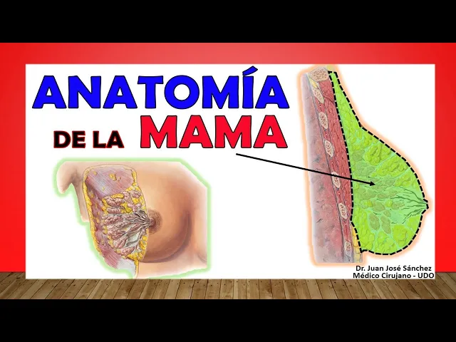
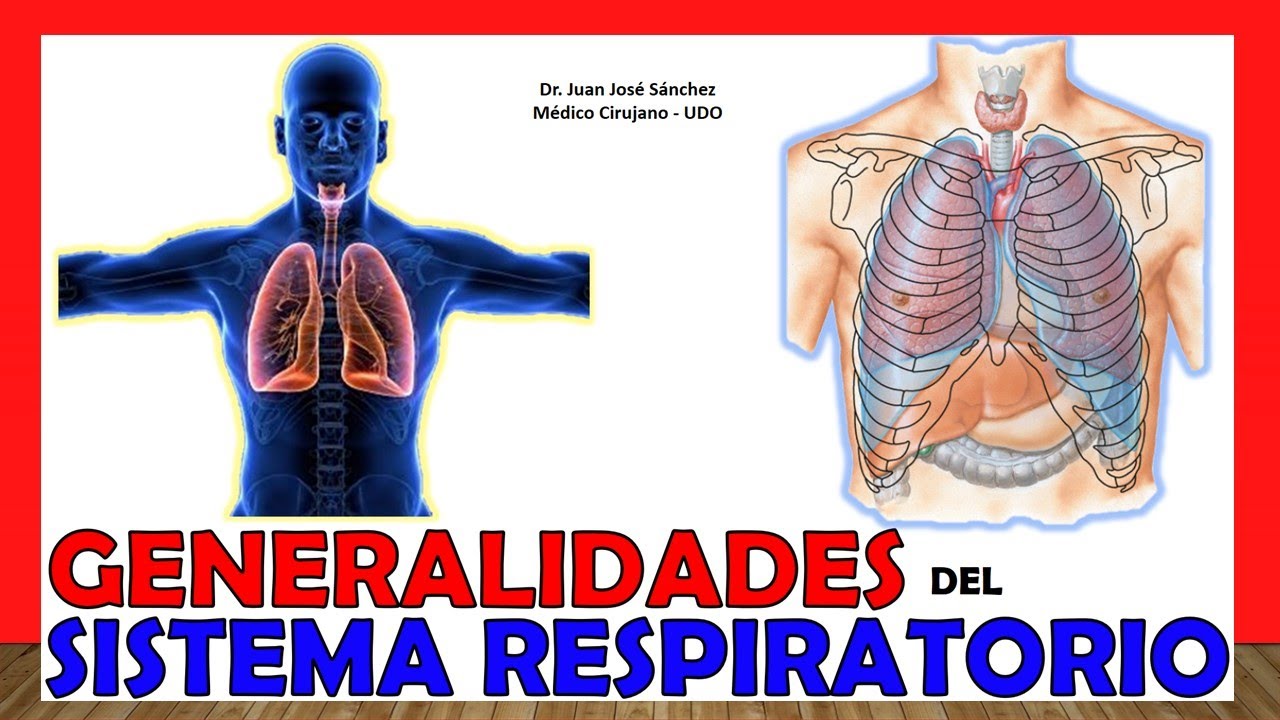
hi how are things? Welcome to the breast anatomy video, today we will touch the breast and everything that is with it, that is, we will touch the mammary glands and we will touch all the structures adjacent to the mother, follow me then in the next video on this channel easy anatomy by Juan José Sánchez, When we talk about the mother, it is a structure that is present in both men and women, but the man has it very little developed because the function of the mother is a function that, let's say, co-helps the reproductive system. because?
because once that product of reproduction that is the baby is born, he needs to suck, he needs to suck to feed on the milk and that is what is found inside the breast, we are going to see that the mother is everything but inside the In the breast, the most important structure is the glands that are found, which are the mammary glands, which are glands of the exocrine system. Why exocrine? because it is a secretion product, it goes through some ducts to the outside as we are going to see right now, some authors like to call the breast, breast, however there are others who say that in anatomy this really is a depression and it is a term that is more or less distorted because the breast would be the space between the two mothers that would be the breast, however almost indistinctly some authors call the breast as breast and that is what makes humans more mammalian animals because we We need the breasts for our development, we need that great product that women excrete there, which is milk, so the anatomy that we are going to describe right now is the anatomy of a woman's breasts, which are the breasts you already develop, in men it is everything is almost the same but it is very little developed in abundant fibrotic or fibrous tissue.
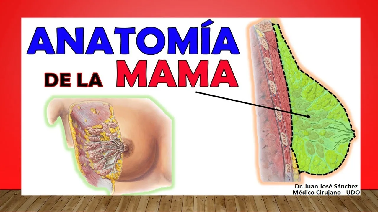
Well then we begin to talk about the breast, in general it is a structure like the gland but these glands that are immersed in a stroma, are immersed in a lot of connective tissue and in many fatty tissues, in fact the breast of some women is more fat than tissue. glandular and in others that are less quantities more glandular tissue than fat, but that structure is how they exist there and that is why the shape is so varied from one woman to another, the size is very varied, the amount of glandular content is very varied. What it will have inside, the shape is also varied, there is a lot of variation in the breast, so this is a more or less conical-shaped structure whose base rests on the pectoralis major at the level of the latissimus dorsi muscle and more or less at the level of the serratus muscles.
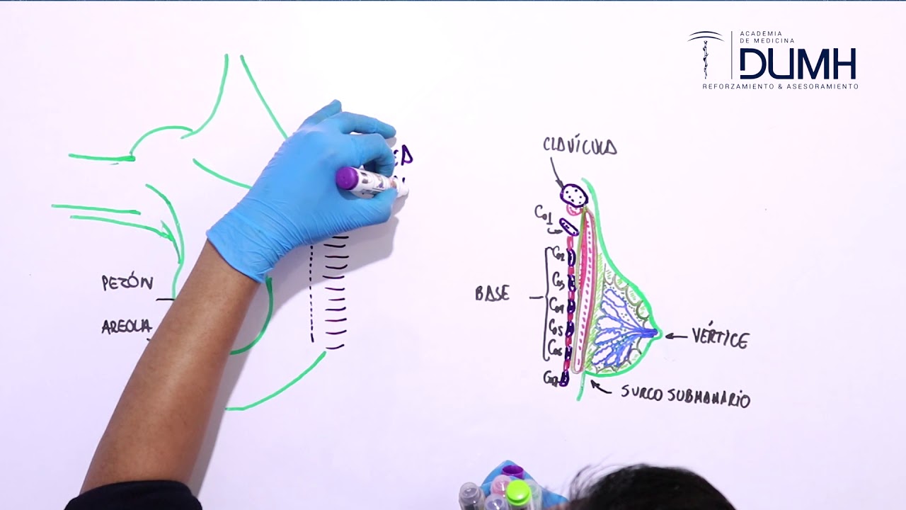
If we have to more or less compare it with the ribs, we say that the breast goes more or less between the second and the seventh rib, although some authors say that it is between the third and the seventh rib , but well, on average, let's say this between the second. and the seventh rib, it depends on the stage of reproduction in which the woman is, if she is a woman who has already given birth or if she is a woman who has already developed, mothers tend to be larger because they already develop greater weight due to the effect of gravity she will tend to fall, the more pregnant the woman is, the older this mother is, the more she tends to fall so that these relationships that I tell you with the ribs can vary enormously, at the level of the midline it is said that the breast or glandular tissue can extend to the lateral edge of the sternum and to the axillary line, those are the common limits of the breast tissue as such, however in anatomy we talk a lot about what the mammary line is, a line that It is also used in embryology which is a line that goes from the clavicle to the groin and theoretically in any part of that mammary line accessory breast tissue can be found, if in any part of that mammary line if everyone knows that area, Now in this sagittal section of the breast we realize what I tell you that this mammary gland is not just glandular tissue but there is also a lot of connective tissue, stromal tissue, tissue in this case it would be adipose that is immersed within them, So after the skin, which is what we find here, this skin from this region is a very smooth skin that really does not have large fields or anatomical variations, but rather it is a skin that is quite stretched, Then we see that here the superficial or superficial subcutaneous cellular tissue is found and then the deeper subcutaneous tissue is found behind the subcutaneous tissues. We find the glandular tissue that would be the mammary gland itself, behind that mammary gland we find the pectoral muscle and the fascia.
What covers it is the pectoral fascia, as well as on the sides we find the serratus anterior muscles also called serratus major muscles, we see that the mammary gland is larger than the breast and how is that, well the breast is everything as I told you and here in this section you see that there is tissue under the breast, glandular breast tissue but see that it has an outward extension that we call the axillary tail, this according to the greatest amount of tissue towards the armpit That is what explains why when you cut into four quadrants in a breast tissue, the malignant locations of the tumors always correspond to more than 70 percent. In fact, at the level of the upper external quadrant, consider that you divide the breast into Four, it may have superior external because there is more glandular tissue, it is the place that is most likely to do some type of damage, neoplasia, a type of proliferative disease, now it gives breast, as I told you, it can extend along the midline It is said that it can pass above, above the clavicle laterally, it can even pass the limits of the latissimus dorsi medially, it can pass the midline and inferiorly we can find glandular tissue even in the epigastric fossa, notice how large the location of a breast, at the level of the parenchyma as such that surrounds that mammary gland we find abundant fibrous tissue and abundant adipose tissue, in addition to the epithelial tissue which is the tissue itself, very well these mammary lobes are going to make 15 to 20 lobes in each breast, Other authors say that there can even be 25 to 30 lobes, it varies greatly from one woman to another. Within those lobes that are in the mammary gland, which are made of epithelial tissue because it is a gland, we will find large cavities where they are produced.
milk, breast milk, of course, this is influenced by many hormones that make that gland prepare for milk production. Now these lobes have to carry their excretion products, which is milk, through some ducts, these are the milk ducts and these milk ducts all begin to converge looking for the nipple because it is the final place where this milk is, which is the product of The secretion of these glands reaches the nipple, now when those lactiferous ducts are converging near the nipple they tend to dilate and this dilation is known as the lactiferous sinuses. After these lactiferous sinuses, others follow in the galactopharynic ducts, which are the They open in the nipple more or less between 15 to 20, finally there is one of them that join together but finally they can reach up to 15 or 20 lactophore ducts at the level of the nipple.
Very well, there are some ligaments that are the ligaments that Cooper described by a doctor that we even call it Cooper's ligament or suspensory ligament, this is very important from a clinical point of view because these are connective tissue ligaments that go from the skin to the parenchyma of the mammary glands of the stroma better said and this explains why the sign exists, look for it, google it, which is the sign of the orange or the orange shell, the orange peel that is seen a lot in cancers of the breast, any tissue that grows in size at the breast level will pull these ligaments, distend them, tighten them and that will cause the place where it is inserted into the skin to retract and form holes that are characteristically seen on the skin of women's breasts, like when they have some intra-breast growth, so this is a sign that without touching the woman, when you see it you have to quickly suspect that the woman must have some growth breast and it must be studied to see if it is malignant or benign, but it is important to know that this is thanks to these Cooper guidelines. Let's continue talking about the breast, let's Let's get into the structures that are in the nipple, but first don't let them get away from the video [Music] I invite you to subscribe here in the lower right corner click [Music] and don't forget to like the video, it's important to like it I remember again that it is important that you subscribe to the channel below, you click and access the more than 150 videos that you will find on this channel, the anatomy of all the muscles of the chest wall, the lung, the heart, the bone of the limb upper limb, the joint of the lower limb, arteries, everything you can imagine, there is another video of axillary lymph nodes, armpit, axillary space and axillary artery, axillary nerve that may be related to the breast and that may be able to help you. interested, just by subscribing to the channel below.
Let's talk then about the nipple, which is a structure that is theoretically located or is said to be in the center of the breast that actually falls below the breast, which is the place or the anatomical site where those galactoferous ducts that would go from the mammary glands themselves to him, something that I forgot to tell you that in the external anatomy of the breast since I see it here in the upper part it continues very well with the skin of the thoracic region, of the antero-upper region of the thorax, but At the bottom if you see a groove that is what we call the submammary groove, a groove that is important from the point of view of surgical anatomy since there are several incisions that are made at the level of that submammary groove, then this The nipple almost always corresponds to the fourth intercostal space, which I told you at the beginning that, as the position of the breast always varies a lot, this nipple can vary, it can be a little higher or generally lower at the level of that fourth intercostal space , I already told you that the nipple is the place where the galactophore ducts open, let's see how abundant smooth muscle fibers are, some arranged longitudinally, some arranged radially, and this is due to the fact that when these smooth muscles contract by the mechanism of the autonomic nervous system, they squeeze, let's say, the milk ducts of the breasts and even these mammary glands as such have smooth muscle around them that when squeezed by that smooth muscle there is nothing left for the milk to do except to be ejected, to shoot out; We are going to see that this nipple, which we say is the part, the central dot measures approximately one centimeter, it is surrounded around by a structure called the areola, which in full diameter measures 1. 5 to 2. 5 centimeters, this area is a more colorful area than You see at the level of the breast and it becomes even more colored after pregnancy and there it remains that way thanks to the stimulation of a hormone that is not relevant to the melanocyte-stimulating hormone.
Let's see what areola is. Let's see mammary glands. accessories, although I distance myself, we are going to find a sweat gland and we are going to find some elevations called the areolar tubercles, also called Montgomery tubercles, which look very marked, especially at the end of pregnancy and during breastfeeding, so that is the nipple that is the point.
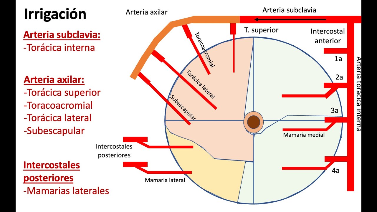
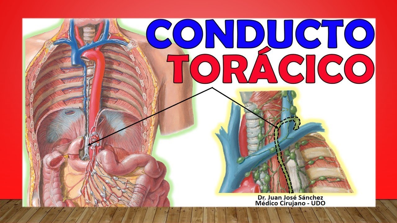
central and the areola what is on the sides, there are patients who may not have areola or nipple that is called an atelia, when they have many it is called a polytheia because in many variations you can even find nipples along the entire mammary line that went from the clavicle to the groin, accessory nipples as well as animals, you see other mammal animals and they have four or five mothers on each side, then the human, as it is supposed to be an evolutionary process, can have breast tissue and nipples even accessories along that entire mammary line, which is what it is called the mammary line. Let's move on then to the irrigation of the breast, irrigation is well known through mainly the two mammary arteries, an internal mammary artery also called the internal thoracic artery, which is a branch, as you can see here, of the first portion of the subclavian artery, also greatly irrigated by the external mammary, also called external thoracic or lateral thoracic, which would be a branch of the third, second forgiveness portion of the axillary artery of venous drainage, also by some superficial veins that drain, let's say the most superficial part of the breast, they drain towards the internal mammary and towards the superficial veins of the neck, there is a deeper vein that drains into the breast tissue deeper the glandular tissue is the one closest to the pectoral that also drains towards the internal mammary vein but also through the axillary vein and the anterior intercostal veins that are here at this level, if you do not know those thoracic and intercostal veins in the mammary I invite you to watch my video on veins of the costal wall, they have the blood vessels of the chest wall so that you can understand well the route of these blood vessels, finally the lymphatic drainage, a large part of the lymphatic drainage which is through the nodes axillaries and this is described in a separate video on lymphatic drainage of the upper limb and the armpit, here he is going to talk specifically about the lymphatic drainage of the breasts, then the skin the skin tissue drains the level upwards towards the axillary nodes, towards the deep cervical lymph nodes that are seen here, some towards the pectoral lymph nodes is that they are in fact part of the axillary lymph nodes and parasternal lymph nodes that you see are parallel to the sternum, the lymph nodes are always looking for the venous tissue in which they are always in relation to the veins, that's just the skin, well those are the deep cervical veins of the pectorals, there we see the parasternal ones, we are going to see the nipple and the glandular tissue as such first through some plexuses that are seen by a network of the lymphatic system, one perilobular and one sub areolar that we are going to see below the areola, let's say this lymph was collected by those plexuses that drain towards two large collecting trunks, let's see one here right now you can see the left one and one right one, a good one maybe the right one The left one is seen in the left breast and these collecting trunks look for the axillary lymph nodes and from the axillary lymph nodes they look for the deep cervical lymph nodes to end up in the structures of the subclavian collecting trunks. The parasternal lymph nodes also help to drain this deep glandular tissue.
Now that nipple, here we see the axillary nodes very well along the entire axillary vein, they are abundant and important from a clinical point of view because the majority of breast cancer metastases seek these nodes, that is why the When you notice a swollen lymph node, it has to catch your attention and that lymph node has to be studied. Here you can see the subaleolar and perilobar plexuses, as they look for the collecting trunks, that the left on the right, see how some look for the parasternal ganglia, look for to the axillary nodes and finally to the deep cervical nodes to finally drain into the collecting trunks, you can look for my thoracic duct video so you understand how this lymph finally ends up in the venous tissue.