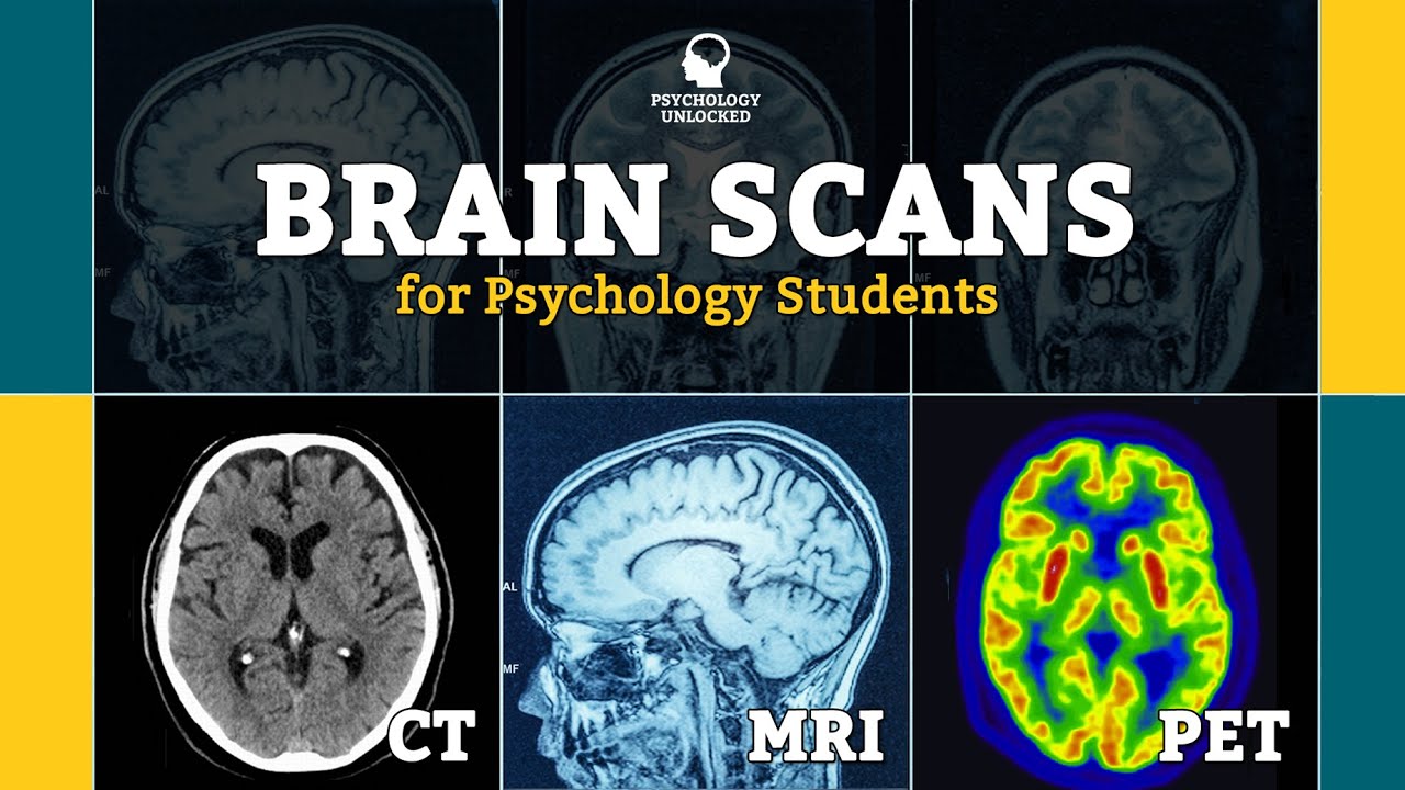
In the field of healthcare there have been major advancements in medical imaging techniques. Their ability to create visual representations of internal structures and organs of the body allow these imaging techniques that have major clinical applications. Today, we discuss three of the most commonly used imaging techniques: the MRI, CT scan and the PET scan.
This is Michael. Michael suffered a knee injury while playing basketball last week. After an appointment with his doctor Michael finds out he will be needing an MRI scan to determine if he needs surgery.
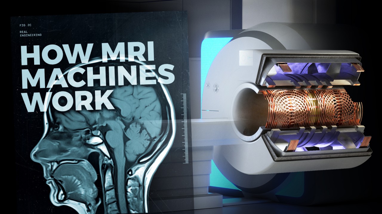
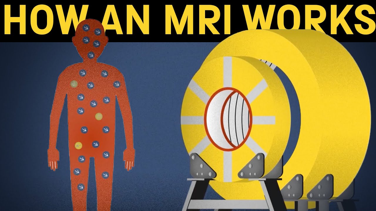
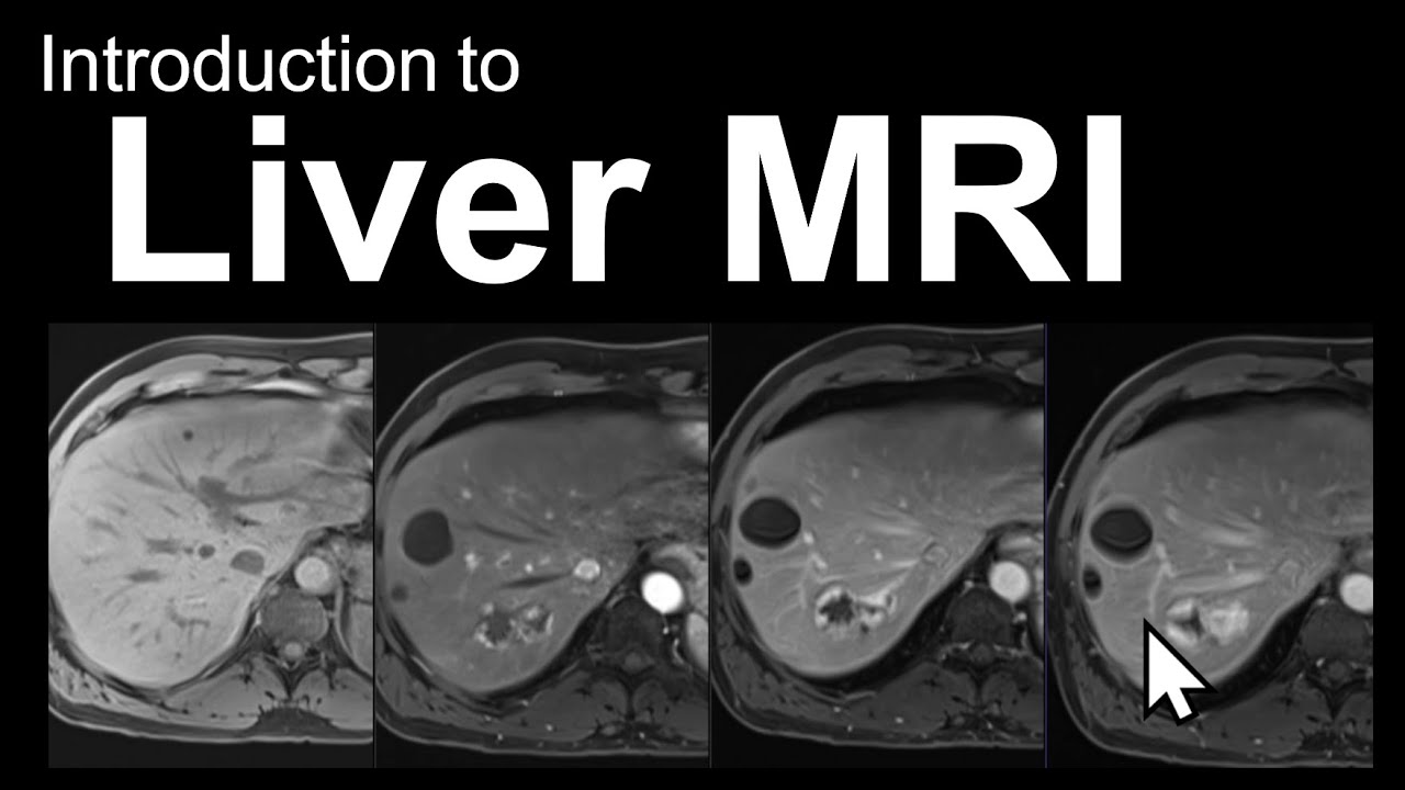
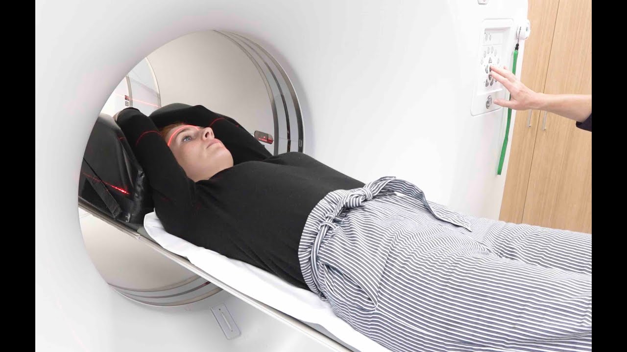
Well what exactly is an MRI? An MRI which stands for magnetic resonance imaging is a technique used to generate an image of one's body non-invasively. When undergoing an MRI scan a patient lies on a moveable bed which is surrounded by the MRI scan and a circular magnet.
Radio waves 10,000 to 30,000 times stronger than the magnetic field of Earth are sent through the body. As we all know, our bodies are mainly composed of water which contain hydrogen atoms these waves cause the nuclei of the atoms to change position. As the nuclei move back into their original positions they emit their own radio waves which the scanner picks up and ultimately turns into a picture that is seen on a computer.
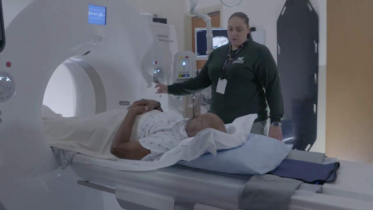
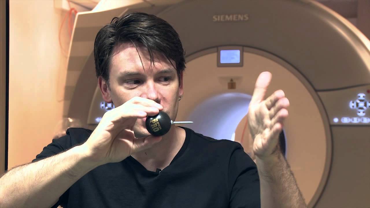
For the most part this procedure is painless and risk-free. It should be noted that pregnant women or anyone with a pacemaker or any sort of metal implant cannot do an MRI scan due to its powerful magnetic force. After finishing his MRI scan Michael meets Sarah who just underwent a CT scan for her severe abdominal pain.
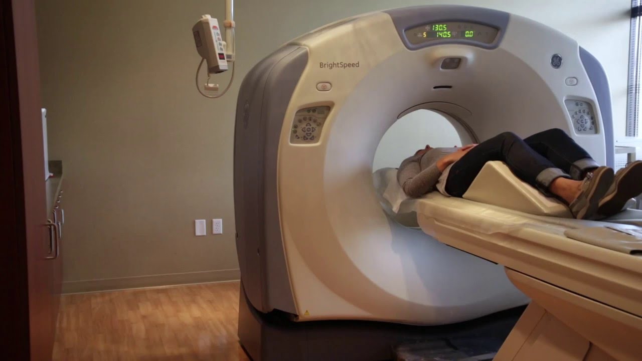
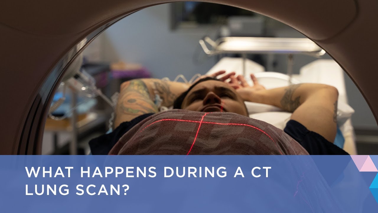
A computerized tomography scan produces detailed images of the body mainly through the use of x-rays. Several beams of x-rays all from different angles are sent through the body during a CT scan. Each angle provides a picture of a thin slice of the area being analyzed.
The data is saved on a computer which takes these two-dimensional pictures and creates a 3d cross-sectional picture of the area. In some CT scans a dye called contrast material is taken by the patient through injection, solution or inserted rectally. This dye makes structures and organs easier to see on the pictures.
Because of the extreme accuracy and detail of these pictures CT scans are used for a myriad of the body's internal situations including internal injuries from trauma, muscle and bone disorders, detecting lung and liver diseases, and also detecting different types of cancer. CT scans provide advantages in comparison to an MRI scan as they provide higher resolution pictures, however they do pose some risks as well. The ionizing radiation from the x-rays has been linked to increased risk of cancer, although it is a low possibility.
The most common risk of CT scans is a reaction to the contrast material which is generally mild and results in hives or itchiness. Similar to MRIs, pregnant women should not undergo CT scans in order to avoid radiation exposure to the fetus. Finally, meet Claire.
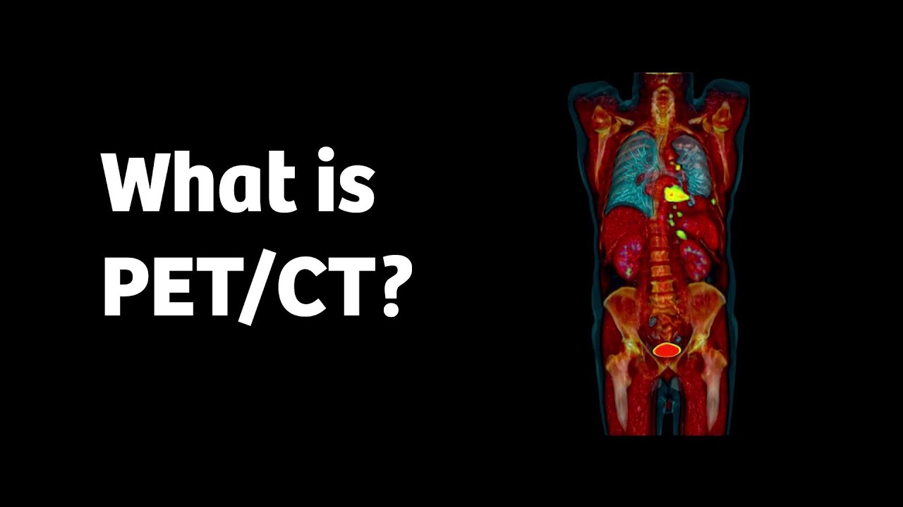
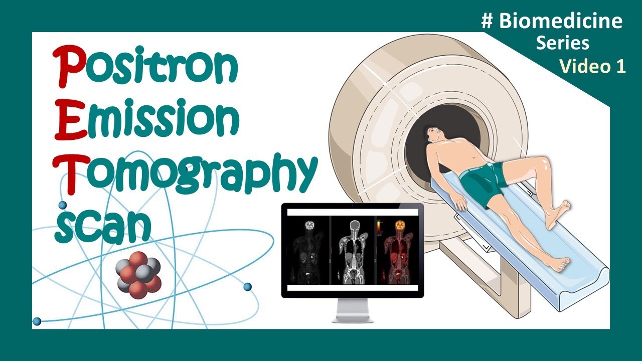
Claire has a history of coronary artery disease so her doctor advises her to undergo a PET scan. A Positron Emission Tomography scan is performed by using a special dye that has radioactive tracers. The patient injects this tracer and it is subsequently absorbed by organs and tissues in the body.
The PET scanner can highlight these traces and see how well organs and tissues are performing. PET scans can therefore measure blood flow, oxygen use, and glucose metabolism. A major difference between PET scans and the two previous scans is the ability of a PET scan to show problems and complications at a cellular level.
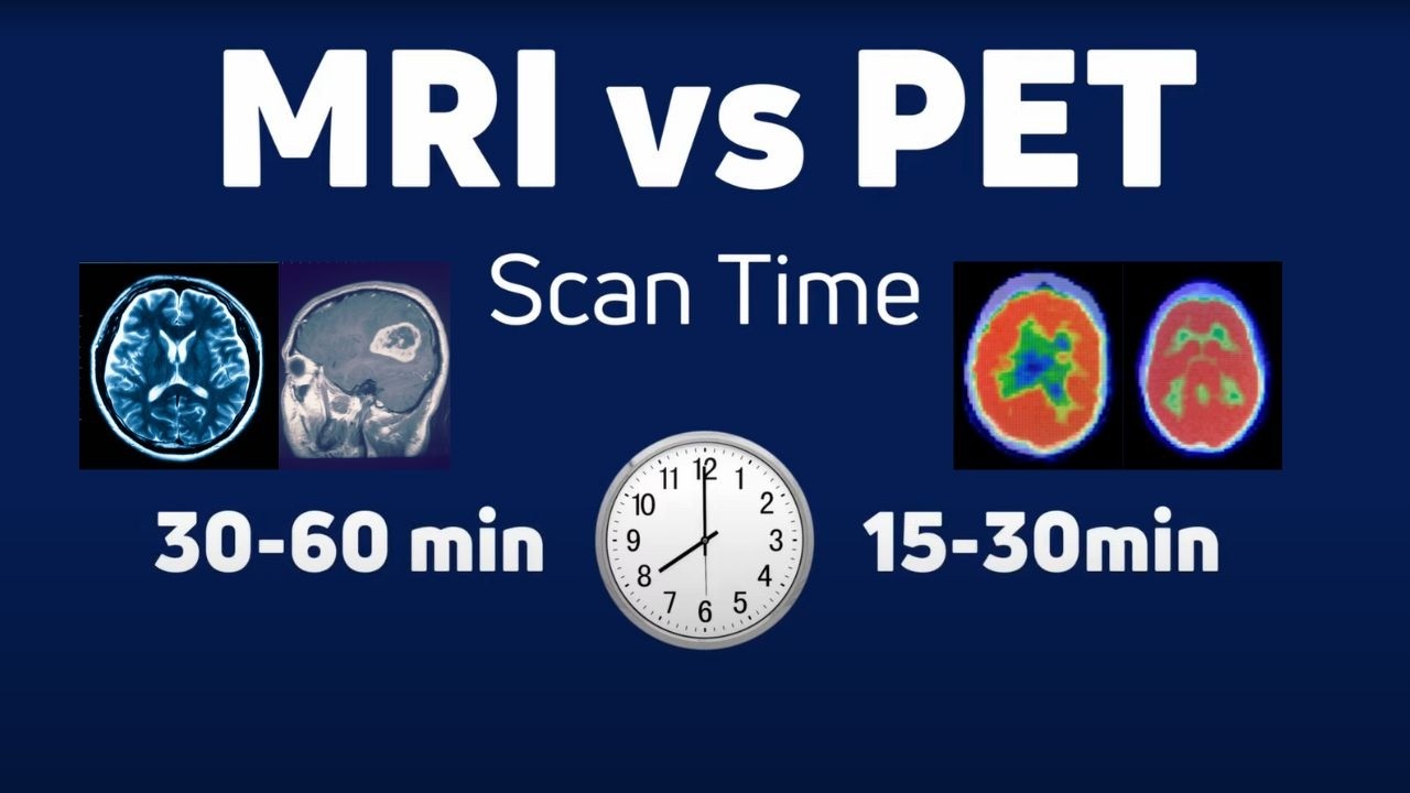
The resolution might not be as clear as an MRI or CT scan, however, it gathers more functional information. To prepare for a PET scan, the patient must notify their doctor about any medical conditions and if they are pregnant. In some instances it is advised that the patient does not eat for 8 hours prior to the scan.
Risks associated with a PET scanner similar to those of a CT scan including exposure to radiation and allergic reactions to the injected tracer. The MRI, CT scan, and PET scan are three of the most commonly used techniques for medical imaging. We learned the benefits associated with each, such as the high-resolution provided by the CT scan or the functional information given by the PET scan.
Furthermore, we also learned about the disadvantages and risks associated with these scans such as the possibility of an allergic reaction to the tracers in a PET scan or the exposure to high radiation in CT scans. In conclusion, the benefits outweigh the risks in all three of these scans as they provide essential information for patients and healthcare providers in need of help. Thank you for watching!