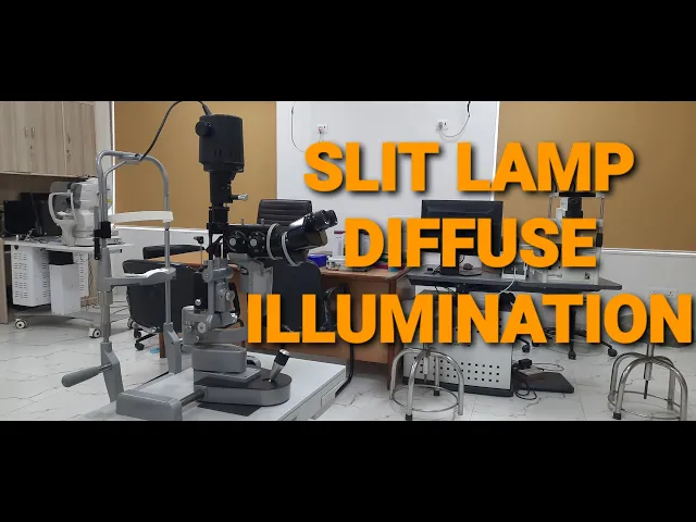
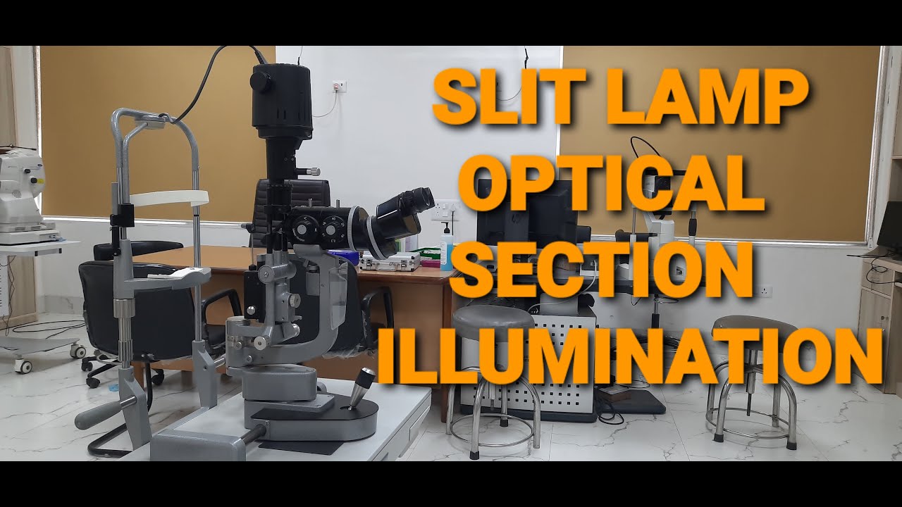
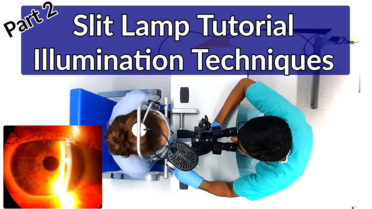
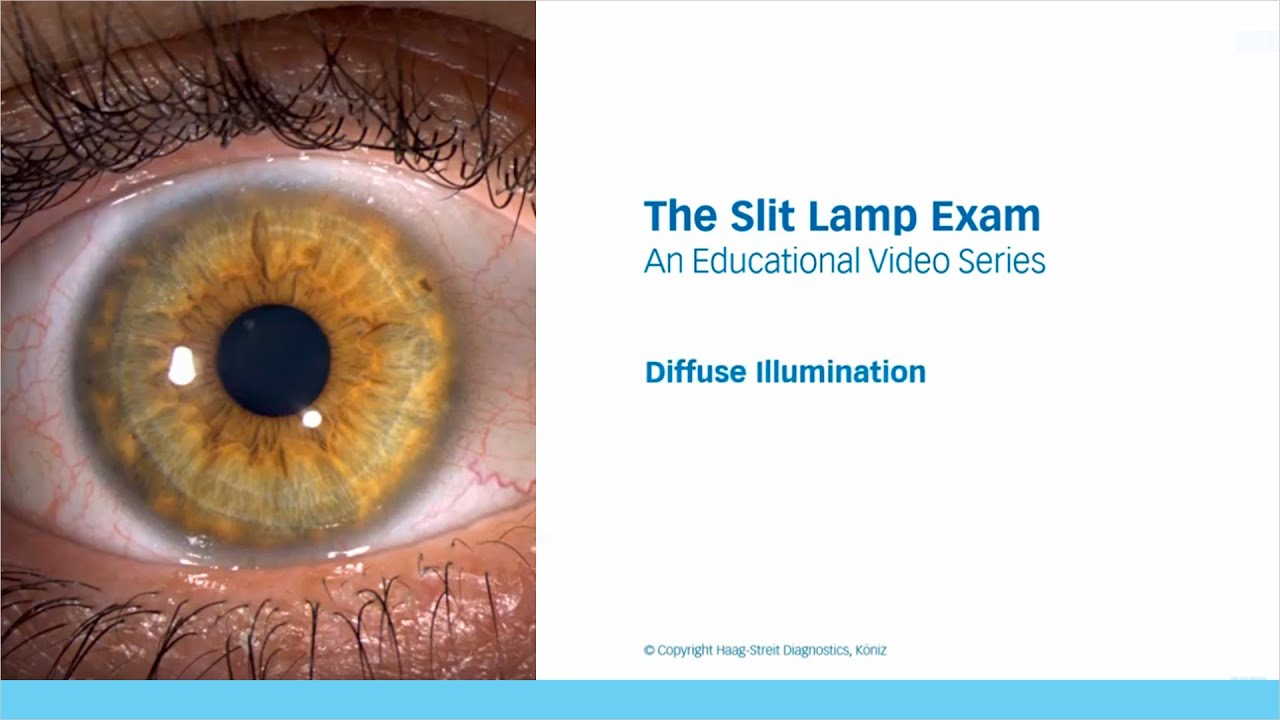
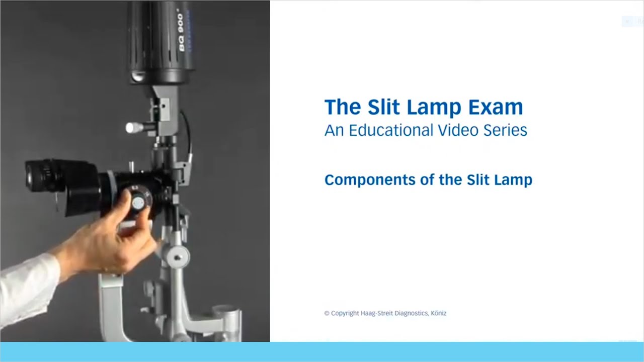
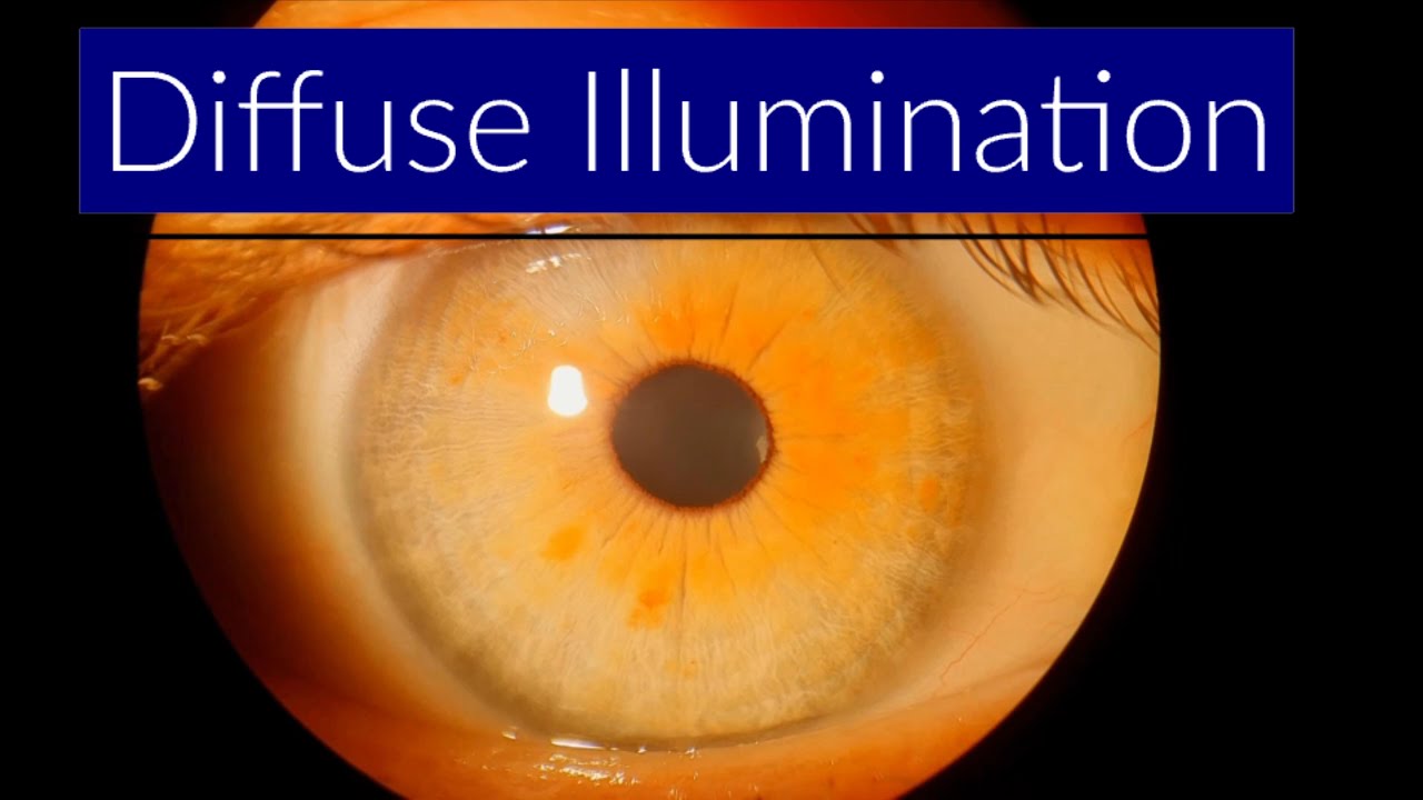
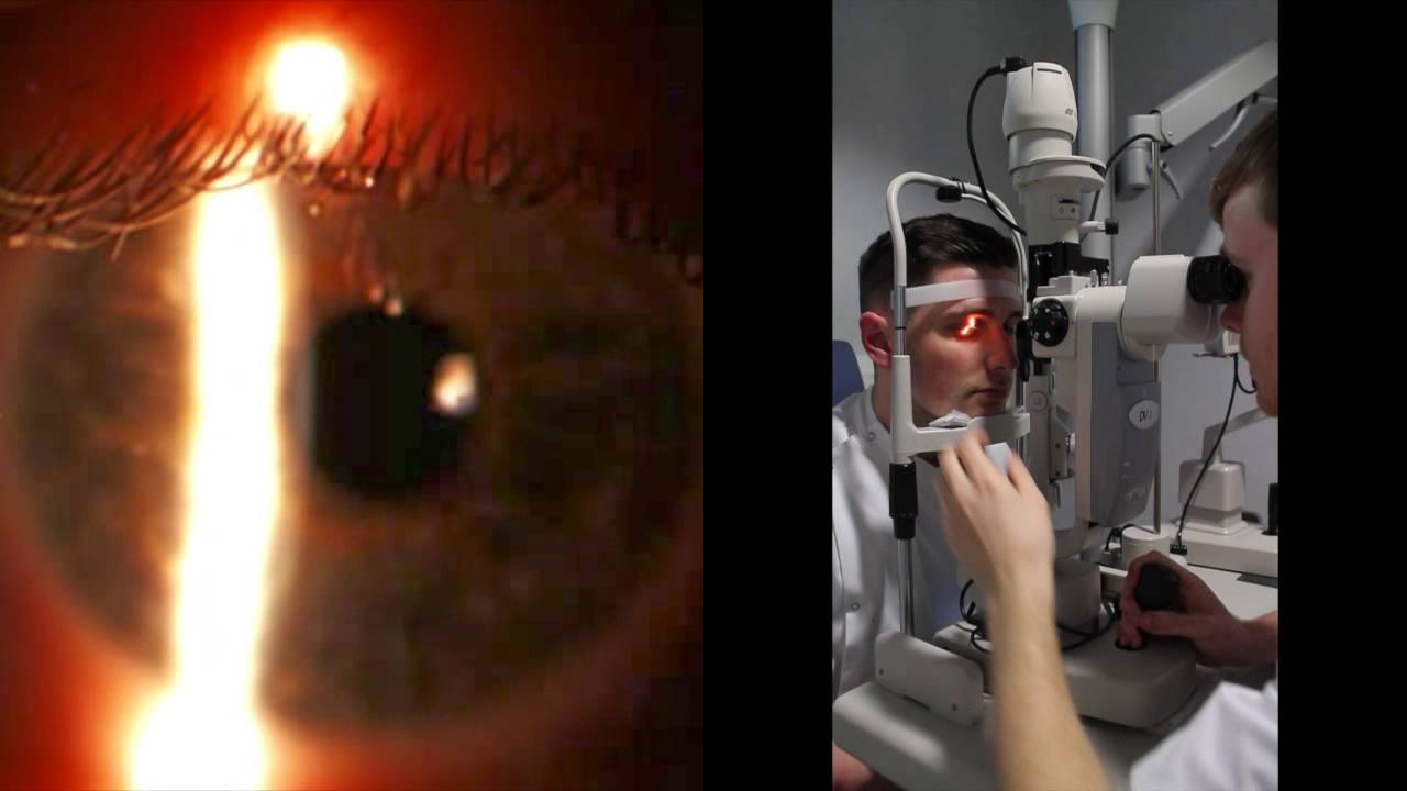
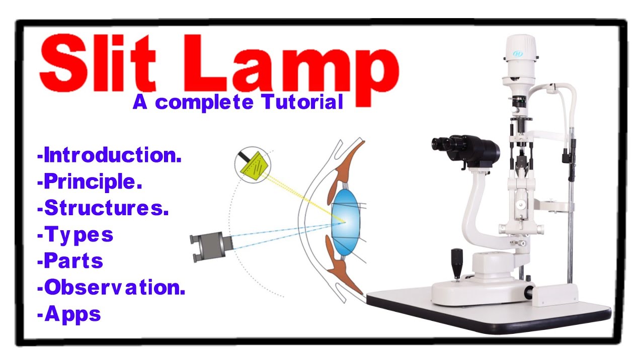
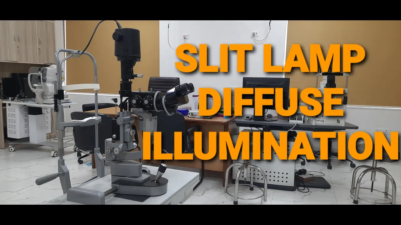
hello friends welcome to the video series lecture of slate lamp this is the third part in which we will discuss about the diffuse illumination and we will cover this video in two parts in first part we will cover the setup and second part the examination of oculus structures to be examined so let's just start both observation and illumination are mounted coexily this is observation and this is the scale which shows the angle between observation and illumination arm now we have to turn the illumination arm to set the angle between both of them to 30 degrees
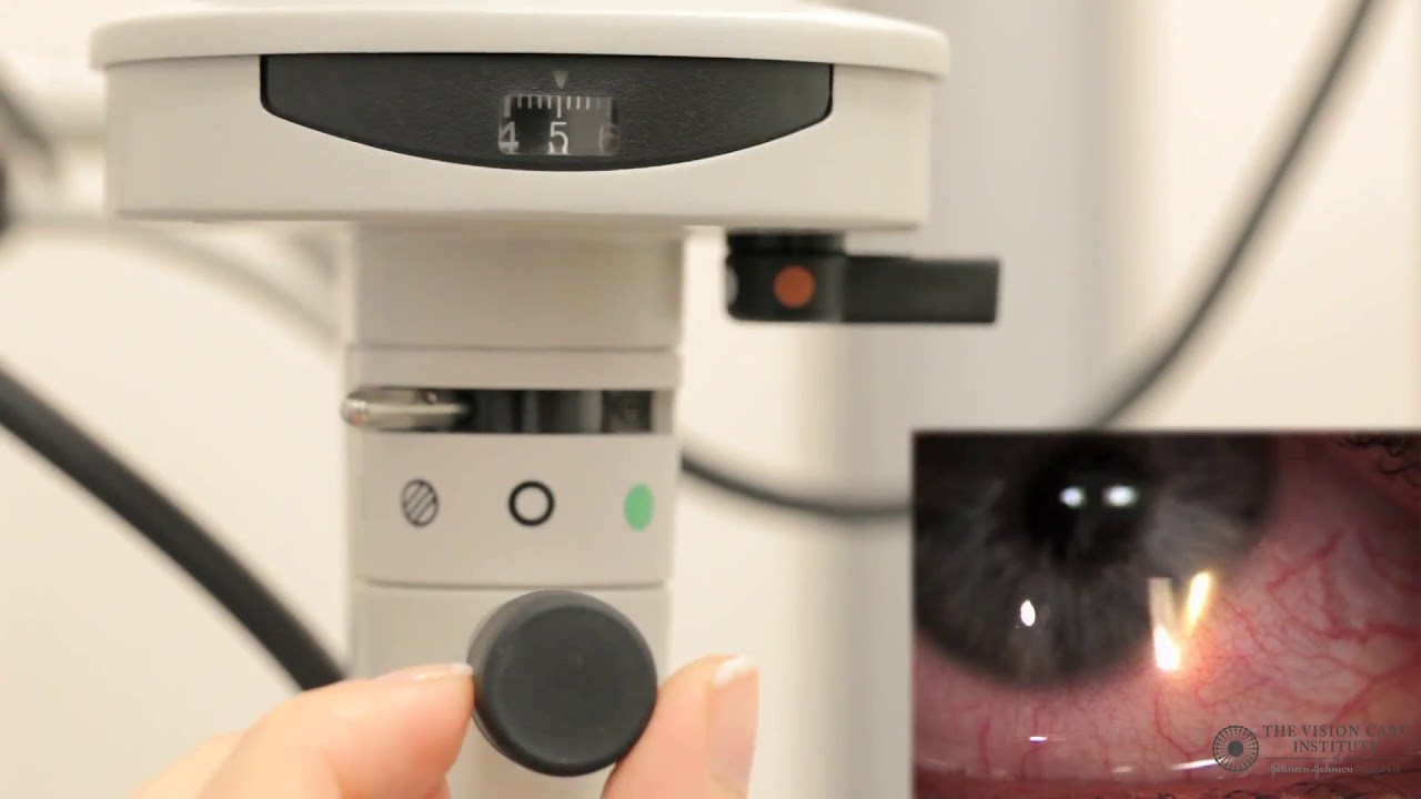
then the magnification should be adjusted on 10 or 16x by rotating the magnification dial then after switch on the slit lamp by this bottom button the intensity of light which can be regulated by this real state should be moderate now it is adjacent to the moderate intensity now the slit lamp should be placed in vertical position by rotating this knob by using the angle as you can see in this part now it is vertical now by rotating this knob the height should be adjusted to the maximum level that is eight millimeter which can be appreciated
on this focusing rod it slowly increase the length of the height then by rotating this knob the width of the slit beam should be adjusted at its maximum level that is 20 millimeter as you can see the width gets slowly increased by rotating the knob now the most important thing is to adjust the filter which is used for diffuse illumination it should be placed on the neutral density filter so the final adjustment is this for the slit lamp setup the first structure to be examined is eyebrows which is examined for its color density and presence
of any scale or any deposits on the eyebrows so which is very basic thing that we have to observe first during the ocular examination and diffuse element the second structure is to be examined is the eyelid skin which is examined for presence of lead crease or any abnormalities or any scales or deposits present on the eyelid skin the third structure is to be examined is the upper eyelashes which is examined for its color direction and the presence of any extra rows of eyelashes this is the normal appearance of eyelashes it is it looks normal in
our subject now the same pattern is also observing lower eyelashes for its color direction and pattern in presence of an extra rose any misdirection of eyelashes that drop the cornea the fourth structure to be examined is the late margin which is examined for the opening of mabovian glen as you can see in the video this small circular white opening shows the wind land opening in the lid now it is more appreciable in the next structure is to examine the tarsal conjunctiva which is examined for the presence of any papillae follicles or any concretions that are
present in the tarsal conjunctiva as you can see it is the clear tarsal conjunctiva and this small circular structure shows the membrane gland opening on the upper eyelid this is the normal pattern the next structure is bulwark nectar in which the blood vessels are examined which shows the presence of any uh chemosis or congestion is there any in case of any abnormalities which are present in double conjunctive that can be easily appreciated in this subject this is the appearance of normal birth injectable as you can see in the video these are the normal blood vessels
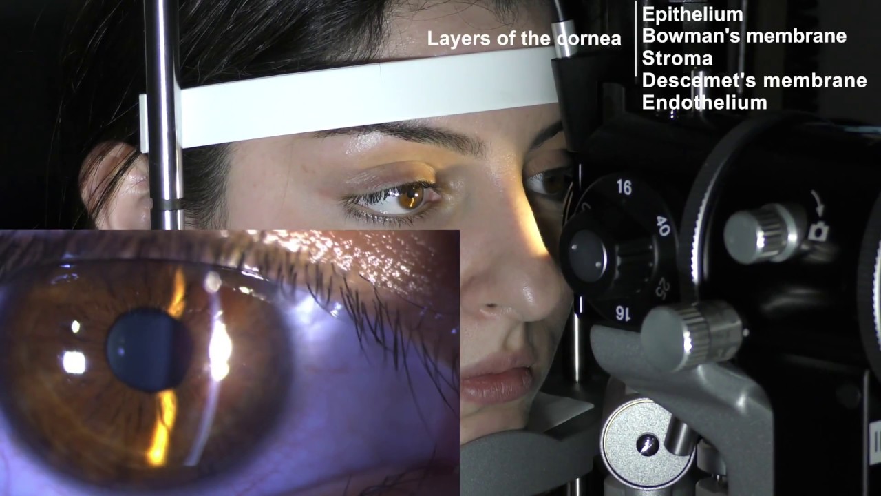
and the normal sign of conjunctiva if there is any dynamic the sign will go off next structure is cornea which is a for its transparency and r is for his normal color pattern now the next structure is medial canthus which is examined for lower punctum which shows as a small pit as you can see in the video it is little magnified and better appreciable in the same way the upper phantom is also examined it is little difficult to avoid the perfunctum but it can be examined on the diffuse illumination so in diffuse illumination we get
overview of interior segment of eyeball now the last structure is to be examined is limbus which is around 360 degree around the cornea which is like grayish white in color which is the transition zone between cornea and sclera so stay tuned please watch like subscribe comment and follow for next video very soon