hi this is melinda eshelman i'm a cardiac anesthesiologist at the university of kentucky and this youtube video focuses on cardiovascular keywords for the ite first and foremost i have to give credit to dr shell for starting this list of keywords that he has updated for many many years and so i updated it in this last year the current list of keywords that we are going to at least present to you contains keywords from 2009 to present i'm not going to review all of these keywords this keyword review is going to focus on keywords that showed
up in the last it's in the ite from 2021 and also keywords that were frequently missed by our residents at the university of kentucky so what's going to follow in the next few slides is a list of all of the cardiac keywords that have shown up on the it exam i will not be covering all of them in these lectures dr shell has videos that he's put out a few i think two or three years ago that go through all of these i'm just or most all of these i'm mostly going to focus on again
the ones that showed up on the 2021 and training exam i split the keywords down into five subsets and these subsets include the pre-op evaluation cardiac physiology heart disease anesthesia for cardiac surgery and then congenital heart disease the key words that have to do with the pre-op evaluation are the actual pre-op cardiac exam the significance of heart murmurs perioperative beta blockade and the risk of perioperative beta blockade perioperate perioperative anti-hypertensive medications and indications for pre-op ekg um the acc aha guidelines when it comes to stents and stents and surgical delay the rcri major adverse cardiac
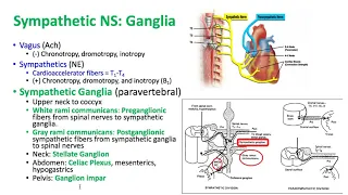
event risk calculator prophylaxis indications for endocarditis and bragata syndrome and the diagnosis of brugada syndrome the 3d keywords that have to do with cardiac physiology include keywords related to the cardiac cycle the ion flux that occurs throughout the cardiac cycle pressure throughout the cardiac cycle ekg and the cardiac cycle electrolyte abnormalities and how they appear on ekg the calculation of cardiac output based on both the fit principle and thermal dilution calculation of svr and pvr mixed venous oxygenation cardiac innervation cardiovascular reflexes including the carotid sinus and the effects of heart transplantation on these various cardiovascular
reflexes and the bain bench reflex and those wonderful ventricular function curves which i actually really like diastolic dysfunction and how they appear on these ventricular function curves chf and how it looks on these curves the effects of various drugs on the shape of these curves including melron and phenylephrine systolic versus diastolic heart failure lucitropy and diastology what happens with lv dilation and how that affects wall stress um the causes of beta 1 up regulation the effects of beta blockers and myocardial ischemia cardiac function the parasympathetic effects on cardiac functions and factors that cause the release
of anp the cardiac keywords that have to do with heart disease include myocardial ischemia coronary blood flow and coronary blood supply supply including to the av node wall motion abnormalities and coronary blood flow and heart block that can possibly occur due to blocked coronary blood supply aortic stenosis aortic stenosis and arrhythmias aortic insufficiency management slash medical treatment of ai acute mr versus chronic mr carcinoid syndrome and the cardiac lesions that you see with carcinoid syndrome cardiac tamponade and the effects on heart rate and cardiac output and how you differentiate between cardiac tamponade and pericardial effusion
the pacific pathophysiology of a pe rv failures rv failure arrhythmias digoxin toxicity torsada point ventricular tachycardias svt and wolf parkinson white so abnormalities that you can see on akg and different arrhythmias afib and the stroke risk of determination management of complete heart block aicd placement indications the treatment for symptomatic bradycardia indications for biventricular pacemaker interop complications of a pacemaker placement and pulmonary hypertension keywords that have to do with anesthesia for cardiac surgery include cures related to a heart transplant talked about it a little bit already but the denervated heart the exercise physiology of the denominated
heart bradycardia and heart transplant the treatment organ transplant cold ischemia times and fluid management of organ donors cardiopulmonary bypass separation from cardiopulmonary bypass management of low scr post cardiopulmonary bypass the oxygenator and gas embolism that can occur while you're on cardiopulmonary bypass the prime of the bypass circuit additives and then the different temperature monitoring sites that we use throughout bypass a minimally invasive cabbage and a single lung ventilation that um is required for that indications for retrograde cardioplegia complications of a tavr contraindications of an intra-aortic bloom pump um and then t-e-e is something that's coming
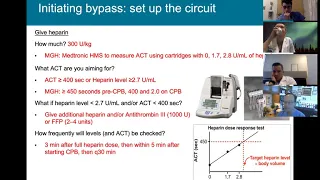
up more and more so basic tea views tea views and cannula placement aortic valve and left ventricular anatomy and left atrial penetrage anatomy and these views on tee um cardiomyopathy onto e the diagnosis and the treatment hit heparin resistance antithrombin 3 deficiency protamine reaction prevention and then sort of not necessarily cardiac but related to cardiovascular physiology bilateral carotid under direct means and the physiology complications of carotid endotherectomy and ischemia monitoring during and after carotid endoderectomies and transcranial doppler not too many key words that have to do with congenital heart disease but it does show up
sometimes specifically tetralogy of fellow and the lesions associated with tetralogy of below fontan single ventricle physiology and circulation and ventilation patients with a fontan hypoplastic left heart syndrome and pge1 use okay that was a lot of keywords that i just threw at you i'm not going to go into depth with a few of the keywords that have shown up more recently on the ite and the ones that seem to be frequently missed both by our residents and residents across the country so the first keyword i want to discuss is the revised cardiac risk index or
the rcri this is a list of six independent predictors of major cardiac complications um i think they make sense logically but they've also been shown in studies to increase the risk of cardiac complications postoperatively the first independent predictor is high risk surgery so the type of surgery itself the major vascular surgery open intraperitoneal surgery intro thoracic procedures all of these are examples of high risk procedures that get you one point on the rcri the second independent predictor is a history of ischemic heart disease this is not limited to just an mi so if you've had
an mi yes you do get one point for this but it also includes a positive exercise stress test or if the patient currently has chest pain that you believe to be due to myocardial ischemia if they regularly use nitrates if an ekg has pathologic q waves all of these are examples of ischemic heart disease and all of these would give you one point on the rcri if a patient had coronary disease and then was revascularized completely you do not get a point with this because they do they no longer have coronary disease because of the
revascularization a history of heart failure gets you one point a history of cerebrovascular disease whether this is a stroke or a tia some sort of cerebrovascular disease gets you one point diabetes specifically requiring insulin so if you're not on insulin therapy you do not get a point for this but if your diabetes requires insulin then you do get one point for this and the serum creatinine greater than two so again to go through this the six predictors that increase the risk of major cardiac complication are high risk surgery history of ischemic heart disease which is
not just limited to an mi history of heart failure history of cerebrovascular disease diabetes requiring insulin and a pre-op serum creatinine greater than two afib and obesity i just wanted to talk about these for a second so actually afib a retrospective study did show an association between a history of prior admission for afib and post-op complications and the risk of post-op complications was actually higher than in patients that had coronary artery disease however this study did not afib did not make it onto the rcri but just something to think about that your patient might be
higher risk if they have afib even though they don't get a point on this particular scoring system obesity in our notes we regularly write obesity complicating all aspects of care it certainly does complicate all aspects of hair care however it hasn't been shown specifically to be an independent predictor of end end-organ damage unlike these other six factors which have been shown to be independent predictors of major cardiac complications so why do we care and what do we mean when we say major cardiac complications so this is using the rcri scoring system they have no if
you have no risk factors one two or three risk factors the top study was done in 1999 the bottom um is from a study that was done in 2005. i think the take-home point is if you have no risk factors you have about half a percent risk of having an adverse cardiac outcome and if you have three or more risk factors your risk is somewhere between five and nine percent and so that's not an insignificant number that's 1 in 20 patients somewhere between 1 and 20 and and 1 in 10 patients um and so i
just think it's important to keep in mind that yes we understand these risk factors but the importance and the reason why we're looking at risk factors is because these are some really bad complications that can happen afterwards cardiac death non-fatal mi cardiac arrest um pulmonary edema v-fib like these are all really bad things and so the when you think about the rcri um i mean a lot of my patients i know have diabetes requiring insulin have had a tia and are having high risk surgery so right there has three or more risk factors and one
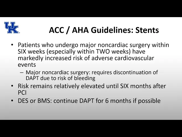
in 20 of those patients or more is going to have a complication so i think just kind of bringing it home and making sure that we look at the reason why we care about all this so the american college of cardiology and the american heart association guidelines when it comes to stents so five to seven percent five to ten percent of patients that have a drug eluding stead placed will require surgery within one year currently the recommendation is for patients that have a drug eluding stent placed to be on one year of dual antiplatelet therapy
which is why it's important that five to ten percent of these patients may require surgery during this one year where they're supposed to be on dual anti-platelet therapy dual anti-platelet therapy is usually aspirin and clopidogrel called pedophile is a p2y12 inhibitor um and the complications of discontinuing this dual anti-therapy prematurely are pretty severe complications we think about are mi death stent thrombosis or the urgent need to repeat revascularization so these aren't small complications that we're talking about premature cessation of dual antiplatelet therapy is the strongest predictor for stent thrombosis and if there is a stem
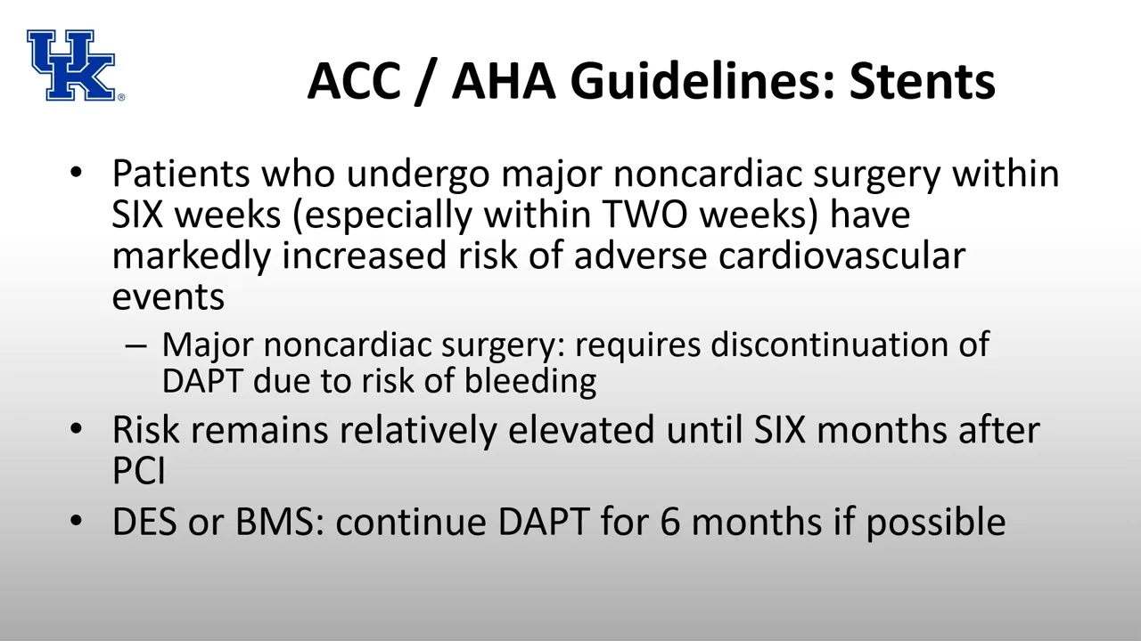
thrombosis that develops the complications of this again are pretty severe so 50 to 75 50 to 70 of these patients will develop an mi and 10 to 40 percent of these patients will actually die because of this this uh thrombosis this stent thrombosis so what do we do with these patients that require surgery within one year of this stent placement so it all kind of depends on the timing of this surgery if it can wait at all um so patients who undergo major non-cardiac surgery within six weeks especially within two weeks have a markedly increased
risk of adverse cardiovascular events so if you need surgery you know within six weeks of having this drug looting step place or really any stent placed it significantly increases your risk of adverse cardiovascular events major non-cardiac surgery is defined as something that that would require or would recommend discontinuation of the dual antiplatelet therapy due to the risk of bleeding so that is what is considered major non-cardiac surgery there are that being said there are better and worse times to do surgery the risk of adverse cardiovascular events remains elevated until six months after a pci so
if you can wait six months for this major non-cardiac surgery it is best to wait six months if you absolutely cannot wait six months um you know wait at least one to three months if possible and interestingly um whether the it was a drug looting scent or a bare metal stent um the recommendation was still to wait six months if possible i know traditionally we thought you know bare metal scent um should be fine within one month but according to the acc and aha it is better if you can wait up to six months so
if you can keep these patients onto a lantern if it's a surgery that can wait for six months it's best to wait six months there are other things that also increase the risk um you think about you know patients non-elective surgery um is going to increase risk so if there's some sort of trauma a fall a head bleed or something and there are certain surgeries that are out of there also you know you have to balance instant thrombosis versus you know if if there's a bleed in the brain um obviously neurosurgeons are going to want
to stop to antiplatelet therapy so it is um you know risk benefit and a discussion with the surgeon that you can have but if you can wait at least six months the answer and the test is if you can wait at least six months the risk remains elevated until about six months it's better to wait a year but really if you can wait six months it's best to wait six months before stop and dual antiplatelet therapy if you can stop just one um you know the i think we think it's better if you're only on
aspirin however just being on aspirin can still increase your risk of bleeding 20 to 50 so it's it's not benign at all um but uh better than you know instant thrombosis and an mi if if the surgeon's okay with continuing aspirin um throughout the perioperative period all right trans esophageal echocardiography hopefully my residents don't react this way when i start talking about the echo uh but as a cardiac anesthesiologist i get excited about the heart i get excited about echo and i get excited about echo findings but this is something that it would behoove you
to learn even if you decide not to do a fellowship in cardiac anesthesia this is something that is showing up on the in-training exam it's showing up on the advanced exam and it's showing up on the osce and aside from it showing up on the exams i think it's a great tool to be able to use if you have a patient that decompensates or if you're trying to evaluate a patient so even if you don't find yourself doing a cardiac fellowship i think it is important an important skill for you to learn okay the first
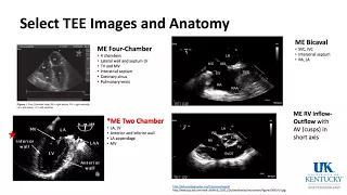
keyword um related to te is left ventricle anatomy so this i think the easiest way to start is with the trans gastric short access view on the left is a pictorial of what you see and on the right is a live intraop echo of a trans-gastric short axis view i think it's easiest to start here because in this view you can see all six walls of the ventricle you can see only one segment of them um the mid-segment of it so you can't see you know obviously the entire wall in this view but you can
identify blood supply from each of the coronaries which we'll talk about in a minute um in the heart the anterior wall is across from the inferior wall so normally we think of anterior and posterior superior and inferior but in the heart we talk anterior and inferior and the way um i remember this is a.i artificial intelligence anterior inferior um and so the a is across from the eye unlike normally when we talk about anatomy so you have your anterior wall and your inferior wall the other four walls of the heart are again i think logically
named they're either part of the septum or part of the lateral wall so if it's interior and part of the septum it's anterior septal if it's inferior and part of the septum it's inferior septal if it's anterior and lateral it's anterior lateral and if it's inferior and lateral it's inferior lateral so again anterior is always going to be across from inferior so anterior lateral is going to be across from inferior septal anterior septal is across from inferior lateral okay and you can see all these walls in this short axis view so the other view that
i think is good to know and is relatively easy to remember the walls in is the mid esophageal two chamber view and if you think about what we were looking at in the stomach what we're basically doing is cutting the heart straight through right between the anterior and inferior wall straight in half so you're not going to see any of the right ventricle in this mid-esophageal two chamber view it's going to be found at around 90 degrees and so you're going to see the anterior wall and the inferior wall now an easy way to remember
which way is the anterior wall and which way is inferior also the anterior wall is close to the appendage so you can see the left atrial appendage and the upper right hand side of the image and so that wall on the right hand side is going to be the anterior wall and the wall on the other side the left side of the screen is going to be the inferior wall so now that we can identify the different walls of the heart we can start talking about coronary artery blood supply to the different walls frequently asked
is there's a change in the echo what graph do you think went down or this graph went down what change can you expect to see in the echo on the left this is a really nice pictorial put out by the journal of the it was published in the journal the american society of echocardiography and it's a consensus statement again it might be a little more detailed than you need for the in training exam uh two of the images that we just talked about are here you can see the two chamber and the trans-gastric short axis
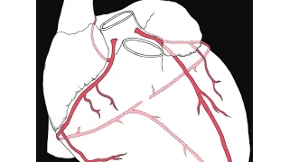
view [Music] and the four chamber and the the long axis you are there as well and so that does a nice job color coding the different blood supplies to those walls the left main so of the two coronaries the left main quickly bifurcates it's a very short artery it quickly bifurcates into the left anterior descending artery and the circumflex artery the left anterior descending led often most often gets the arterial graft the lima graft and the blood supply the led supplies the anterior two-thirds of the septum the anterior wall of the left ventricle and the
apex of the left ventricle the circumflex artery supplies the anterolateral wall and part of the inferior septal wall so the intralateral wall is supplied both by sometimes the led and sometimes the circumflex but it's usually the left side the inferior septal wall can be also supplied a little bit by the posterior descending artery but we'll get to that in a second the right coronary supplies the inferior third of the septum the right side of the heart and the inferior wall of the left ventricle and as well most often the av node so there is an
av nodal artery that comes off usually the right coronary artery 90 of the time and that supplies the av node that's important to know because if you do lose supply blood supply to the av node you can go into complete heart block um so that again that av nodal artery often comes from the uh right coronary artery the blood supply to the various parts of the heart that i just reviewed is what is generally common in a quote normal person there are people that have anomalous coronary circulation and you have to look at their cath
in relation to their effort to kind of figure out what's going on but i think that's beyond the scope of the ite what does show up sometimes is left versus right dominance so left versus right dominance is determined based on where the posterior descending artery arises from most of the time 70 of the time the pda arises from the right coronary artery and so that is what is referred to as right dominance 20 percent of the time there is dual blood supply and so from the circumference and the right coronary and that is considered co-dominant
and then 10 of the time you get blood supply from the left side only from the circumflex only and that is considered left side dominant so it's somewhat anomalous if you have you know your pda arises from the circumflex but there are other anomalies that are much less common that can affect your echo findings so kind of bringing things back home we've talked about the coronary blood supply and the coronary anatomy and then identified all of the walls of the ventricle this was my case that you're seeing in in this echo uh and which wall
can you identify which looks more hypokinetic than the rest i would say there's global hypokinesis but which was the worst the worst wall in my mind would be the inferior wall which is the wall closest to the top of the screen which is closest to the tee probe now bringing it all together which coronary artery usually supplies this wall of the heart this inferior wall i really hope that you said the pda which 70 of the time comes from the rca so this is an example of how it might uh i was a useful clinically
and then b might show up in a test question i promise i'm almost done talking about tee this is the last image that we'll look at the last view that we'll look at um i do want to say that this the key there were quite a few keywords on last year's ite that were related to dee and that is something that again is an emerging concept so i know i spent a fair amount of time on it but i do think it'll keep showing up more and more so this view that we're looking at here
is the mid-esophageal sword excess view it's around 30 to 35 degrees and again is in the mid esophagus anytime you're in the esophagus the most posterior thing is usually the left atrium so for the most part what you're going to be looking through your viewing window it's going to be your left atrium so that's what's going to show up at the top of the screen okay and then to the left of that you can see is your right atrium i didn't do the greatest job drawing that and then on the bottom of the screen is
your right ventricle but what we're looking at really in addition to the free wall of the right ventricle in this view are the three cusps of the aortic valve the three cusps are named after the coronary arteries that should come off of them so the first cusp we'll talk about is the non-coronary cusp which yes there are no coronary arteries that come off of it so this is located on the upper left hand side it's the upper left hand cusp and it is located in between the intra-atrial septum so again you see the left atrium
at the top of the screen and then you see the septum there and and the cusp closest to that is going to be your non-coronary cusp on the bottom of the screen again is the right ventricle and the turns out the right coronary comes from very close to the right ventricle and so that bottom cuff is going to be your right coronary cusp and that leaves on the upper right hand side of the screen is going to be your left coronary clasp it is closest to the left atrium and is where your left main coronary
artery comes from so in the vein of focusing on mostly what was tested in the last year and what was most frequently frequently missed i'm going to talk about aortic insufficiency but i'm actually not going to talk about aortic stenosis i would encourage you to read and review aortic stenosis but i'm just going to focus on aortic insufficiency there are both chronic and acute causes of aortic insufficiency like most lesions a chronic aortic insufficiency is better tolerated than acute aortic insufficiency causes of acute aortic insufficiency include infective endocarditis destroying the valve and aortic dissection sometimes
in a dissection the dissection flap comes down and and results in an incompetent aortic valve so that can result in aortic insufficiency chronic causes of aortic insufficiency include connective tissue diseases including marfan syndrome and ehlers-danlos syndrome sometimes there's dilation of the aortic root and this changes the anatomy and the leaflets cannot come together and they cannot collapse and so you end up with aortic insufficiency bicuspid aortic valve i know traditionally we often think of aortic stenosis but it can also result in aortic insufficiency different inflammatory diseases of the aorta and rheumatic heart disease so romantic
heart disease affects the mitral valve but it can also affect the aortic valve as well it more frequently affects the mitral valve and it's something that we're seeing less and less of over time but it can still result in aortic insufficiency so cardiac lesions are thought of as either pressure overload lesions or volume overload lesions aortic insufficiency is a volume overload lesion there's an increase in stroke volume there's an increase in end diastolic volume an increase in end systolic volume and an increase in left ventricular radius while both end diastolic and systolic volume increase the
end diastolic volume increases more than the end systolic volume this long-term volume overload and increase in volumes eventually leads to eccentric hypertrophy so on the left you see a picture of concentric hypertrophy where the lv size gets smaller the wall gets thicker the lv mass increases but the diameter is the same eccentric hypertrophy you also have an increase in left ventricular mass but this is because you have a a larger diameter so you have the same wall thickness but a large like a normal wall thickness but a larger diameter and because you have that larger
diameter you have an increase in ld mass this is seen with volume overload lesions and so this is what you see in aortic insufficiency so why is this increased left ventricular radius bad why is this eccentric hypertrophy bad um it goes back to the law of laplace the law of laplace can actually both be applied to um aortic stenosis and aortic insufficiency when you're looking at it through the lens of aortic insufficiency you have a dilated left ventricle this dilated left ventricle at the bottom you see the law of the plaster the the wall stress
of the left ventricle is equal is is directly proportional with both cavity pressures so the pressure inside the left ventricle and the radius so if the cavity pressure increases or the radius increases you have an increase in left ventricular wall stress and when you have an increase in left ventricular wall stress this increases your myocardial oxygen demand so both in aortic stenosis where you have an increase in cavity pressure and an aortic insufficiency where you have an increase in left ventricular radius you end up with an elevated left ventricular wall stress as far as the
hemodynamic management of a patient with therapeutic insufficiency your heart rate goal is high normal so 80 to 100 not tachycardia not tachycardic but definitely not a heart rate slower than 60 is not good the slower your heart rate is the more time that is spent in diastole and this allows for more time to um for regurgitation so a slower heart rate is definitely not good you also want to reduce afterload because if your afterload is too high this is going to encourage reversal flow of flow and encourage insufficiency so if your afterload is a little
bit low this is going to encourage forward flow you also need to maintain preload it is a balance though because this is a volume overloaded state and so if you give the patient too much preload too much volume um excessive left ventricular preload can lead to pulmonary edema and the pressure to stall back up so an ideal pressure for a patient with aortic insufficiency going to maintain or augment the contractility left ventricular contractility in order to again encourage forward flow you don't want something that's going to purely result in arterial vasoconstriction because again this discourages
forward flow and encourages backward flow and so an increased afterload is just going to increase regurgitation so phenylephrine is not great you can use it in small bits but it's not great because not only is it pure arterial vasoconstriction it also causes reflux bradycardia and again bradycardia isn't great for insufficiency so ephedrine is a really great choice you have some alpha activity some beta activity and so you maintain your contractility have a little bit of an increase in svr and are able to increase your coronary perfusion so a note about the coronary perfusion in a
patient with the aortic insufficiency because their diastolic pressure is so low because of all the insufficiency they may have normal coronary circulation with no blockage but they may suffer from poor coronary perfusion just because their diastolic pressure is so low and remember these are patients where the diastolic pressure becomes low and their left ventricular end diastolic volume and pressure is elevated so they have extra volume within their lv and they have decreased delivery because of the low diastolic pressure so their their oxygen delivery their transmural pressure between um means and uh intraventricular pressure is low
and so you can actually have like symptoms of coronary ischemia in a patient with aortic regurgitation and completely normal coronaries anti-aortic balloon pump in my mind this is connected to aortic insufficiency because when i have a patient with aortic insufficiency i think of some of the things that may or may not be possible if i were into trouble for example delivering anti-grade cardioplegia is going to be difficult in this patient because of the insufficiency the cardiovascular will leak through that incompetent valve um they're probably going to be volume overloaded and it is a contraindication for
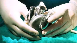
an injury aortic balloon pump so that's why in my mind aortic insufficiency and intra aortic blood pump are keywords that are that are related um so intranetic balloon pumps are used in patients with pretty bad coronary disease they have a couple different ways that they function so they are used to decrease the myocardial oxygen oxygen demand while also increasing the myocardial oxygen supply so this balloon pump is placed through the femoral vessels and it is placed distal to the arch to the aortic arch into the vessels to the head and it inflates during diastole and
so inflation during diastole in increases perfusion it improves your diastolic pressure and so it augments coronary perfusion during diastole it then deflates during systole and this allows for forward flow but in addition to allowing for forward flow it also decreases afterload which decreases cardiac work and thus decreasing myocardial oxygen requirements so by inflating during diastole it increases coronary perfusion so it increases myocardial oxygen supply and by deflating during systole it decreases the afterload thus decreasing the myocardial oxygen requirements so contraindications ii intra-aortic bloom pump placement as we talked about moderate to severe aortic insufficiency uh
it will by inflating during diastole it will just encourage more regurgitation and so it will just it make the problem worse it will not augment coronary blood flow instead more of that blood will end up inside the ventricles so uh moderate to severe insufficiency is a contraindication to intra placement other entre contributions include aortic dissection as you can imagine uh the balloon may make the flat porous and it might might also result in pulsations in the false lumen it could expand the dissection it is a contraindication to introverted balloon pump aortic aneurysm uh it's the
risk is too high there's a risk of embolic complications uh it's difficult just because of the size of the aorta so it's contraindication severe sepsis you don't want to place a device in the body in a patient's septic or coagulopathy so if this patient is already bleeding and already coagulopathic patients that have bloom pumps do need to be anticoagulated so if you're already coagulopathic it is a contraindication to a balloon placement plus you're cannulating fairly large vessels patient refusal they don't want it we can't force them to do it inability to place device so if
the arteries are very tortuous and and it's difficult uh you know there's there's risk of damage to the artery risk of dissection sometimes a risk of placement due to uh arterial tortuosity is just not not worth it so uh again uh oregon sufficiency aortic dissection narrative aneurysm sepsis gregor the patient refusal or inability to place are the contraindications to enter aortic balloon pump placement these are a few of my references i get a lot of help and input from these two guys here uh this is miller and mac miller's in the foreground clearly editing a
slide that i'm preparing mac is in the background overseeing the entire process i am not cool enough to have named them after the rapper mac miller um but i am an anesthesiologist and so i did name name them after the macblade and the miller blade so uh thank you so much for listening i hope everyone have a fantastic day