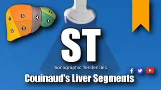
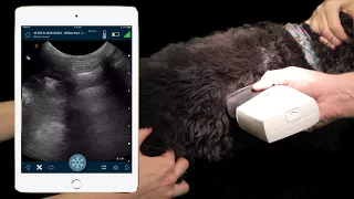
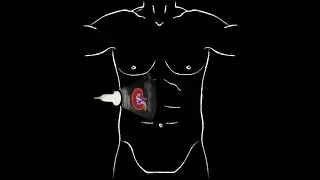
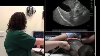
[Music] do the liver is located under the costal arch between the diaphragm and the quadrilateral portion of the lungs and the stomach the combination of acoustic shadowing artifacts and reverberation caused by these different structures can sometimes limit our ultrasound axis the head of the spleen is close to the liver on the left side this proximity sometimes enables us to compare respective echogenicity on the same image the right kidney is buried in the renal fossa of the caudate lobe of the liver dorsally on the right whereas the right limb of the pancreas and the descending duodenum
are located just laterally the rest of the small intestine and more dorsally located colon can also extend under the costal arch abdominal vascularization comes from three sources the arteries branching out from the aorta the veins that flow into the caudal vena cava and the ramifications of the portal vein that drain most of the abdominal organs toward the liver the ability to recognize these different vessels helps in identifying many other smaller abdominal structures such as lymph nodes pancreas and adrenal glands the hepatic lobes or the interlobar fissures are not easily distinguished unless significant peritoneum effusion is
present the gallbladder is visible in all patients and it's filled with variable amounts of bile you can sometimes follow the common bile duct all the way to where it empties into the major duodenal papilla the intra hepatic bile ducts are not normally visualized but will become in case of outflow obstruction a convex probe with a small footprint is most appropriate for a complete evaluation of the liver when placing the probe slightly cuddled to the xiphoid process in a transverse plane to the patient and oriented cranially the anechoic gallbladder is identified the gallbladder is located to
the right of the midline but on the left of the image since the probe marker is placed to the right the gall bladder is usually round and smooth and its wall is either invisible or less than one millimeter in thickness the probe should then be smoothly rotated ventrally and dorsally and in different oblique planes to cover the entire hepatic parenchyma liver parenchyma is hypoechoic with respect to the fat of the phosphor ligament observe the aquasic enhancement artifact deep to the gallbladder observe the edges of the liver where the diaphragm starts with the stomach located just
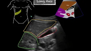
cuddly the scanning zone must include the area under the ribs which will come into view in cast shadows this enables us to confirm the lateral edges of the liver [Music] do in the longitudinal plane or sagittal plane with respect to the patient the gallbladder is drop shaped with the body located towards the diaphragm cranially and laterally to the right the probe is moved to the right then to the left to cover the entire liver parenchyma the stomach is located just cuddled to the hepatic margin and the falsiform ligament fat is ventral you can see the
heart beating on the other side of the diaphragm again the probe needs to be inclined at an angle to reach the most lateral portion of the liver passing by the stomach once you see the ribs you can tell you've reached the side of the liver the liver volume can be assessed by placing the probe vertically at the caudal edge of the last pair of ribs and determining how much of the liver extends cuddled to these just like you would do with a lateral radiograph in most dogs and cats the liver extends as far as the
end of the cost large but in deep-chested dogs it's often located more cranially and in some small dogs and puppies it may extend more cuddly the shape of the cuddled tip of certain lobes such as the left lateral lobe provides information on the size of that lobe and the whole liver if the infiltration appears diffuse in this illustration the normal lobe as a pointed end where it touches the spleen in hepatomegaly the edges of the liver are rounded and extends even further cuddly with respect to the cost large it is also important to know the
position of the stomach it can be deviated cuddly by an enlarged liver as with the kidneys and spleen [Music] as with all the other organs one of the main parameters to assess is parenchymal echogenicity the liver is generally less echogenic than the spleen but more echogenic than their mental cortex in all dogs and cats the kidneys often have similar echogenicity than the liver and can sometimes even be hyperechoic kidneys of obese cats are often more echogenic therefore the echogenicity of the kidneys is not always reliable when it comes to conclude on the one of the
liver in these two images from an healthy dog the liver is hypoechoic compared to the spleen and isoechoic compared to the right kidney also the grainy texture or echo texture is more pronounced in the liver than for the spleen the size of the gallbladder varies in dogs and cats according to the phase of digestion its shape also varies according to the plane of the scan the gallbladder wall is normally fine smooth and hyperechoic when viewed from a 90 degree angle the neck of the gallbladder and the cystic duct can sometimes be detected and the common
bile duct that empties into the duodenum is more difficult to image in dogs in cats the common bile duct is usually more accessible and can be found next to the portal vein adjacent to the duodenum identifying the portal vein at the liver hyalus is useful in locating the bile duct which runs along the portal vein for several centimeters on the ventral side before continuing on the right close to the gastroduodenal vein and ending in the duodenal wall in a few moments we will take a closer look at how to recognize the portal vein at the
liver hylus biliary sludge is sometimes identified in the gallbladder of dogs and cats this modification of the bile content shows up as moderately echogenic amorphic areas or foci that are normally mobile and sediment over time even though this finding has been considered non-significant in dogs there is growing evidence that this process is probably associated with cholestasis in cats and perhaps also in dogs approaching eventually the cranial abdominal aorta is located to the left of the midline with the carlovine cava on the right and ventrally the liver is ventral to the aorta and has a first
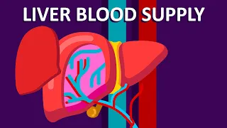
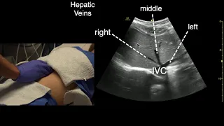
group of vessels the hepatic veins which carry the venous drainage from the liver to the caudal vena cava a second group of hepatic vessels emerges from the celiac artery which turns into the hepatic branches these arteries provide approximately 25 percent of the liver's blood supply the remaining hepatic blood flow comes from the portal vein which drains most of the abdominal organs the portal system includes the gastroduodenal vein and the pancreaticoduodenal veins which drain those organs the cranial mesenteric vein that drains the small intestine in particular and the spinach vein which drains the spleen and part
of the stomach and the gastroepiploic vein along the greater curvature of the stomach venous drainage from the organs moves from causality cranially and from the periphery of the abdomen toward the liver hyalus color doppler is a rapid way to identify the two types of veins in the liver the hepatic veins and the portal veins the hepatic arteries are normally too small to be seen because the blood inside the portal veins flows from the principal portal vein at the center of the liver to be distributed peripherally in the lobes which is in direction of the ultrasound
transducer the flow has a red signal note also the hyperechogenicity of their walls regardless of the angle of the probe conversely the hepatic veins show a blue signal due to their flow that is directed away from the probe and toward the center of the liver and caudal vena cava the wall of the hepatic veins is not consistently hyperechoic as it is for the portal veins unless there is a 90 degree insulation angle causing ultrasound specular reflection after examining the liver vascularization the examination is completed with an assessment of the main portal vein and its abdominal
branches the main portal vein is identified by following the intra hepatic portal veins all the way to the hepatic ileus the portal vein can then be measured at that location and compared to the other major abdominal vessels the same scanning route is followed in the transverse plane to locate the body of the pancreas next the main portal vein is examined in the longitudinal plane using color doppler the signal is blue due to the fact that the flow is toward the liver and away from the probe in the transverse plane the probe is moved cuddly along
the caudal main portal vein which becomes the cranial mesenteric vein using this technique allows to identify potential porosity shunt as well as locating the many mesenteric lymph nodes along that large mid-abdominal vein [Music]