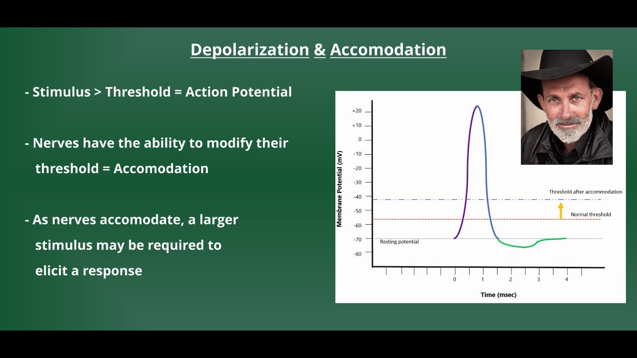
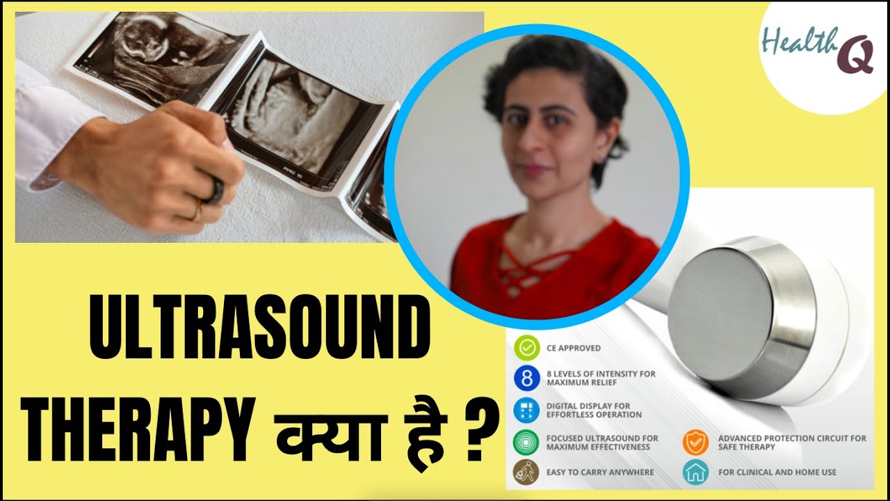
welcome today we want to talk about what is ultrasound and how does it work if you've ever wondered how that music gets from the speaker to your ear it's by sound waves by creating vibrations in the air it transmits that energy over a great distance it's also no surprise that moving air can carry a great deal of power and energy in physical therapy we harness the power of moving air through the technology called ultrasound before we can jump into how ultrasound works we need to review a little bit about waves a wave is defined as
a repeating pattern that moves energy from one place to another either through space or through matter waves come in two different types a longitudinal wave or transverse wave there are some characteristics that are common to both types of waves those include things such as frequency wavelength and amplitude let's take a quick look at how longitudinal and transverse waves are different in a longitudinal wave the particles of the medium move parallel to the direction the wave is transmitting energy this leads to alternate areas where the material is compressed and where it's refracted or spread out in
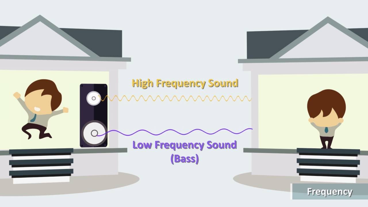
a transverse wave the particles move perpendicular to the direction that the energy is being transferred this leads to alternating areas of peaks and valleys there's some terminology used to describe waves that will be helpful in understanding ultrasound anyway if we refer to one cycle as the distance from one point on a wave to that same point again when discussing waves we often discuss the term frequency frequency is defined as the number of cycles that occur within one second the unit used to represent frequency is the hertz one hertz equals one cycle per second so for
the right side of the graph we have two cycles occurring in one second so our frequency would be two hertz waves are also referred to by their amplitude and their wavelength amplitude can be defined as the maximum displacement of a particle from its position of equilibrium in general amplitude refers to the amount of energy a wave is carrying finally we have wavelength wavelength is the measure of distance from one point on a wave cycle to the same point on the next wave cycle the typical unit for wavelength is meters now let's return to the concept
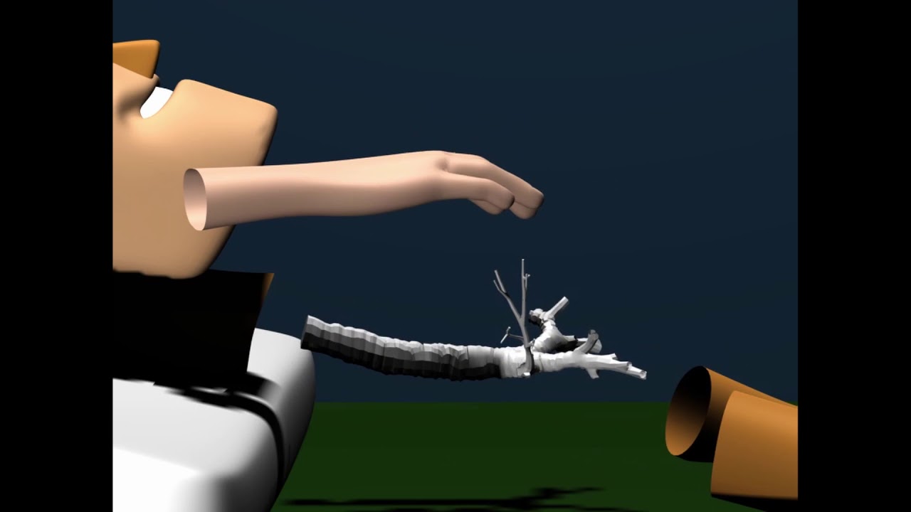
of ultrasound the definition is easy if we break the word down ultra typically means beyond while sound is a longitudinal wave that carries energy so in this case ultrasound refers to sound waves that are beyond our range for hearing in pt we create ultrasonic waves using an ultrasound unit the ultrasound unit consists of a control unit and a transducer the control unit determines the amount of energy that's sent to the transducer the transducer converts electrical energy into sound waves the sound wave is created by a vibrating crystal inside the transducer it's important to note it's
the crystal inside the transducer head that determines the effect of area producing energy we refer to this area as the effective radiating area or era of the crystal the expansion and contraction of the crystal creates a longitudinal wave that travels out from the transducer head it's the area created by this longitudinal wave that affects tissues in the human body here you can see alternating areas of compression and rarefaction of our sound wave most ultrasounds create sound waves in the megahertz range or a million cycles per second when we apply that ultrasound to the human tissue
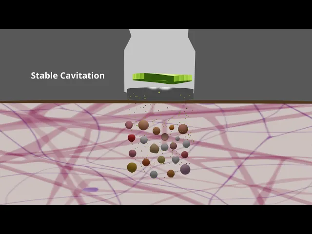
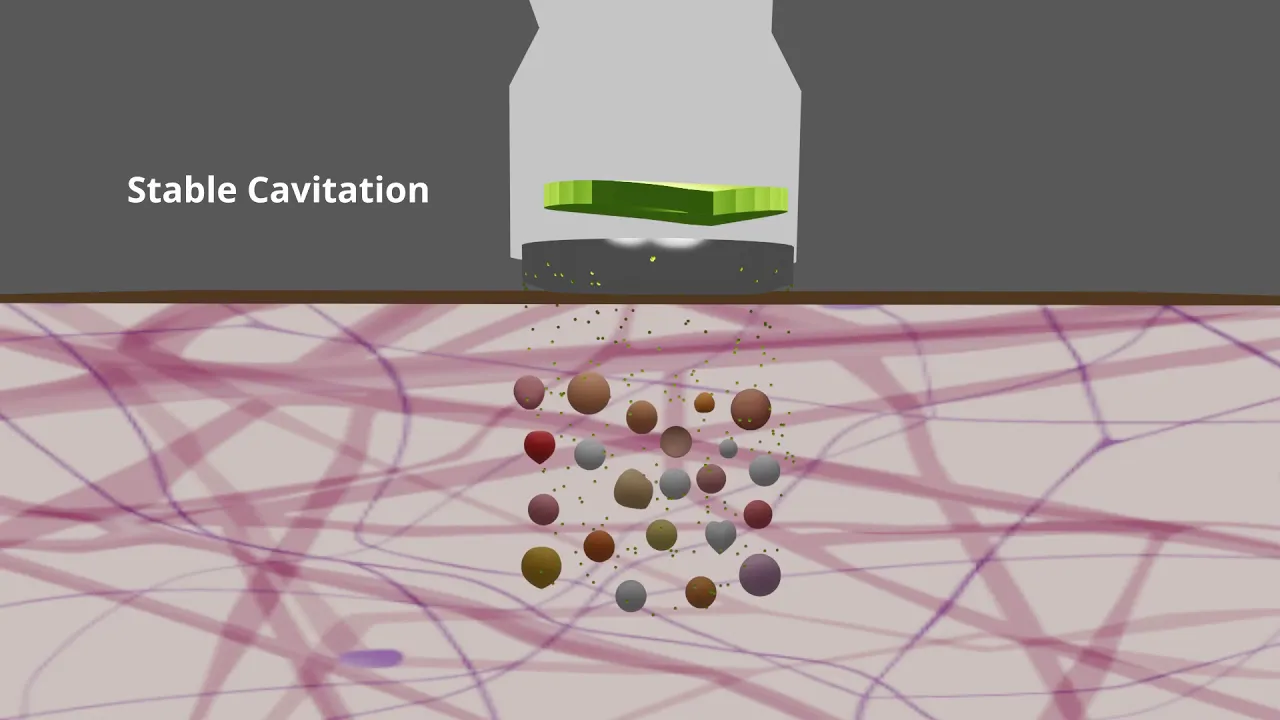
we get a vibration of molecules inside ultrasound is capable of penetrating up to five centimeters inside the human body so what happens when that energy reaches human tissue while all the effects of ultrasound are not completely understood current theory suggests that ultrasound achieves its physiologic effects through cavitation and acoustic streaming cavitation refers to the formation of small bubbles that result from the rapid pressure changes created by the ultrasound wave the ultrasound waves also cause other molecules to vibrate as a result microcurrents are created in the fluids that surround the vibrating tissues we refer to these
small currents as acoustic streaming an example of cavitation as energy is transferred to the patient small gas bubbles form and vibrate affecting the surrounding tissues if too much energy is applied those gas bubbles can become violent and burst this is what we call unstable cavitation the bursting of those bubbles can cause tissue damage here we have an example of acoustic streaming surrounding one of the vibrating gas bubbles again the small currents that are created have an effect on the surrounding tissues cavitation and acoustic streaming result in the non-thermal effects of ultrasound ultrasound also has thermal
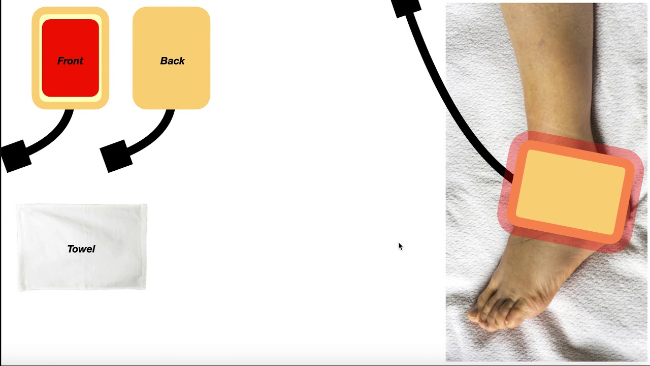
effects the use of higher ultrasound energies can result in the transfer of thermal energy from the ultrasound head to the patient this transfer of heat can provide therapeutic benefits as well unfortunately the transmission of ultrasound energy from the sound head to the patient is not a very efficient process therefore we try and improve the efficiency of the process by use of a conducting medium the ones used most often include ultrasound lotion or gel water or the use of a gel pad so what's the result of transferring all that energy to our patients with non-thermal ultrasound
we get an increase in many of the cellular processes needed for tissue to be able to heal with the use of thermal ultrasound we get the additional effects of increased blood flow reduction and pain and increased tissue extensibility to make the best clinical decisions it's important to understand that the ultrasound waves do not affect every type of tissue equally tissues with higher collagen content absorb the ultrasound energy more efficiently therefore tissues that receive the most benefit include ligaments tendons and other connective tissues such as joint capsules now let's take a look at the different ultrasound
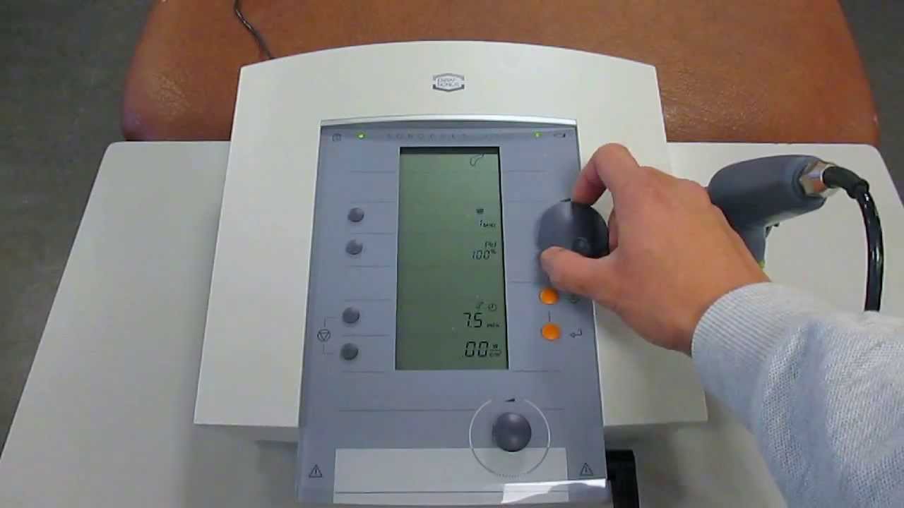
parameters that need to be determined before treatment the control unit of the ultrasound allows us to modify a variety of different parameters which parameters are chosen are determined by the size and depth of the target tissue and your clinical goals for the use of ultrasound our clinical goals are often dictated by which stage of the healing process the patient is in let's take a closer look at our different parameters and discuss how they affect our ultrasound treatment and clinical goals the parameters we have control over include frequency duty cycle intensity duration of the treatment and
the size of our treatment area let's look at these one at a time first up is frequency most ultrasound units have the ability to switch between two different frequencies usually around one megahertz and three megahertz when deciding which to use you should consider the depth of the target tissue one megahertz ultrasound has the ability to penetrate deeper up to five centimeters ultrasound waves at three megahertz will not penetrate as deep only up to one to two centimeters so if you're trying to target deeper tissues you would want to use one megahertz ultrasound if your tissue
is more shallow you can use three megahertz as your ultrasound frequency our next parameter is duty cycle duty cycle is defined as the percentage of time that the ultrasound transducer is emitting energy the duty cycle can range from 100 percent or continuous ultrasound where it is always on to zero percent where ultrasound is off most units will have a few presets you can adjust using a lower duty cycle setting is a way to decrease the amount of energy being transmitted to the patient as a general rule continuous duty cycles are 100 are used for thermal
ultrasound settings below that are used for non-thermal the intensity determines the amplitude of the ultrasound wave or the amount of energy being transferred to your patient choosing an ultrasound intensity is usually determined by the clinical goals for the ultrasound and the patient's stage of healing to take advantage of the non-thermal effects of ultrasound a lower intensity should be used if the thermal effects of ultrasound are desired then a higher intensity should be used it's important to point out that at higher levels of intensity there's an increased risk for tissue damage it's common to change the
intensity during treatment to ensure that the desired effect is being achieved the duration refers to the length of the ultrasound treatment treatment duration is most directly affected by the size of the treatment area and the amount of energy you want delivered to your patient as a general guideline in five minutes of duration you can treat an area twice the size of the effective radiating area of the transducer head so if the ura is 10 centimeters squared you can treat an area that's 20 centimeters squared over a 5 minute treatment period a typical time for ultrasound
treatment runs between 5 and 10 minutes with larger areas getting longer treatment times the last parameter we want to consider is the size of the treatment area typically the size of the treatment area is determined by the type of tissue involved and the condition being treated while the size of the treatment area may be predetermined it's an important factor when determining the other ultrasound parameters to be chosen it's important when performing ultrasound to keep the size of the treatment area manageable if ultrasound is performed over too large an area then the tissue within that treatment
area doesn't get enough ultrasound energy to benefit therapeutically as we just discussed you can treat an area about twice the size of the era in a five minute period so if treatment time is increased to 10 minutes you can treat an area roughly four times the size of the era treating areas larger than four times the ura should be avoided so it's important to consider the size of the treatment area when determining the other ultrasound parameters so when will we choose to use ultrasound and physical therapy we use ultrasound to achieve one of the following
clinical effects thermal ultrasound can be used to increase tissue extensibility for connective tissue that is adaptively shortened this can be very beneficial ultrasound can also be used to help treat pain this can be done by either directly affecting the tissue causing the pain or by altering its transmission ultrasound is also commonly used to help improve tissue healing for the acute stages of injury only non-thermal ultrasound should be used to facilitate tissue healing for more chronic injuries both thermal and non-thermal ultrasound can be used finally ultrasound can be used through a process called phonophoresis to help
deliver topical compounds into the skin it is believed that ultrasound changes the permeability of the skin which helps deliver the compound to the tissues beneath it knowing when not to do ultrasound is just as important as knowing when it's beneficial the contraindications or reasons to not do ultrasound are listed here while there's a general consensus for most contraindications for ultrasound there will be slight deviations based on your reference resource before considering doing ultrasound it's important that the patient has been screened to ensure they don't have any contraindications for treatment the clinician should also consider the
precautions for the use of ultrasound listed here when determining if ultrasound will be completed if after reviewing the precautions you can't be 100 sure the patient will be safe then ultrasound should not be performed hopefully you have a better understanding now of what ultrasound is how we create it and how it's used clinically for more guidance on how to properly apply ultrasound and choose the correct parameters you can refer to our other videos that cover those specific topics [Music] you