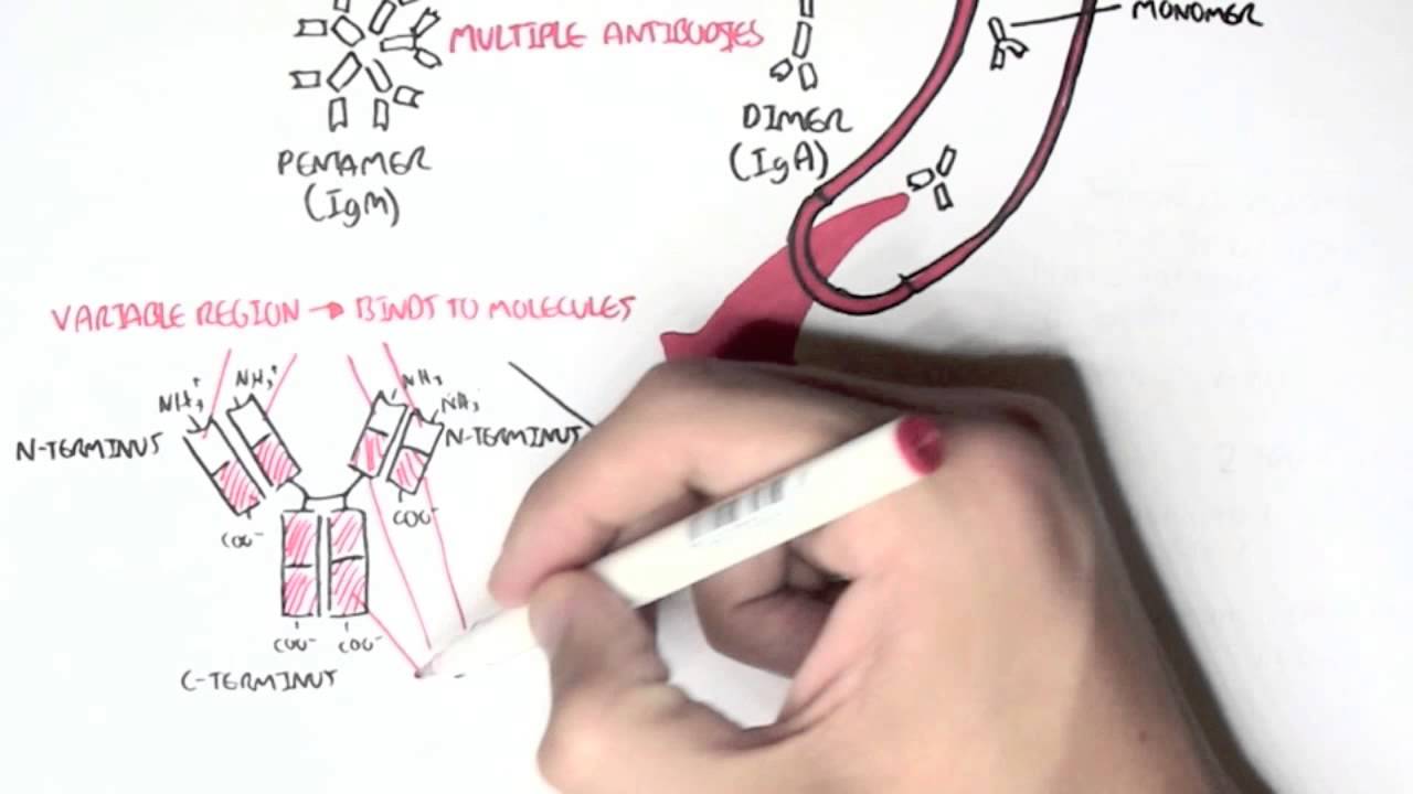
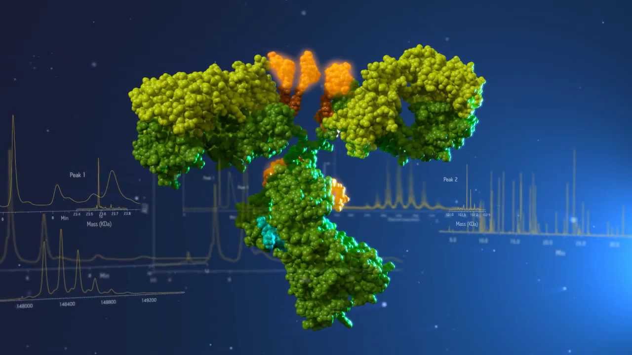
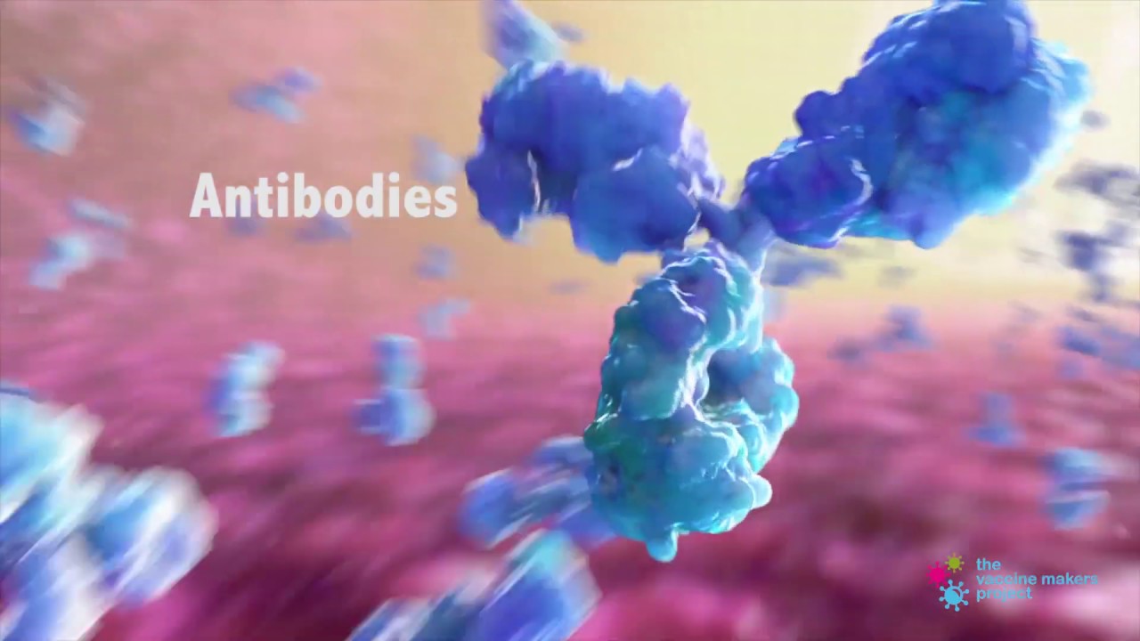
antibodies are made of several different peptide chains bound together in a Y shape an antibody is made of two identical immunoglobulin heavy chains and two identical immunoglobulin light chains roughly speaking the structural features of an antibody can be split into two parts the domains that recognize a target known as the variable region and the domains that determine the antibodies biological function known as the constant region the heavy chain constant region of an antibody determines where in the body the antibodies are located what kind of immunological response an antibody is able to mediate and what the
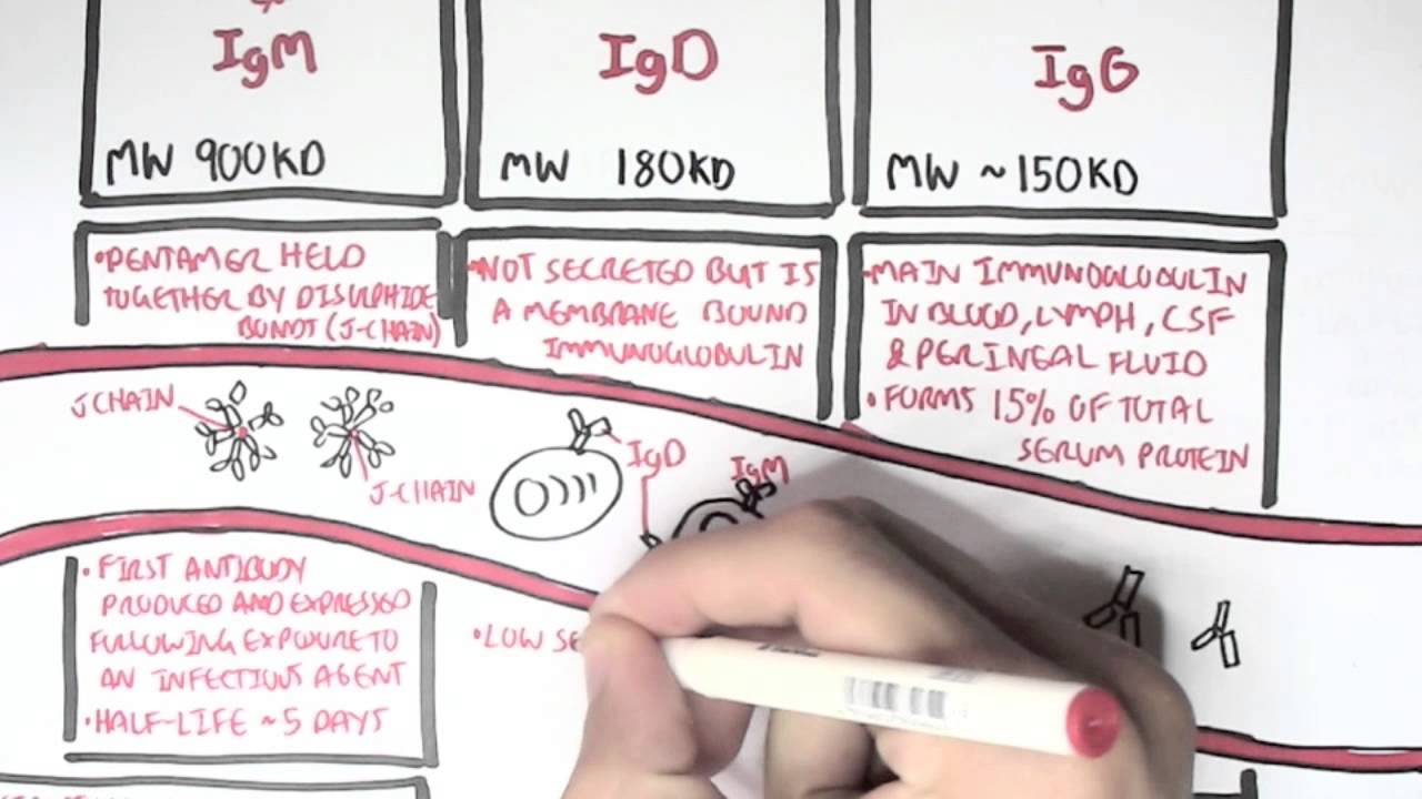
oligomer ik states that the antibody is carbohydrate chains are attached to the heavy chain constant region and fit between the two heavy chains they are associated with stability and affective function of the antibody heavy chain constant regions also determine the class of the antibody known as the isotype different ISO types can have different oligomeric states with immunoglobulin a or iga forming a dimer IG d IgE and IgG forming monomers an IgM forming a pentamer the two heavy and two light chains of an antibody are held together through disulfide bonds within the constant regions a set
of two disulfide bonds connects the two heavy chains in the hinge region while the heavy and light chains are bound together with a disulfide bond that bridges opposing cysteine residues within each constant region a second way of classifying structural regions of antibodies is based on how antibodies can be cleaved by proteolytic enzymes the enzyme Patt ain please just above the disulfide bonds within the hinge region leading to a constant region or FC fragment and two antibody binding or fab fragments another proteolytic enzyme pepsin cuts just below the disulfide bonds within the hinge region which gives
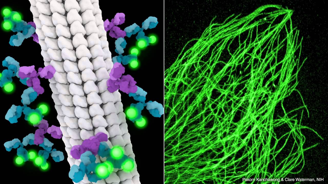
a single fab two fragments with two arms still connected generating these separate antibody fragments in the lab has enabled the function of each region to be determined while each antibody fragment can be useful for a variety of biological assays an antibody finds it epitope through the variable regions in the tips of the heavy and light chains there is an enormous amount of diversity in the variable regions so that different antibodies can recognize many different targets epitopes each of these chains contains three complementarity determining regions or CDRs located at the tips of each variable domain parts
leading up to those tips are called the framework region most of the diversity between different antibodies is generated within the CDRs together the three CDRs form the epitope binding site and determine the specificity of individual antibodies