Hello. Welcome to Byte Size Med. This video is on the cardiac cycle and in part three, we're going to look at the atrial pressure changes.
The cardiac cycle has a series of events that happen on repeat between heartbeats. The contraction and relaxation of the atria and the ventricles circulates blood through the chambers and then through the lungs and the rest of the body organs. The contraction is systole and the relaxation is diastole.
If we start with atrial systole, that lasts for a very short duration. It's followed by atrial diastole. The ventricular systole starts after atrial systole and it's longer.
It's then followed by ventricular diastole. That's one cardiac cycle. That's going to repeat.
So the phases overlap between the chambers. During this cycle, there are volume and pressure changes. The pressure changes happen in the atria, the ventricles and these two big vessels.
In this video, we're going to be looking at the atria. This is my schematic heart and I'm going to be using this to explain some of the changes. It's got four chambers.
The right and left atria and the right and left ventricles. The superior and inferior vena cava open into the right atrium. The pulmonary veins into the left atrium.
Emerging from the right ventricle, we have the pulmonary trunk and from the left ventricle, the aorta. These are guarded by the semilunar valves. That's the pulmonary valve and the aortic valve.
While between the atria and the ventricles, are the atrioventricular valves. That's the tricuspid valve on the right and the mitral valve on the left. During the cardiac cycle, the atria fill and then empty into the ventricles.
Now we'll look at the pressure changes in the atria during the cycle. We have atrial systole, followed by ventricular systole and then ventricular diastole. Atrial systole repeats starting the next cycle.
Let's start with atrial systole. That's when the atria contract. They do this to push the last bit of blood into the ventricles.
A contracting muscle means the pressure in these chambers rise. The atrial pressure rises during atrial systole when they contract. Once the atrioventricular valves close, the atria start relaxing and the pressure starts dropping.
Now it will continue dropping, but something happens in between. The ventricles contract with the atrioventricular and the semilunar valves closed. This is isovolumetric contraction.
The pressure in the ventricles is much higher during systole than the atria. The atrioventricular valves bulge back into the atrial chambers. That causes the pressure in the atria to rise a bit.
But once the semilunar valves open, the ventricles eject blood out. And during this ejection phase, the valve gets pulled back down. The atrial pressure now continues to drop.
The atria are filling now during atrial diastole, with the valves closed. This causes the pressure to slowly rise as the ventricles are relaxing in isovolumetric relaxation. At the end of this, the ventricular pressure which was going down goes lower than the atrial pressure, and the atrioventricular valves open.
Now the atria empty as the ventricles fill during the filling phase. The pressure in the atria drops until the last 10 to 20ml of blood is pushed into the ventricles by contraction of the atria. This happens during atrial systole, causing the rise in pressure and that's where we started.
The pressures in the right atrium and the left atram are similar, but the pressure in the right atrium is slightly lower than the left. So the curves for both would be similar with minor differences. The right atrium is continuous with the superior vena cava and then with the big veins in the neck.
Those are the jugular veins. The pressure changes in the right atrium get reflected back here as waves called the jugular venous pulse. So the JVP is a reflection of right atrial pressure, and it rises and falls with peaks and descents.
Let's bring back the events of the cardiac cycle. Now at the start of isovolumetric contraction, the atrioventricular valves close and at the start of isovolumetric relaxation, the semilunar valves close. At the end of isovolumetric contraction, the semilunar valves open.
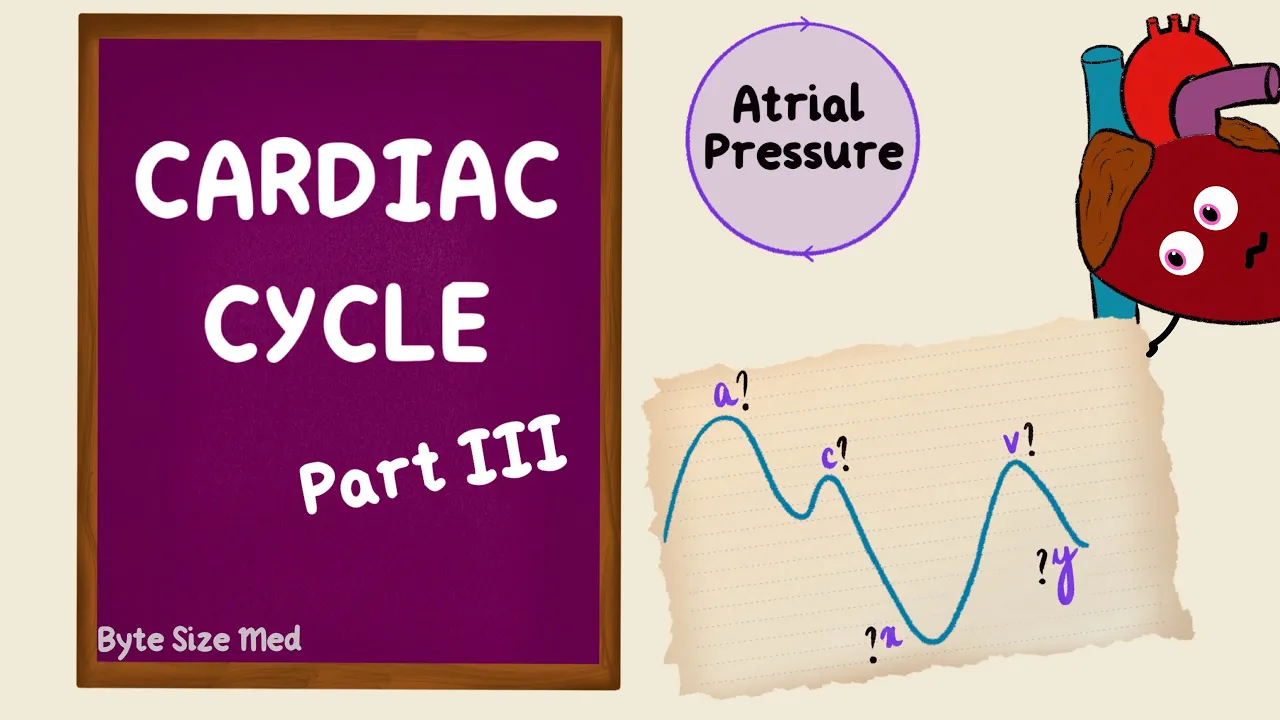
At the end of isovolumetric relaxation, the atrioventricular valves open. The JVP waveform has three peaks and two major descents. The peaks are labeled a, c and v.
The descents are the x and the y descents. Now let's put this together with the atrial pressure changes. The first peak is during atrial systole, when the pressure in the atrium rises from atrial contraction.
Then the pressure starts falling. So it starts descending. This is because the atria are now relaxing.
But it's interrupted by the tricuspid valve bulging back into the right atrium. That causes a pressure rise and that's the 'c' peak. The 'a' was from atrial contraction, the 'c' is from the valve.
During the ejection phase, the valve gets pulled down, and atrial relaxation makes the 'x' descent. The atrial pressure rises as they fill against closed valves, until it peaks just before the valves open. The pressure rise was from atrial filling during atrial diastole.
That peak is labeled with the letter 'v'. Opening of the valve starts ventricular filling and that's from atrial emptying. That pressure drop creates the 'y' descent.
Then when the atria contract, the cycle repeats. Now I'm sure you'll find lots of places showing you ways to remember what the waves are, but I'm going to show you how I remember them. Three peaks, that's a, c, and v, and two descents, x and y.
The x and the y descent. The 'x' is when the atria relax and 'y' is when the atria empty. The last letters of relax and empty.
Now this 'x' descent gets interrupted by the valve bulging back into the atria. The shape looks like a 'c' and that's the 'c' wave. Since it's split, sometimes this portion gets labeled x and this is x'.
Then x' would be when the valves get pulled back down. To simplify it a little, I'm going to remove the x'. What's left?
The 'a' and the 'v'. The 'v' is when the atria fill. What empties into the atria?
Veins. Venous atrial filling. That's how i remember it.
The last is the 'a' wave, when the atria actively contract. Atrial contraction. If this doesn't work for you, you can look around.
There are lots of different ways to remember these waves. But try to understand why the peaks and descents happen, before resorting to memorisation aids. Once you understand why they happen, it becomes easier to figure out the pathological appearances.
Like the 'a' waves, they could be absent or they could be giant 'a' waves. The 'x' descent could be prominent, reduced or absent. Same thing with the 'y' descent.
The 'v' wave could be large. And all this happens in different pathological conditions. On Wiggers diagram, which is the giant diagram that summarizes everything that happens in the cardiac cycle, you'll see a left atrial pressure curve usually and it's lower down with all the peaks and descents.
Now the reason it's this far down is because it's being compared to the ventricular and aortic pressures, and they reach much higher systolic pressures. So we'll add those in in part four. But that's atrial pressure.
I hope this video was helpful. If it was, you can give it a like and subscribe to my channel. Thanks for watching and I'll see you in the next one!