hello everyone and Welcome to our video on down syndrome if a fertilized egg has three copies of one of the chromosomes that's called a triom and most trisomies are not compatible with life and will lead to a miscarriage however there are a couple of triom disorders where the baby can potentially be carried to term and sometimes survive after birth and the three main disorders for which that's the case are down syndrome which is triom 21 Edward syndrome which is Tri 18 and Pau syndrome which is triom 13 we'll talk about these other two in another
video but in this video we're going to focus on down syndrome which is the most common triome disorder and something that you're likely to see in clinical practice Down syndrome is the most common liveborn chromosome abnormality and the word liveborn is important because among fertilized eggs in which there is a miscarriage there are many other chromosomal abnormalities which can be found Down syndrome is also the most common form of intellectual disability intellectual disability was formerly called retardation and down syndrome is the most common cause of this among children other key features of down syndrome which
we'll go through on the next few slides include what's known as dismorphic features these are basically abnormal features of the face and extremities this includes the face hands and also the stature of the child with Down Syndrome congenital malformations are also common in Down syndrome we'll talk about those these include the heart and the GI tract people with Down Syndrome develop early Alzheimer's disease and they also have an increased risk of malignancy and you you should be aware that the clinical phenotype of down syndrome is highly variable there's a range of features from mild to
severe some people with Down Syndrome are highly functional and other patients are severely limited let's start by talking about the dysmorphic features of down syndrome children with Down Syndrome are often described as having a flat facial profile if you look at this picture on the screen here you can see that this baby's face just looks relatively flat relative to normal in addition they have a flat nasal Bridge so this is the nasal Bridge it's the portion of the nose between the eyes and if you feel your own it's probably kind of risen up a little
bit it's flat in babies with Down Syndrome Down syndrome children also have low set small ears like this drawing on the screen and a short neck and then finally they have what's described as Breaky sephy this is the posterior skull being flat so you can't see that in this picture of this child but if you could look at this child in profile you'd see the front of the face is rounded but the back is relatively flat you can google and find pictures of this and that's brachy sephy which is common in Down syndrome there are
several classic abnormal ities of the eyes that can be seen in children with Down Syndrome the first one are prominent epicanthal folds so the epicanthal folds are the skin of the upper eyelid that covers the inner portion of the eye I'm marking my pen with it on this zoomed in image on the screen here and these are prominent in children who have Down syndrome also in children with Down Syndrome the palpal fissures are upslanting the palpal fissures are the separation between the upper and lower eyelids normally they are horizontal however as you can see in
the zoomed in picture the outer corner is higher than the inner corner and therefore they are upslanting in this picture of a person with Down Syndrome brush field spots are another characteristic finding of the eyes and down syndrome these are white spots on the iris this baby has it in this picture here I've zoomed in on the right side of the screen so you can see the white spots on the iris those are brush field spots children with Down Syndrome often have short broad hands and then there are two classic findings in the hands and
the feet that you should know about the first one is called a transverse Palmer crease this is a crease that across the Palm I'm circling one on the right side of the screen with my pen this is a classic finding in children with Down syndrome in the feet the classic finding is a sandal Gap this is a space between the first and second toes as shown in this picture here this is the separation that would occur if you were wearing sandals that's how it gets its name other physical features you should be aware about are
hypotonia so the muscle tone is often decreased and this can often be identified shortly after birth and children with Down syndrome and then as children with Down Syndrome grow up they often have short stature and are short for their age intellectual disability is another Hallmark of down syndrome almost all patients are affected although there's a wide range of cognitive impairment a normal IQ is 100 or greater children with mild Down syndrome can have an IQ of about 50 to 70 children with a more severe case can have an IQ of 20 to 35 and this
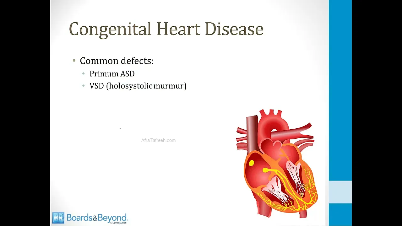
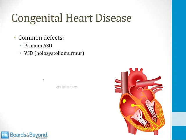
will obviously have a big effect on the type of services these children need as they grow up congenital heart disease occurs in 50% of patients who have Down syndrome most commonly these are defects that result from from abnormal formation of the endocardial cushions in the Cardiology section in the embryology video I talk about the endocardial cushions in detail they are embryologic structures that form the septum between the two atra and the two ventricles if you look at this picture on the screen here this is the right atrium here and the left atrium here we have
the left ventricle here and the right ventricle here the endocardial cushions form this section of the heart right in here so if they are abnormal you can get an ASD at the bottom of the interatrial septum that's called a premium a SD you can also develop a vssd right here and in addition the endocardial cushions contribute to the formation of the mital valve so if there's an endocardial cushion defect you can potentially develop mitro regurgitation and I've summarized that here the endocardial cushions involve the atrio ventricular septum they form the base of the interatrial septum
and the upper interventricular septum so common defects that can be seen in Down syndrome as I mentioned before are defects that involve the atrioventricular septum that comes from the endocardial cushions so this includes a prima S D or a vssd and remember that a vssd can cause a holos systolic murmur and this is high yield for you to remember especially for the boards if they describe a baby with a holosystolic murmur they may be describing a vssd that can be seen in Down syndrome gastrointestinal anomalies are also common in children with Down Syndrome they occur
in about 5% of patients the most common congenital anomaly is duodenal atreia or stenosis I talk about this in more detail in the GI section in addition children with Down Syndrome are more likely to develop hrung disease than the general population most patients with Down Syndrome will eventually develop Alzheimer's disease and in addition to being common it occurs earlier in patients with Down Syndrome than in a general population in patients with Down Syndrome the average age of onset of Alzheimer's disease is in the 50s in the neurology section I discussed the pathophysiology of Alzheimer's in
detail but I just want to mention that amalo precursor protein which is a very important pathologic element of Alzheimer's disease is found on chromosome 21 and patients with Down Syndrome have three iies of chromosome 21 amalo precursor protein breaks down to form beta ameloid and amid plaques are a pathologic element of Alzheimer's disease children with Down Syndrome also have an increased risk of malignancy especially leukemia the lifetime risk of leukemia is about 1 to 1 and a half% it often occurs in childhood children with Down Syndrome can develop acute lymphoblastic leukemia the risk is 10
to 20 times higher in Down syndrome children than in the general population they can also develop acute myoid leukemia and they develop a very unique subtype it's called the M7 subtype it is a mega Carol blastic leukemia made up of leukemia cells that resemble megga caros sites let's talk about the genetics of down syndrome in other words why do babies with Down Syndrome develop with three copies of chromosome 21 well the most common reason is a myotic non-disjunction and I talk about this in detail in the video on meiosis when this happens the baby receives
two chromosomes from one parent and one from the other this is is the most common cause of down syndrome it's responsible for 95% of cases and in 90% of cases the error is in meiosis 1 in addition the extra chromosome comes from the mother in 90% of cases and for this reason Down syndrome is associated with Advanced maternal age in the video on meiosis I discuss how that process is protracted in females and often Frozen in place for many years of time for this reason the more advanced the mother's age the longer it takes takes
for meiosis to complete in the egg and therefore the greater risk that there can be a problem in meiosis that leads to triom 21 in cases that are not associated with myotic non-disjunction one of the potential causes is a robertsonian translocation I also talk about this in the video on meiosis this is responsible for 2 to 3% of cases of down syndrome in a robertsonian translocation chromosome 21 is fused with another chromosome most commonly chromosome 14 or 10 and because the two chromosomes are fused two copies of chromosome 21 can be passed to the fetus
from one parent now this problem is different from myotic non-disjunction in that it is not associated with Advanced maternal age what's unique about a robertsonian translocation is that the fused chromosome can be passed through families and therefore there is a recurrence risk for Down syndrome within families so this is especially important for a board question if you see a description of a child born with Down syndrome and there are a number of family members in the extended family who also have Down syndrome you should think about the possibility that a robertsonian translocation may be the
underlying genetic cause of down syndrome in that family another rare cause of down syndrome is a mitotic error so this is an error in mitosis not meiosis in this situation the egg is fertilized normally with two copies of chromosome 21 however in early development during mitosis a cell develops which has three copies of chromosome 21 and it will continue to replicate making more cells with three copies of chromosome 21 this will result in somatic mosaicism mosaicism is when some cells have different numbers of copies of chromosomes compared to others so in this situation when there's
a mitotic error the child will have some cells with three copies of chromosome 21 and other cells will be normal this can lead to milder features of down syndrome as you can imagine because some cells in the body have the normal numbers of chromosomes and this is also not associated with Advanced maternal age this is a sporadic error I'll finish this module by talking about prenatal screening in other words ways to screen pregnant women for the possibility that their baby may have Down syndrome the definitive test is to obtain a sample of the fetal cells
and do a fetal kype which means determining the number of chromosomes in the baby the problem with this screening test is that it is invasive and Carries risk so you only want to do it when there is a high probability that the baby may have Down syndrome there are two ways a fetal kype is commonly done one is called chonic Villa sample Ling this requires taking a sample of the placental tissue the other way is an amniocentesis which involves inserting a needle into the amniotic cavity and taking a sample of the fluid which contains some
cells from the baby so when you do a carot type you create a kogam which is a picture of the chromosomes from one of the cells and this is a normal cariogram you can see that there are two copies of all the chromosomes and here is a cariogram from a baby that has a triome you can see that for one of the chromosomes here there are three copies and that is abnormal so because karot typing requires an invasive test there are several non-invasive tests which are usually done to determine the likelihood that the baby may
have Down syndrome and only in cases where the likelihood is high is the non-invasive test followed up with a kot type the two non-invasive tests that are commonly used are an ultrasound of the baby and maternal blood testing or serum testing in the first trimester there are several findings on fetal ultrasound that are suggestive of down syndrome the first one is the presence of small poorly formed nasal bones we talked before about how children with Down Syndrome have a flat profile and a flat nasal bridge and you can see some of those features on a
fetal ultrasound the second characteristic finding is nucal translucency this is fluid under the back of the neck this is a picture showing nucle translucency on the screen this is the back of the neck here and there's this black space that represents fluid and if that is seen in a first trimester ultrasound that is suggestive of down syndrome in addition to fetal ultrasound there are several pregnancy Associated substances whose level can be measured in the maternal blood and if the levels are high or low compared to normal this can suggest Down syndrome is present in the
fetus there are different substances that are associated with Down syndrome in the first trimester and the second trimester in the first trimester the two substances whose levels can be measured to screen for Down syndrome are the level of pregnancy Associated plasma protein a or Pap a and the free or total level of beta HCG Pap a is a glycoprotein produced by the placenta and lower levels compared to normal are found in pregnancies with fetal Down syndrome beta HCG as you may know is a hormone produced by the placenta and levels are higher in pregnancies that
are associated with fetal Down syndrome in the second trimester there are four substances that can be measured in the maternal blood to screen for Down syndrome this is often called the quad screen the first two are alpha fetal protein and estriol or it's sometimes called ue3 levels of these two substances are reduced in pregnancies with fetal Down syndrome AFP is a protein produced by the ysac in liver and estral is an estrogen and just a note here increased levels of AFP are associated with neural tube defects which I talk about in the neurology section so
this is a very useful screening marker in pregnancy low levels suggest Down syndrome high levels suggest the neural tube defect the other two components of the quad screen are beta HCG just like is used in the first trimester and inhib and a levels of these two substances are increased in pregnancy with fetal Down syndrome in the second trimester inhibit a is synthesized by the placenta and in pregnancies associated with Down Syndrome more of that substance is secreted and therefore MERS found in the maternal blood and that concludes our module on down syndrome






![Upbeat Lofi - Power and Energize Your Workday - [R&B, Neo Soul, Lofi Hiphop]](https://img.youtube.com/vi/ONcY0BM5EAg/maxresdefault.jpg)