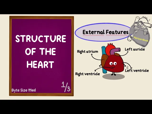
Hello. Welcome to Byte Size Med. This video is on the structure of the human heart, and in part one, we're going to look at some of the external features of the heart.
The heart is a muscular organ that sits in a space in the thoracic cavity called the mediastinum. If we look at it from the side, this would be the front, that's anterior, with the sternum. And this would be the back, that's posterior, with the vertebral column.
This mediastinum extends from the superior thoracic aperture to the diaphragm, and from the sternum to the vertebral column. But it's divided. The superior mediastinum above, and the inferior mediastinum below.
The inferior part is further divided into anterior, middle and posterior parts. The boundaries of the middle mediastinum are formed by the pericardial sac. This is the sac that surrounds the heart.
So the heart, along with the pericardium, are in the middle mediastinum. The pericardium is the outer covering of the heart, and it has two layers, the fibrous pericardium and the serous pericardium. The fibrous pericardium is the connective tissue on the outside, and the serous pericardium is on the inside.
Now it has two layers, a parietal layer and a visceral layer. The parietal layer is attached to the fibrous pericardium, and the visceral layer is stuck to the heart, so it's also called the epicardium. The visceral layer of the serous pericardium is the epicardium.
These two layers are continuous at the roots of the great vessels. Between the parietal and the visceral layers is a potential space. That's called the pericardial cavity, which has a little fluid to help movement of the heart.
The heart itself has three layers. The outermost is the epicardium, which we saw earlier is the visceral pericardium. This is simple squamous mesothelium.
The next layer of the heart is the myocardium, that's the muscle layer. It's the thickest layer. Beneath the myocardium is an endocardium, which is simple squamous epithelium.
There are thin layers of connective tissue that connect the layers. So from inside out, we have the endocardium, the myocardium, the epicardium or the visceral layer of the serous pericardium, the pericardial space, then comes the parietal layer of the serous pericardium, and finally the fibrous pericardium. Those are the layers of the heart from inside out.
The serous pericardium, which is stuck to the heart, reflects near the vessels. That creates spaces called sinuses. These are called pericardial sinuses, and there are two important ones.
Transverse and Oblique. This is the posterior pericardial sac, after removing the heart. So we just see the vessels, with the pericardial reflections.
These are the pulmonary veins. This is the pulmonary trunk, which is gonna bifurcate, the aorta and its arch, superior and inferior vena cava. The oblique pericardial sinus is where the serous pericardium reflects around the pulmonary veins.
So that would be here. It forms a little cul-de-sac. The transverse pericardial sinus is up here.
Now it goes behind the aorta and the pulmonary trunk, and in front of the superior vena cava. It's like it separates the arteries from the veins. Those are the two sinuses.
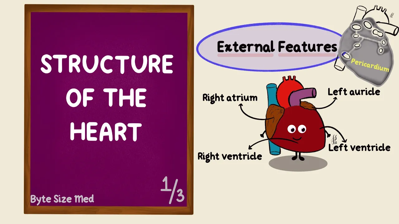
So we've got the transverse and the oblique pericardial sinus. Now coming to the heart itself, it's got four chambers: the right atrium, the left atrium, the right ventricle, and the left ventricle. This is the front of the human heart, and this is the back.
This is the right atrium, along with its ear- shaped projection called the right auricle. Here we have the right ventricle, and next to that is the left ventricle. Peeking up here we have the left auricle, because turning over to the back is where the left atrium is.
So on the back of the heart, this is the left atrium. Next to that, this would be the right atrium. Most of the left ventricle is here, along with a bit of the right ventricle.
The superior and inferior vena cava enter the right atrium. The aorta emerges from the left ventricle. The pulmonary trunk emerges from the right ventricle, and then divides into the right and left pulmonary arteries.
The four pulmonary veins enter the left atrium. Those are the chambers of the heart. The heart has an apex that's directed downwards, forwards and to the left.
It has a base, but it doesn't sit on the base. The base is the posterior surface. So it faces behind.
What it sits on is the diaphragmatic surface, which is inferior and obviously faces the diaphragm. The anterior surface would face the sternum and the costal cartilages, so it's also called the sternocostal surface. There are also surfaces on the sides that face the lungs.
These are the right and left pulmonary surfaces. So you have an apex, a base, a diaphragmatic surface, a sternocostal surface, and right and left pulmonary surfaces. The four chambers of the heart, that's the two atria and the two ventricles, right and left, contribute differently to the surfaces.
The apex of the heart is mostly the left ventricle, the base is mostly the left atrium, the diaphragmatic surface, again, mostly the left ventricle, and the sternocostal surface is mostly the right ventricle. The left pulmonary surface is mostly the left ventricle, and the right is mostly the right atrium. Now i say mostly because other chambers do contribute, these ones just make up a major part of each surface.
The surface of the heart is marked with grooves or sulci. There are three important ones. The deepest one is the coronary sulcus, which is between the atria and the ventricles.
The other two are between the ventricles, they are Interventricular. Down the anterior surface, we have the anterior interventricular sulcus, and down the diaphragmatic surface is the posterior interventricular sulcus. Now all these sulci house the vessels that supply the heart, the major arteries and veins of the heart, along with some epicardial fat.
Those vessels are obviously super important, and that's why they're in a separate video in part three. But in part two, we're going to take a look at what's inside the four chambers. And that was all about the outside of the heart.
I hope this video was helpful. If it was, you can give it a like and subscribe to my channel. Thanks for watching I'll see you in the next one!