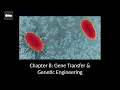
thank you in this chapter we're going to take our knowledge of genetics sort of the basics and apply it to two major Concepts the first concept called Gene transfer is where one cell transfers genetic information to another cell in fact you can see a picture of that here between two cells and the second concept we're going to talk about is called genetic engineering where we are manipulating DNA for some purpose let's go and get started I'd like to introduce the concept of transformation by showing the first demonstration of Gene transfer of horizontal Gene transfer that
is and this was carried out by a scientist named Frederick Griffith in the 1920s and I kind of want you to put yourself in his shoes here and try to imagine interpreting the results that we see here you know the results that he got trying to make sense of them he was dealing with an organism called streptococcus pneumonia which causes respiratory pneumonia and he was injecting different strains in mice so let's look at the four experiments that he carried out so we have our first control panel a here where we have a pathogenic strain of
streptococcus ammonia it's called the S strain and these cells have a capsule so you can see the capsule around the red cells there when injected into a mouse the mouse dies and then we can isolate isolate these bacteria from the mouse grow and grow them on an agar plate and see that they are living encapsulated streptococcus cells all right so that's first experiment second experiment which was another control we take he took a different strain it's still streptococcus pneumonia but this strain has no capsule inject That Into The Mouse and the mouse is fine Mouse
survives and just to double check uh we we take a culture from the mouse and sure enough streptococcus is in the mouse it is alive but these are strain cells just don't kill the mouse okay so that was control number two experiment number three on the upper right here Griffith took the S strain cells the encapsulated the encapsulated version and then heated them up so warm that the cells died so here we have heat killed s strain cells injected into a mouse the mouse survives we culture the mouse and no colonies presence right so growing
none of these bacteria and it makes sense because they were dead in the first place all right now here's the most important one in panel D the bottom right we have a mixture of harmless horse trained cells and pathogenic s strain cells but the pathogenic s strain cells have been killed with heat so we have heat killed s strain and harmless R Str strain mixed together injected Into The Mouse and the mouse dies which is odd because the r cells we know are harmless and the heat killed s cells are also harmless by themselves so
something happened here we culture uh to you know take a sample from the mouse and culture that sample on an aggro plate you can see that we have um live s cells and live R cells so it almost looks like the S cells came to life but what really happened was that the Dead s cells in the process of sort of being recycled and being broken down the cells released their DNA so the DNA was exposed in the environment and the r cells were able to take it in and that's a process called transformation when
these R cells took in the genes from the S strain they gained the genes to make a capsule and so some of these cells sort of turned into s strain cells because they were able to make a capsule now so transformation was introduced on the previous slide with the Griffith experiments and here we're going to get into some more detail in a nutshell transformation is a process of a live cell taking in extracellular DNA so imagine some DNA kind of hanging out in the surrounding environment and the cell takes it in now that's not a
very easy process and so it requires the cell to be equipped uh to to do this and that term to describe a cell being equipped or or able to take in extracellular DNA is called competence so in other words a cell needs to be competent in order to transform so a competent cell is kind of shown on the right side of the screen here so in actually a non-competent cell is shown at the top and this is not able to take in extracellular DNA but let's compare that to a competent cell below all right so
I'm circling it right now this particular cell is equipped with binding proteins or enzymes on the on the cell surface so that it can bind to DNA for example and then take it into the cell now there are a wide variety of mechanisms through which this occurs and so you can see several of them listed here um we may have pylie involved specific translocase enzymes I'm not so worried about those details I'm just trying to give you the the gist of the process um but what's important here is that if the cell takes in this
genetic material and it keeps it in the cell perhaps even changes its chromosomal genes then the genotype has changed of the cell and we call it transformed now this does occur naturally in bacteria so out in nature but it's a it's a rare process so less than one percent of the time this is occurring and often it's induced by some sort of stressful situation stressed environment for example starvation so even though less than one percent looks like a very low number keep in mind the population of bacteria for example can get huge so in one
particular culture of bacteria we could have millions and millions of cells and so with a population that contains millions of cells one percent of that population now we're talking about thousands of cells that actually undergo this process the last thing I'll mention here is that we can do this in the lab so we can induce competence of specific strains of cells and we can induce transformation in fact it's going to be one of the experiments that we do in lab the second type of horizontal Gene transfer I want to talk about is called conjugation this
is quite a bit different than transformation conjugation is where we have two live cells as you can see in this figure that make contact and a donor cell sends genes to a recipient cell and something called the F factor is going to be involved the F factor is a a plasmid called the F plasmid and F stands for fertility factor or just for fertility and the F plasmid I want you to think of it as kind of a selfish plasmid it really wants to spread itself it wants to replicate itself and so the F Factor
contains a couple dozen genes that codes for uh the the stuff and the steps necessary for that transfer so as we're going to see in upcoming slides a cell that contains the F Factor the F plasmid floating around in its cytoplasm is called an f plus cell a cell that does not contain that F plasma is called the F minus cell now as an alternative in kind of a more complex situation we have some cells that have the F Factor integrated into its chromosome and this is called an HFR cell or high frequency of recombination
because it's more likely to generate these recombining events than one that does not have an F Factor so let's go ahead and look at the specific examples next here's the easiest example of conjugation this is where we have an f plus cell donating the f f Factor transferring the F factor to a recipient F minus cell so in this figure we have a the donor cell on the left this is our f plus cell and how do we know it's f plus cell well we see the F Factor floating around in the cytoplasm all by
itself separate from the chromosome that's what determines that this is an f plus cell the F factor is going to be able to code for the proteins necessary to to transfer itself and to replicate itself so it's going to code for the formation of this conjugation pilus as you see here so conjugation pilus where it's going to the cell is going to reach out and grab onto a recipient cell notice the recipient cell is an F minus meaning it has no F Factor floating around in cytoplasm all right so once connection has been made the
conjugation pilus brings the recipient cell closer and then a conjugation bridge is formed this bridge is kind of like an elongated pore through which stuff can move from one cell to another and in this case it's going to be the F Factor all right so in the third step here you see that the F Factor has started to replicate itself through this rolling Circle mechanism and so it's simultaneously doing two things it's making a copy of itself so that the f plus cell keeps a copy and it's transferring the other copy the other strand over
to the recipient F minus cell so the entire plasmid is copied and eventually transferred over to the F minus and so we end up with two f plus cells at the end okay the the the recipient cell is now considered f plus because it has the F plasmid by itself outside of the chromosome floating around the cytoplasm a second type of conjugation is shown here so in this situation notice that there is no F Factor as a separate plasmid floating around in the cytoplasm in in the donor cell therefore we do not call the donor
cell an f plus cell in instead the F Factor has been integrated into the chromosome and so you see that part of the circular chromosome here is green and I try to outline it here and so the F Factor genes have sort of inserted themselves the F Factor has has inserted itself into the chromosome so this type of cell this type of donor has a different name it's called a a cell an HFR cell HFR stands for high frequency of recombination and that's basically a fancy way of saying it likes to undergo conjugation it likes
to share its genes with high frequency okay so we have our donor cell it's not called f plus because it does not have the F plasmid floating around separate from the chromosome instead the F Factor has integrated itself into the chromosome and so it's called an HFR cell that's our donor the recipient is just a plain old F minus okay it doesn't have anything fancy about it it's an F minus cell all right so just like the F plasmid the integrated F Factor just because it's integrated doesn't really change its personality so the integrated F
Factor just like the plasmid is still selfish and it contains genes for its own replication it contains genes for its own transfer over to a recipient's cell and so that's we're going to see here so there is the formation of a conjugation pilus formation of a conjugation Bridge the recipient cell is pulled close and there is this origin of transfer sort of right in the middle of the integrated f f Factor so the origin of transfer is basically where this copying begins so instead of just copying over the F Factor because it starts in the
middle of the integrated F Factor the cell actually ends up copying or trying to copy the entire chromosome as well and so the net effect is that we get a piece of the integrated F Factor transferred over and we get some chromosomal genes transferred over there too now even though it's going to try to copy the entire chromosome including the integrated F factor it's not going to happen completely so in nature these conjugation Bridges don't last all that all that much time and those conjugation bridges are going to break and so the whole chromosome and
the entire integrated F factor is not going to be copied over so just some of it is going to make it over so what we end up with is a recipient cell that contains a portion of the F Factor notice that there is no F factor or F plasmid floating in the cytoplasm therefore this is still an F minus cell if it was f plus it would have the F plasmid floating around in the cytoplasm but it doesn't so it's still an F minus cell but because it received some chromosomal genes from the donor this
recipient cell has a new combination of genes and that's what the word recombinant is referring to so a new combination recombinant a new combination of genes and then the still the cell is still F minus and so all together this is called an A recombinant F minus cell now if we sort of just pull back and think about the big picture here what just happened is the donor cell was able to copy over some chromosomal genes to a recipient cell and perhaps those chromosomal genes are are relevant are important for example maybe a gene for
antibiotic resistance our third and final type of horizontal Gene transfer is called transduction and the big difference here is that transduction involves a virus so we don't have two cells contacting each other we don't have extracellular DNA floating around in the environment being taken in by a competent cell here we have DNA from one cell to another cell and the virus carries it there so when we're talking about viruses that infect bacteria talking about what's called a bacterial phage you can see an example a tem of bacteriophage in the the figure here so everything you
can see kind of all this white stuff the tail the head Etc is composed of protein and then inside of the head we have all balled up in here we have some DNA we have it the viral genome so the goal of the bacteriophage is to inject its genome into a host cell and once the genome is in the host cell The genome basically does everything um everything else so it takes over the cell it kills the cell but before it kills the cell it results in the production of a lot more viruses so as
we're going to see there are two types of transduction um so without getting into too much detail here let's just go ahead and look at the next figure so there's a lot of detail here a lot of information on this slide but you can use the zoom tool to kind of zoom in and scroll around if that helps um this shows the two types of transduction but everything starts from the same place so here's our starting point up here where we have a a donor cell right so the the original cell here it's going to
be infected with a bacteriophage and you can see that the bacteriophage injects its DNA into the donor cell right into its host cell so you see that happening right there notice that the cell and its chromosome just for the sake of example we're going to say that it has three genes of Interest has Gene B Gene a and Gene D so we're going to kind of refer to those genes in the future all right let's take the left Fork first let's talk about generalized transduction and that Branch first so this is where the the bacteriophage
goes immediately into what's called its lytic cycle it's going to end up with the cell lysine and a bunch of new viruses produced so the virus we think like a virus the virus wants to do two things it wants to make more genomes and it wants to make more viral proteins and then it wants to assemble those things to make a new virus so that's that's what's happening right here we have a bunch of viral genomes being copied and what we can't see are viral proteins being made and notice also that the Genome of the
host cell is being cut up into pieces all right so then the next step we have assembly of the viruses so the genomes that were just made assembled with the viral proteins that were just made to make a a new virus particle you can think of them as like little viral babies and that's great but notice that there was a mistake made so we have a defective particle where instead of packaging a viral genome accidentally a chunk of the host chromosome was packaged into the the viral protein so this defective particle has Gene B in
this example okay so it has bacterial DNA no viral DNA just bacterial DNA packaged into this viral head so it's called a defective particle because it's going to be able to infect another cell but when it does infect another cell it's not going to be able to make new viruses so at this point the viruses are going to cause lysis of the host cell so now we're here and those viruses will go off and infect new cells and repeat the process but let's follow the fate of our defective particle it's going to infect a new
cell and when it does that notice that the phage has transferred gene B but it's a capital B and the host cell has Gene B as well but it's a little B so they both have Gene B both pieces of DNA have Gene B but just different versions of Gene B so another way to say that is they have different alleles just different Gene versions so it's certainly possible because they're the same gene just different versions that we can have a recombination event and that's what you see at the bottom here so we've effectively changed
the genotype of the recipient cell of this second cell from Little B to Big B and in the case of this example on the upper right you see that Gene capital B gives resistance to an antibiotic called streptomycin again this is just sake of example but you get the idea of how that's going to be advantageous all right that's generalized transduction now let's follow specialized transduction the Right Fork this involves a different phase of the viral life cycle so bacteriophage can also do What's called the misogyny or the lysogenic cycle and this is where the
viral DNA integrates itself so trying to indicate this here in red you see that the viral DNA has integrated itself into the bacterial chromosome and it can hang out there for a long time and this is this is to the advantage of the virus because it's it's going to be there undetected let's see it's protected basically and as the cell replicates itself and as the cell replicates its genome it's unknowingly replicating viral genomes as well right because the genome is integrated into the the host's chromosome at some point though the virus will exit or can
exit the lysogenic cycle and enter the lytic cycle and start making new viruses and when it does that it's going to cut out it's going to remove its genome from the bacterial chromosome but oftentimes that removal is not very precise so in this example the red part was taken out but it accidentally took some bacterial chromosome with it specifically Gene D is coming with it so when you look at the new particles that have been made um there is some you know viral DNA in there but there's also some bacterial DNA in there specifically gnd
in this example so after the cell is lysed and the the particle infects the new cell the same type of thing happens as as it did during generalized transduction we have a recombination event in this case Little D is changed to Capital D and in our example capital D confers resistance to penicillin and again that's just for the sake of example but you can see how this can be advantageous to the recipient cell so again zooming out big picture we have effectively transferred DNA from cell number one up here in the upper left to these
cells down here and change their genotype and a virus was the vehicle to carry that Gene okay so now that we're done talking about horizontal Gene transfer let's finish by talking about some examples of biotechnology and genetic engineering so how can we use microbes and how can we use our knowledge of genetics to um make something of interest to change something of Interest now of course this can be used therapeutically like gene therapy applications you know trying to change uh the Genome of a cell for therapeutic reasons uh someone with a disease and so on
we can use microbes to make protein products of Interest we can insert useful genes into plants you know these all sound great but of course there are downsides there are consequences uh potentially some bad things that can happen um you know being able to manipulate the instructions for life is a pretty powerful thing and so then therefore it's going to be controversial and and for good reason so let's keep that in mind as we talk about some specific techniques just to demonstrate that we are all impacted by genetic engineering I wanted to show this graph
where you can see the number of FDA approvals on the y-axis and the last few decades on the x-axis and so all these pharmaceutical drugs Etc and products have been approved each year and the portion that's in yellow is thanks to recombinant DNA technology okay so many years it's a significant portion maybe half maybe even over half so this is just kind of a glimpse into how much out there is actually tweaked by us how much is is engineered by us so for sure we are all exposed to it impacted by it whether or not
we realize it so because genetic engineering is so common and because it's so powerful and because it's so controversial knowledge is power and it's going to be useful for us to know it's going to be useful for you to know some Basics about how it works so we're going to go into uh some specifics here and talk about kind of build up to how we are going to make human insulin as an example so all of this kind of started with a major breakthrough the discovery of restriction enzymes also referred to as restriction endonucleases and
what a nuclease does is it cuts DNA or RNA so it cuts up nucleic acid and Endo means within so these are enzymes that cut Within a a DNA strand specifically they cut at palindromic sequences so what a palindrome is a sequence that reads the same on either DNA strand so let's just look at some examples down here I have what five examples so let's look at the top example So reading five to three on the top strand it's agct on the bottom strand 5 to 3 agct let's look at the third example reading five
to three on the top strand GG a TCC reading five to three on the bottom strand GGA TCC so you get the idea they read the same 5 to 3 Prime on either DNA strand so these enzymes cut at palindromes and they can cut either straight through or with the zigzag so we're going to focus on not the blunt end formation but we're going to focus on the zigzag here that creates what are called sticky ends so we have these we have these overhangs and these overhangs will re-anneal so you know imagine this cut occurring
and we kind of pull apart the two DNA strands from each other well G still pairs with C and A still pairs with T so if they come back together you know in any sort of proximity they can certainly hydrogen bond again so we can pull them apart we can stick them back together so they're called sticky ends now on this figure on the right we can see kind of a molecular model in red and yellow of a restriction enzyme and it's embracing the DNA Helix in blue and so it's kind of holding on to
that DNA Helix is going to find a palindromic sequence and it's going to make a cut so each specific restriction enzyme cuts at a very specific restriction site so by the way the specific sites specific palindromes that restriction enzymes cut at are called restriction sites all right let's look at some more details here I'm gonna I'm gonna hold off on the word Vector until we see it on the next slide here okay so what good are restriction enzymes and sticky ends well what we want to do is cut and paste we want to cut open
a plasmid that we have already mapped its sequence we have already highly engineered it it's a small circular DNA molecule that's really easy to use and manipulate so we're gonna have restriction enzymes in that plasmid that we have engineered in there and then cut it open so here is the plasma that's been cut open you can see these sticky ends you can see the overhangs now of course this gray this great piece of DNA this plasma that's been open can certainly come back together into a circle because those sticky ends are complementary to each other
but now let's say that we take this red piece of DNA a foreign piece of DNA that we are interested in making more copies of we're interested in doing something with that piece of DNA in red so we're going to cut it with the same exact restriction enzyme as we cut the plasmid with so that means that it has the same exact sticky ends as the gray one does in other words these sticky ends will stick to each other so the red now sticks to the gray and we can form one big circle so the
final molecule that we hope to get is in the bottom right and so we have this red piece which is the foreign DNA that of Interest this is called the insert and it's been inserted into the gray part which is our plasmid now because this plasmid is going to serve as a vehicle to transfer and manipulate that red insert the gray plasmid is called the vector so we have a vector that's opened up we stick an insert in there and now we have our final recombinant plasmid now one little detail that I left out is
that when the sticky ends come together the hydrogen bond but hydrogen bonds are weak so we can pull those sticky ends apart again so if we want this to be kind of a permanent solid recombinant DNA molecule we need to seal the phosphate backbone sugar phosphate backbone here so not just the hydrogen bonds are important but if you recall from the structure of DNA we have this sugar phosphate backbone and so we want to seal those Nicks I want to seal those holes and we do that with an enzyme called DNA ligase so that's an
essential part of this process is sealing the final bonds with DNA ligase to give us this recombinant DNA molecule so now what are we going to do with this recombinant DNA molecule let's see at the top here we have kind of the same thing that was shown on the previous slide except now it's showing it coming from cells so we have a plasmid Vector that's been opened we have a gene in a human cell the gene of Interest we're going to cut that out it doesn't have to be human cell could be a plant cell
or whatever but we have we have a gene of Interest going to cut it out and we're going to form our recombinant DNA molecule a recombinant plasmid I'm going to stick it into E coli so what is that called we have an extracellular plasmid an extracellular piece of DNA that's brought into us a cell specifically a competent cell so in other words that's transformation so we transform E coli with a recombinant plasmid and now now we can do things with it so we can let E coli grow and grow and grow and multiply and multiply
and in the process it's going to multiply the plasmid giving us lots and lots and lots of copies of our recombinant plasmid so in the process that means we're copying the insert we can cut the insert out in this clay in this example we have kind of a test tube of inserts here the test tube of that insert DNA and it's been Amplified we've made a whole bunch of it and we need a lot if we're going to do some genetic engineering applications in this case sticking genes into these plants to make them resistant to
insects and herbicides and prevent spoilage and prevent them from ripening too fast and make them juicier seedless and whatnot another option is we take our transformed E coli let them grow and replicate and replicate and multiply and multiply and in the process the plasmid is multiplying and also in the process gene expression is occurring so don't forget gene expression and do a horrible job of writing here is DNA to RNA to protein we're just going to stop at Pro how about that so we have DNA to RNA to protein so that means that the red
insert here is transcribed and translated we get a bunch of the protein product that's great maybe we want that and we can use that protein for a variety of things so depending on what it is it might be a blood clotting Factor it might be human insulin or human growth hormone or or antiviral proteins to fight viral infections and so on and so on so the last thing we're going to do is look at a very specific example so we're going to kind of do this same thing and make some insulin so let's see how
we would do that so let's take all of our knowledge now of genetics genetic engineering and apply it to a real situation where we want to grow a bunch we want to to make a bunch of human insulin and human insulin is a protein okay so in order to get a lot of it we're going to use E coli okay and this is the process so we're starting with a plasmid highly engineered cloning vector and remember we're going to open it up and stick an insert in there but before we do that let's look at
the genes present so we have this green region right here this Gene in green codes for ampicillin resistance so that means that this Gene codes for a protein product that's going to make whatever cell contains that plasmid resistant ampicillin in red we have a gene called Lac Z as I have in the writing here Lac Z codes for an enzyme so DNA to RNA to protein this is our enzyme or protein enzyme called beta galactosidase or just be gal and to summarize what B gal does it won't mean much to you yet but it'll come
in handy later B gal breaks down a molecule called X gal into a blue product so that's going to come in handy shortly and then finally notice that we have this is really important we have a restriction site stuck right in the middle of Lac Z okay so we're going to cut our plasmid with a restriction enzyme and open it up that's what you see right here notice that that cut occurred right in the middle of the Lexi Gene at the same time we take our human DNA genome we cut out the insulin Gene with
the same exact type of restriction enzyme so these have complementary sticky ends and we can make a recombinant plasmid as you see here containing the human insulin Gene in black as the insert now keep in mind that during this process when we mix the human insulin Gene with the plasmid vector it's not always going to work out perfect where every single plasmid vector gets an insert because some of these vectors because they have sticky ends they'll just kind of stick back together with no insert and so that's certainly a possibility and we're going to have
a mixture of of these two possibilities the recombinoplasmid with no insert and the recombinant plasmid with an insert we're going to have a mixture of those two in the same test tube okay now what we're going to do is transform E coli so that's our next step you see E coli being transformed so here is our we really have three possibilities during the transformation step and we're going to have a mixture of all three occurring remember that transformation is an inefficient process so on the far right we have a cell or a population of cells
in our beaker that don't take up the plasmid at all and that's very very likely to happen on the far left we have an E coli cell population that that takes in the recombinant DNA plasmid with no insert and that's a very real possibility and then of course the ones we want are those cells that take in the recombinant plasmid containing the human insulin inserts okay so the middle ones this is what we want but like we've talked about in lab if this is in broth and they're all mixed together that's basically kind of like
a mixed culture and there's no way to pluck out exactly just the one we want so we're going to have to use an agar plate to do that so in our lab we did a four-way Street plate and here we're going to streak this cell suspension onto an agar plate let the colonies grow and we're looking for not only growth but also their color in order to determine determine which one we want so let's take it Case by case so any bacteria that do not take in a plasmid are going to die as soon as
they contact the ampicillin on the plate so that reminds me let's look at let's look at what's in this growth medium here we're going to have nutrients of course but we're also going to have two important things we're going to have ampicillin added to the growth medium ampicillin is added and we're going to have X gal added as well so the growth medium contains nutrients ampicillin and xcal so that means when any cell like this on the far right does not contain the plasmid as soon as it's plated on the agar plate the ampicillin will
kill it however the cells that did take in the plasmid contain the green Gene the ampicillin resistance Gene and so they will survive and be resistant to ampicillin so these two types of cells these two populations of cells will grow on a plate okay so at least we've eliminated the far right because we don't want those now we have to figure out how we're going to tell the difference between the two transformed bacteria the two types of transformed bacteria okay so let's take the example on the far left first so this cell right here contains
the recombinant plasmid with no insert so if we look at at the top of the screen again remember that the red Gene codes for Lac Z this is the Lac Z Gene which codes for beta galactosidase which breaks down X gallon to something that's blue so the cells here that grow on the plate are going to break down x-cal which is in our nutrient medium and make the colony blue so we're going to be able to tell that those are the the cells that don't have the insert they do have the plasma but they don't
have the insert so we don't want those we want the middle case so the middle case right here these cells have the insert and the insert has interrupted the Lac Z Gene so when the Lexi Gene is transcribed and translated there's no way that we can have a functional beak out because we stuck this giant hunk of DNA right in the middle of the black seed Gene so no functional be gal means no blue product and so these cells grow as a white colony so the take-home message is at the end of all this we're
going to be able to tell the difference between the two because not only did they grow so we know they were successfully transformed but we can tell the difference between insert or no insert by their color now once we pluck off a white Colony let's think about what we would do next we're going to grow it up in a fresh nutrient medium okay so we have the a pure culture basically and we let those cells just grow like crazy and make boatloads of human insulin for us and then we can purify the protein and use
it as a drug use it as a treatment