hi everybody and welcome to our module on muscular distrophy the muscular distrophy are a group of genetic disorders there are more than 30 different types that have been described they all result from defects in genes that code for proteins important for muscle function and therefore the main symptom of all these disorders is progressive muscle weakness most of the muscular distrophy are very rare I've listed on this Slide the three variants of muscular distrophy that you should know for the USMLE step one exam the first one is duchaine muscular distrophy this is the most common variant
the second one is Becker muscular distrophy this is a milder version of duchan but it shares many genetic features with duchaine and for that reason these two are often discussed together and then the third one is a rare type of muscular distrophy called myotonic muscular distrophy but the reason you should know this one for the step one exam is because it is an example of a trinucleotide repeat disorder and I won't discuss it in this video but I do discuss it in detail in the video on trinucleotide repeat disorders so like I said before duchaine
and Becker muscular distrophy are often discussed together one of the reasons for this is that they're both x-l in fact they're often called the x-l muscular distrophy in addition they both involve a gene called the DMD Gene and they both involve a protein called the disten protein and you shouldn't confuse either of these muscular distrophy with myotonic distrophy that disorder has a different Gene a different protein and it is not exlink it is autosomal dominant so we'll start by talking about duchaine muscular distrophy which is the most common form of muscular distrophy it's an ex-link
recessive disorder and it has all the usual features of xlink recessive disorders that I discussed in the video on pedigree so all male carriers are affected a third of cases are due to new mutations in the fertilized egg in other words there is no parental carrier that passes on a mutated X Gene in two-thirds of cases the X Gene that is abnormal in the sun is inherited from a carrier mother the gene that is abnormal in duchan muscular distrophy is the DMD Gene DMD stands for duchaine muscular distrophy and this Gene is famous for its
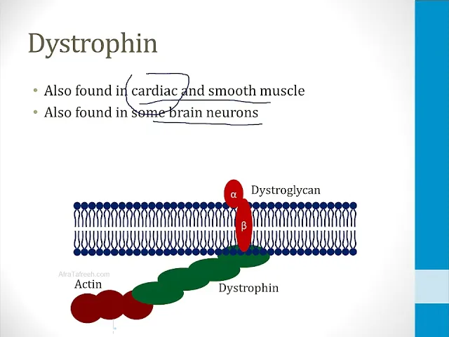
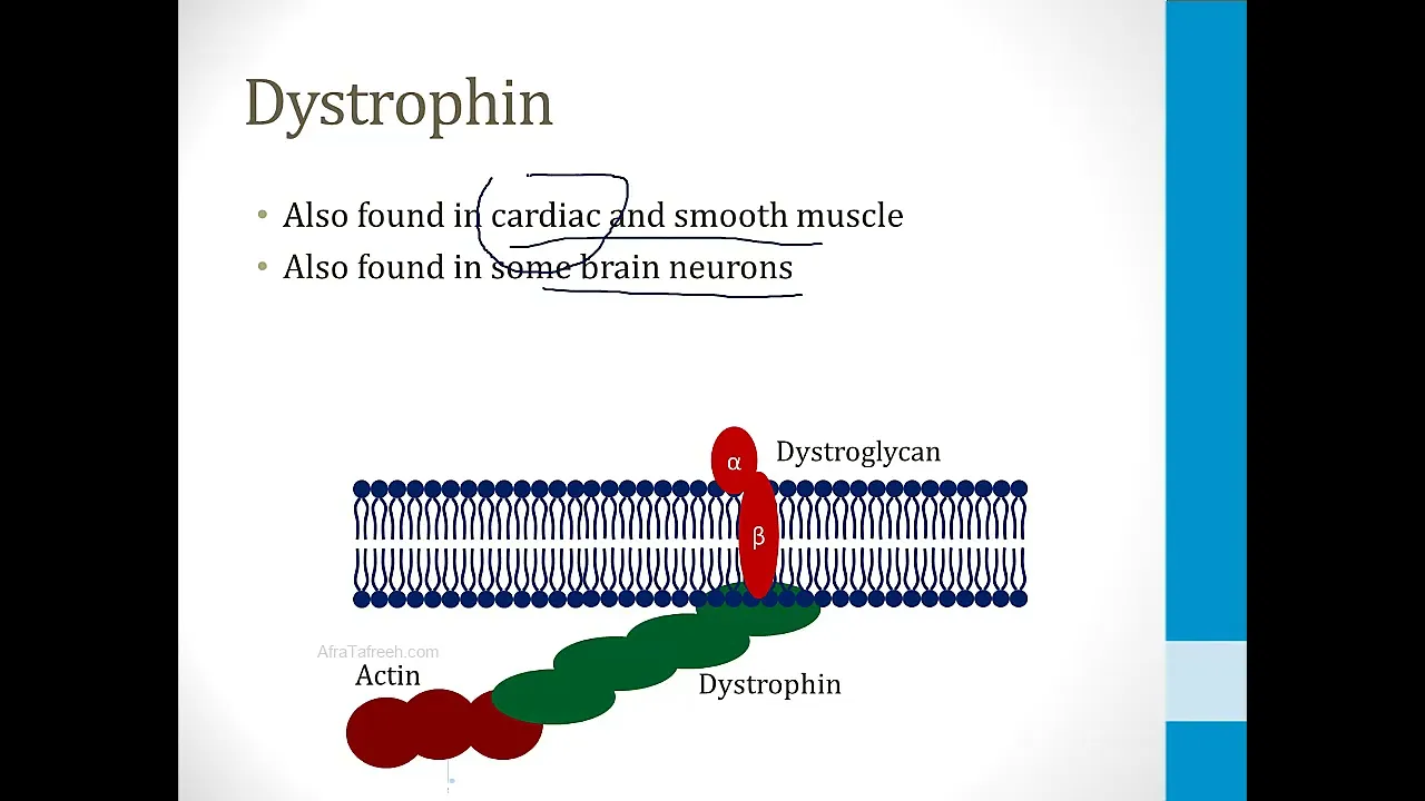
massive size it's 2300 kilobases in size it takes up 1.5% of the X chromosome it is among the largest known genes and because it is so large there are lots and lots of nucleotides that have to be replicated to copy this Gene and therefore there is a high chance that a mutation will occur and this Gene codes for a protein called disten and that's what abnormal into Shane muscular distrophy disten is a protein that's important for maintaining muscle membranes it connects intracellular actin proteins to transmembrane proteins specifically it connects actin to proteins called Alpha and
beta drog glycan in the membrane and these two proteins in turn are connected to the extracellular Matrix to a protein called laminin so in this way disten plays a role in linking actin inside the muscle cell to proteins in the membrane and outside the cell and I've drawn a picture of this at the bottom of the screen to help you understand better we have actin protein which is connected via disten to drr glycan in the cell membrane and in turn Alpha and beta drog glycan are connected to extracellular Matrix proteins like laminin disten is important
in skeletal muscle and that results in many of the muscle symptoms of muscular distrophy however it's also found in cardiac and smooth muscle and also in some brain neurons and as we will see the fact that it's found in cardiac muscle is important because many patients with muscular distrophy develop heart failure and cardiac comp applications one of the most important things for you to understand for the step one exam is what I've listed on this slide about the disten gene mutations related to Duan and Becker muscular distrophy most of the mutations in both of these
disorders are deletions however in duchaine muscular distrophy the deletion results in a frame shift mutation in Becker muscular destrophy the deletion results in a non-frame shift mutation I discuss frame shift mutations in detail in the video on DNA mutations but by way of Brief Review in duchaine muscular distrophy the frame shift mutation results in A disruption of the reading frame this leads to encountering an early stop codon and thus children born with duchaine muscular distrophy have a truncated or absent disten protein in their bodies in contrast children born with Becker muscular distrophy have a non-frame
shift mutation thus they typically have some functioning protein and therefore this tends to be a less severe Disorder so even though both disorders involve deletions of the same gene what distinguishes them and why one form is more severe than the other has to do with whether or not the deletion is a frame shift mutation just to make this very clear let's look at the drawing I've made on this slide under normal circumstances the disten gene has a series of codons that I've numbered one 2 3 four and five and these lead to synthesis of a
normal disten protein in a child born with duchaine muscular distrophy the disten gene has a deletion that occurs after codon 2 and in this case it is a frame shift mut and therefore none of the normal codons after codon 2 are encountered this leads to either no synthesis of disten protein or a very truncated protein in contrast in children with Becker mutation the disten gene has a non-frame shift mutation in this example I've shown a mutation that leads to loss of codon 3 thus what you get is an abnormal Protein that's missing some of the
amino acids but it still has many of them intact because the mutation did not shift the reading frame therefore Becker tends to be a less severe disorder even though it involves the same gene and the same protein when there is loss of disten protein this leads to my necrosis this means that enzymes found inside of muscles can spill into the blood and be detected by blood test so creatin and kinas elevation in the blood is common in the early stages of duchaine muscular distrophy it's released from diseased muscle other muscle enzymes can also be elevated
in the blood including alalas a and ALT when children initially present with this disorder the proximal muscles are typically affected first before the distal limb muscles in addition the lower limbs are affected before the upper extremities this means that affected children will have difficulties with activities that involve the lower extremities things like running jumping and climbing stairs a classic sign of duchaine muscular distrophy is when children use their hands to push themselves up from a chair because their lower extremities are so weak this is called Gower sign in addition they are classically described as having
a waddling gate basically they are using the lower parts of their legs to Waddle because the upper parts of their legs have become weak and then finally the muscle eventually becomes replaced with fat and connective tissue and this can result in apparent enlargement of the calf this is called pseudo hypertrophy many children with duchaine muscular distrophy eventually develop a cardiomyopathy that's why I take care of a few children with this problem they will develop a depressed left ventricular ejection fraction and systolic heart failure and they can have mardial fibrosis seen on biopsy they can also
develop conduction abnormalities including AV any rhythm is the classic finding on a muscle biopsy which is rarely done in the modern era is degeneration of muscle fibers and replacement of muscle by fat and connected tissue this is an example of what a biopsy slide can look like here the muscle fibers have been replaced with these clear spaces and that represents fat a western blot test used to be done for diagnosis but this has been replaced by genetic testing I talk about Western blot in the video on blotting but it is a technique that's used to
detect proteins in a western blot in a patient who has has duchaine muscular distrophy there's usually absence of the disten protein in a patient who has Becker there's usually an altered dropin protein so if you look at this picture on the right side of the screen a normal patient is in the middle and that patient has a band about this far down the gel a patient with Becker muscular distrophy also has a band on the gel but it's at a different location and that's because the protein has been altered by a non-frame shift mutation in
the patient with duchaine muscular distrophy there's no functioning disten protein and therefore there's no band scen on the gel in the modern era the diagnosis is usually made with genetic testing usually with variations of the polymerase Chain Reaction test this test can identify the most common duchaine muscular distrophy Gene abnormalities basically it turns out that most children with this disorder have one of a small number of typical deletions so PCR can be used to test for those deletions and if one of them is found then the diagnosis is made I'll finish by talking about Becker
muscular distrophy it is also an exlink recessive disorder just like duain in Becker muscular distrophy 90% of cases are inherited from carrier mothers the reason for this is because this is a less severe disease so more males live to pass on the gene to female Offspring and thus there are more female carriers of this abnormality in terms of symptoms this is basically a milder form of muscular distrophy it has a later age of onset usually children are older some patients remain ambulatory during their entire lives and patients often survive into their 30s or Beyond with
Becker muscular distrophy and that concludes our video on muscular dist