hello everybody and welcome to our module on cardiac physiology on the screen is an echo cardiogram of the left ventricle of a patient's heart you can see there are QRS complexes rolling along the bottom of the screen and each time there's a QRS complex the walls of the left ventricle thicken and move towards the middle when this happens it shrinks the volume inside the left ventricle and pushes blood out into the aorta so in this module we're going to talk about the changes in volume and the other physiological changes that occur as the heart goes
through the cardiac cycle and moves blood out of the left ventricle as we saw in the moving image on the last Slide the volume in the left ventricle is changing during the cardiac cycle however there are two important volumes when it comes to understanding cardiac physiology the first one is the end diastolic volume or the edv this is the volume at the end of diaso that's how it gets its name this is the point in the cardiac cycle where filling of the left ventricle has completed this is the largest volume of blood the left ventricle
hold this is also the point where contraction is about to begin the other important volume is the n systolic volume or the ESV this is the volume at the end of syy that's where it gets its name this is the point in the cardiac cycle where emptying has been completed this is the smallest volume that the left ventricle will hold this is also the point in the cardiac cycle where relaxation is beginning so now that we understand that we can understand some other important terms as related to cardiac physiology and the first one is the
stroke volume this is the difference between the n diastolic volume and the N systolic volume it's the amount of blood pushed out of the left ventricle with each heartbeat it's the difference between the largest volume the left ventricle holds and the smallest volume it holds another important term is the ejection fraction sometimes called the EF this is the stroke volume divided by the enddiastolic volume in other words it is the percentage of blood pushed out of the left ventricle with each heartbeat and a normal ejection fraction is about 55 to 60% our hearts don't normally
eject 100% of the blood present in the left ventricle with each heartbeat they only eject about 60% under normal circumstances and then finally the cardiac output is the stroke volume times the heart rate this is the volume of blood pushed out with each heartbeat times the number of heartbeats per minute when you multiply these two terms together you get a number called the cardiac output which has units of volume per time for example liters per minute is a common way to report the cardiac output another important term is the Venus return this is the amount
of blood returned to the left ventricle via the Venus system it should be equal to the CC output the amount of blood that is returned to the heart from the body should be equal to the amount that goes out and then another important term is the total peripheral resistance this is the resistance to blood flow out of the left ventricle that comes from peripheral structures for example the organs of the body and the arteries of the body importantly Vaso constriction of peripheral arterials leads to a rise in the total peripheral resistance when those vessels constrict
it's harder to push blood through them and there's greater resistance to flow and when the peripheral arterials vasod dilate this leads to to a fall in total peripheral resistance there are some blood pressure terms that are important for understanding cardiac physiology you may know that we all have a cystolic and a diastolic blood pressure and a normal systolic blood pressure is about 120 millim of mercury a normal diastolic blood pressure is about 80 millimeters of mercury you should also know that the systolic pressure is largely determined by the stroke volume and the diastolic blood pressure
is largely determined by the total peripheral resistance to understand this let's imagine that we're tracking the blood pressure as the left ventricle begins to contract and push blood out into the arterial system what we will see is that the blood pressure will rise and the height of that rise will be determined by the stroke volume and other words when the left ventricle stops Contracting and stops pushing blood into the arterial system the blood pressure will Peak and that Peak will be determined by how much blood is pushed out by the left ventricle the blood pressure
will then Begin to Fall steadily and the depth to which it falls will be determined by the peripheral resistance if peripheral vessels are very vaseo dilated then the pressure will fall greatly so that there'll be a very low low diastolic blood pressure on the other hand if the peripheral vessels are relatively vasoconstricted then will only fall to a lesser degree and thus we will have a higher diastolic blood pressure so in this manner the peak of the blood pressure meaning the systolic blood pressure is largely determined by the stroke volume and the depth or the
nator of the blood pressure is largely determined by the total peripheral resistance one other important blood pressure term is the pulse pressure this is the difference between the systolic and diastolic pressure so for example for a patient with a normal blood pressure of 120 over 80 the pulse pressure would be 40 and this is also proportional to the stroke volume remember what I told you before the height of the blood pressure meaning the maximum value or the systolic pressure is determined by the stroke volume this means that the difference between these two numbers or the
pulse pressure is also determined by the stroke volume another important blood pressure term is the mean arterial pressure or map this is equal to the diastolic pressure plus 1/3 of the difference between the systolic and diastolic pressures which you may recall is called the pulse pressure the reason it's not halfway between the two pressures is because our blood pressure spends more of its time in the diastolic range if you chart the blood pressure you will find that it briefly goes up to the systolic Peak then falls down to the diastolic range and stays down there
for a much longer period of time before it climbs again this means that in order to calculate the mean you don't simply take the Midway point between the bottom and the top you take onethird of the way from the bottom coming closer down to the bottom number because you spend more time down in this range here at the bottom so for example if a person had a normal blood pressure of 120 over 80 the mean arterial pressure would be 80 plus 1/3 of the difference between those two which is 40 and that would work out
to 93.3 of the cardiac physiology variables we've been discussing one of the most important is the cardiac output that's because the cardiac output must rise to meet the demands placed upon the Heart by the body and in many disease States the disease is caused because the cardiac output is insufficient also remember that the more cardiac output the heart produces the more work it must do and the more oxygen that is required the cardiac output is equal to the heart rate times the stroke volume so when the heart rate goes up there are more beats per
minute and this is more work the heart must do in addition when the stroke volume goes up there's more volume per beat that the heart must pump and that also means more work that the heart must do these are very important principles of cardiac physiology for you to understand so the four classic determinants of the cardiac output are the preload the afterload the contractility and the heart rate and we'll talk about these one at a time in the next few slides but I just want to make a general Point here that changes in these four
factors affect the cardiac output and that means that these four factors also affect how much work the heart has to perform the preload is the amount of blood loaded into the left ventricle prior to each contraction that's how it gets its name the preload if you've watched the video on the Starling curve you're aware that the more preload that goes into the left ventricle the harder it will contract and for that reason when there is more preload there's more cardiac output in addition when there's more preload there's more work the heart must do and more
oxygen that is required and because of the Starling mechan in preload is sometimes referred to in textbooks as the amount of stretch on the fibers prior to contraction some books say length instead of stretch but all these are ways of referring to the preload and the fact that the Starling mechanism dictates that the more the left ventricle is loaded and the fibers are stretched the more vigorously the left ventricle will contract and if you find the concept of preload confusing a way I've always explained it to students is to think of a room full of
people and the left ventricle job is to push all those people out into the hallway through a small door the preload is the the amount of people put into the room and you can imagine that if there are more people in the room then there's more work that must be done in order to shove them all through the door and into the hallway outside so if preload is the amount of blood preloaded into the left ventricle to be pumped how could you increase the preload to the left ventricle well the easiest way is to add
volume to the body patients who receive a blood transfusion or intravenous fluids will have increased preloads these two interventions deliver more fluid to the Venus system of the body which goes to the left ventricle this preloads the left ventricle with more fluid that must be pumped and thus they increase the preload this means that both of these things will raise the cardiac output they will also raise the amount of work that the heart must perform another way that's not so obvious is to slow the heart rate when you slow the heart rate there's more time
for filling of the left ventricle thus it fills with more volume and therefore there is more preload and then a final way which is very important physiologically is to constrict the veins the veins are like large storage basins for fluid inside the body they contain a sign significant amount of fluid that can be pushed into the left ventricle if needed so when the sympathetic nervous system is stimulated alpha 1 receptors in the veins will react and constrict and they will push lots of blood into the left ventricle this is very important for the response to
blood loss Venus constriction increases the amount of volume available to the heart and helps to maintain the cardiac output in the setting of blood loss to decrease the preload you do the opposite of the three mechanisms I just discussed you can remove volume patients who are bleeding or dehydrated have decreased preload on the left ventricle you can raise the heart rate this allows less time for filling the opposite mechanism we talked about before in addition you can pull blood in the veins and this is the mechanism of action of drugs called nitrates which are sometimes
used in heart failure and an angena they dilate the veins and they pull blood in the Venus system this pulls blood away from the heart this lowers the preload there's less work for the heart to perform and for this reason it can relieve angen a couple of important terms regarding preload preload is often not called preload it usually referred to by one of the two terms I've shown on the screen here the first one is the lve DV that's the left ventricular end diastolic volume this is the volume I talked about at the beginning of
this module it's the volume of blood in the left ventricle when it's completed its filling phase it's the largest volume of blood that the left ventricle will hold during the cardiac cycle that is an indication of how much the left ventricle has been preloaded and thus the lvedv is usually used to represent the preload now in clinical practice it's very hard to measure the LV DV however it's much easier to measure the pressure at the end of diast the left ventricular end diastolic pressure so instead of using the volume we often use the pressure clinically
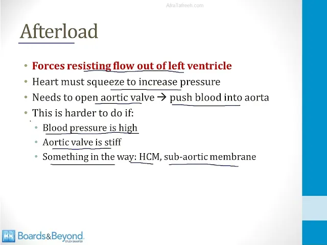
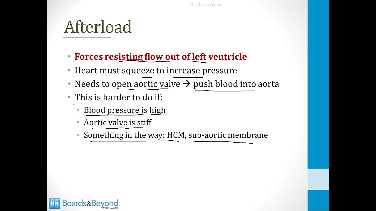
because it's simply an easier number to obtain this is the pressure in the left ventricle when filling is completed this is also a measurement of the preload on the left ventricle the afterload are the forces resisting flow out of the left ventricle remember that the heart must squeeze in order to raise the pressure in the left ventricle it needs to raise the pressure enough to pop open the Artic valve so that blood can be pushed into the aorta this is harder to do if for example the blood pressure is high blood pressure is one of
the determinants of afterload this is also harder to do if the aortic valve is stiff aortic stenosis is a classic condition where the afterload increases there are also some rare forms of high after load such as when things are in the way of blood moving out of the left ventricle this can occur in hypertrophic cardiopathy it can also occur in rare conditions like when patients have a sub aortic membrane obstructing the outflow of blood but all of these things raise the amount work that the heart must do in order to move blood out of the
left ventricle and just like with preload an easy analogy to understand after load is to think of a bunch of people in a room and it's the left ventricle job to push them through a doorway into the hallway outside the afterload is high if there are already a lot of people in the hallway outside it's obviously going to be harder to move those people out of the room and into the hallway that's what happens when your blood pressure is high there's already a lot of high pressure and molecules outside of the heart and therefore it's
harder to move blood out of the way it's also harder move those people out of the room and into the hallway if the door is stiff and won't open and that's what happens in aortic stenosis so these are ways you can think of the concept of afterload so just like we did with preload let's talk about ways we could increase or decrease the afterload so one of the ways to increase the afterload is to raise the mean blood pressure the mean blood pressure is one major determinant of the afterload of the left ventricle another way
to increase the afterload is to obstruct the outflow of the left ventricle in some manner this is what happens when patients have aortic stenosis or hypertrophic cardiopathy to decrease the afterload we could do the opposite of the two things I mentioned on the last slide we could lower the mean blood pressure we could treat the TIC valve disease or the hypertrophic cardiopathy and the principle here is that more afterload means more work the heart must do and therefore more oxygen that is required now let's talk about the third determinant of cardiac output and that is
the contractility this is how hard the heart muscle squeezes and the ejection fraction is one potential measurement of contractility when the ejection fraction goes up it means that the left ventricle is Contracting more vigorously and pushing more blood out of the left ventricle with each heartbeat and the major regulator of contractility is the sympathetic nervous system this is the major regulator under physiologic conditions of both contractility and also heart rate which we'll talk about in a minute the main physiologic mechanism by which contractility is increased is via sympathetic nervous system activity there are two mechanisms
by which the sympathetic nervous system can increase contractility first of all the sympathetic nervous system Direct directly inates the heart in addition the sympathetic nervous system can stimulate the release of catac colomines from the adrenal gland those can circulate and reach the heart those include substances like epinephrine and norepinephrine when either of these mechanisms activates the heart there's increased calcium release from the sarcoplasmic reticulum and this will increase the contractility so some classic triggers are stress also exercise which we'll talk about later anything that increases sympathetic nervous system activity a non-physiologic way by which contract
ility can be increased is via the use of sympathomimetic drugs these are drugs like dopamine dobutamine epinephrine and norepinephrine they all stimulate sympathetic nervous system receptors in the heart and they exert the same effects that the sympathetic nervous system normally does under physiologic conditions and then finally the drug dexin which I talk about in some of the other Cardiology modules inhibits the sodium pottassium atpa pump this leads to an increased calcium level inside of myocytes and this is another non-physiologic way to increase contractility the main way that contractility is decreased is by dialing back the
sympathetic nervous system this can occur naturally when patients enter States where there's less sympathetic activity it can also occur via the use of drugs so the main mechanism of sympathetic nervous system blocking drugs like beta blockers on the heart is to decrease contractility and also heart rate which we'll talk about in a minute some calcium channel blockers especially Verapamil and deltm also affect contractility they lead to less calcium being pulled into mosites and therefore there's less calcium available for muscle contraction and decreased contractility and then finally heart failure especially systolic heart failure is a disease
of myocytes that results in decreased contractility that's the defining characteristic of the disease our fourth determinant of cardiac output and the work of the heart is the heart rate when the heart rate goes up this increases the cardiac output under physiologic conditions and the main Regulators of heart rate the factors that cause it to increase or decrease are the same ones that affect contractility in general heart rate and contractility go together they are both mainly regulated by the sympathetic nervous system and they are both also affected by some pathomimetic drugs and blocked by drugs like
beta blockers there is a confusing concept about heart rate and stroke volume that's shown in many cardiac physiology textbooks and I'll explain this to you in the next few slides in a way that hopefully makes sense so in laboratory settings when you insert pacemakers into the hearts of animals like dogs and you artificially increase the heart rate you see a fall in stroke volume this is because there's less time for filling of the left ventricle so the relationship between heart rate and stroke volume experimentally is like what I've shown on the screen now this is
very counterintuitive most of us think of our output of our heart going up when our heart rate Rises and this slide indicates the opposite but keep in mind this is under artificial settings in the laboratory when you're increasing the heart rate without also raising the contractility even in artificial settings like the laboratory where you're pacing the heart of a dog an increase in heart rate over physiologic I ranges of heart rates still leads to a rise in cardiac output that's because even though the stroke volume Falls when the heart rate goes up like I showed
you on the last Slide the heart rate Rises to a greater degree and remember cardiac output is equal to the product of stroke volume times heart rate thus if you have a small drop in stroke volume but a greater rise in heart rate the net effect will be to increase cardiac output so thus even in the laboratory when you're studying animals and you're pacing their heart as you increase the heart rate you still see a rise in cardiac output even though the stroke volume is is going down as you raise the heart rate and that's
a confusing concept to make sure you understand that all of those relationships I just showed you however are artificial because under physiologic circumstances the sympathetic nervous system controls heart rate and contractility and it never raises the heart rate alone without also raising the contractility what this means is when the sympathetic nervous system raises the heart rate the stroke volume goes up even though there's less time for filling there's more contractility such that overall there is a rise in the stroke volume under physiologic conditions when the sympathetic nervous system raises the heart rate the one clinical
situation where a rise in heart rate can lead to a fall in cardiac output is under pathologic conditions in the setting of some cardiac arrhythmias there are some cardiac arrhythmias where the heart rate can get very very high sometimes as high as 300 beats per minute in this setting there isn't enough time for the left ventricle to fill and thus the stroke volume Falls and the cardiac output Falls in other words at these very high heart rates even though the heart rate is increased the stroke volume has dropped dramatically thus the product of stroke volume
times heart rate begins to fall that means the cardiac output gets low what this means is that some patients with cardiac arhythmia can develop hypotension and shock and this usually happens when the arhythmia results in a very rapid heart rate what this means is the true relationship between cardiac output and heart rate is like what I've shown on the screen here under the physiologic range of heart rates when the sympathetic nervous system drives an increase in the heart rate there is an increase in cardiac output this is because the sympathetic nervous system is increasing contractility
in addition to heart rate and thus the cardiac output is going up however if the heart rate gets very very high for example in the setting of your arhythmia you can reach a point where the cardiac output begins to fall and that's because of the dramatic drop in stroke volume seen when the heart rate gets very very high so to summarize the work of the heart is determined by four key components the first is the preload which we often estimate by the Left entric End diastolic volume or pressure the second is the after load which
is often estimated by the mean or Aral blood pressure the third variable is the contractility which is often estimated by the ejection fraction and the fourth is the heart rate and this will be important when we talk about disease States because hearts that are starved for oxygen need to have these factors modified so that there's reduced oxygen demand in addition hearts that are generating a low cardiac output need to have these factors modified so that they increase the work of the heart and increase the cardiac output and that concludes our module on cardiac physiology