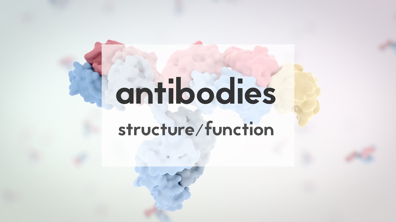
In 2019, you’d probably have to stumble across a group of scientists to hear people casually talking about antibodies. Now, discussions of antibodies and immune response are dinner table conversation. You might have seen little Y-shaped representations of antibodies in news articles, but what do they /really/ look like, and how do they help us to fight off infection?
In this lesson, I’ll touch on the importance of antibodies, examine their structure in detail, and visualize how they interact with and neutralize a viral protein. Let’s get started! Antibodies are proteins produced by B-cells that are part of our immune systems.
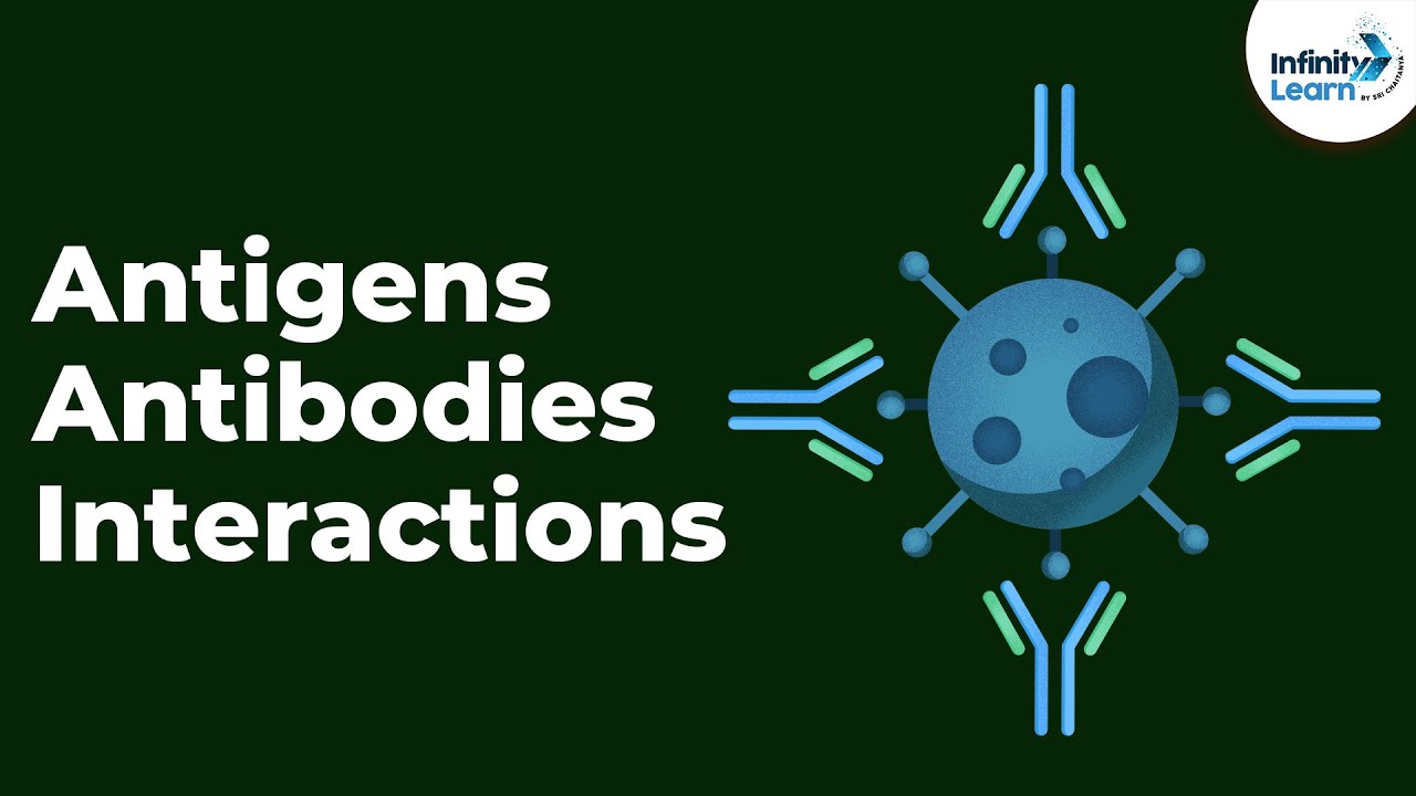
Antibodies bind to antigens, which is something the body recognizes as foreign, like a bacteria, virus, or allergen. The threat doesn’t even need to come from outside. On a small scale, antibodies can bind to a tumor cell and facilitate its destruction, and this is the basis for some cancer treatments.
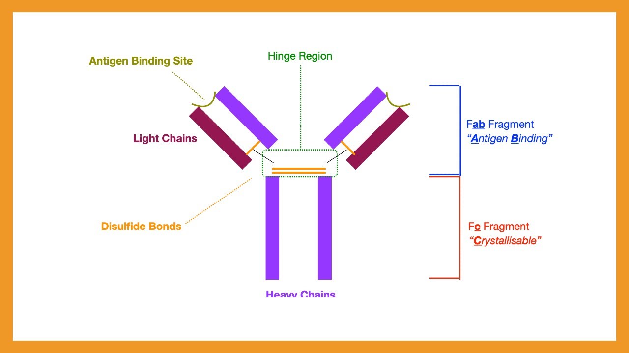
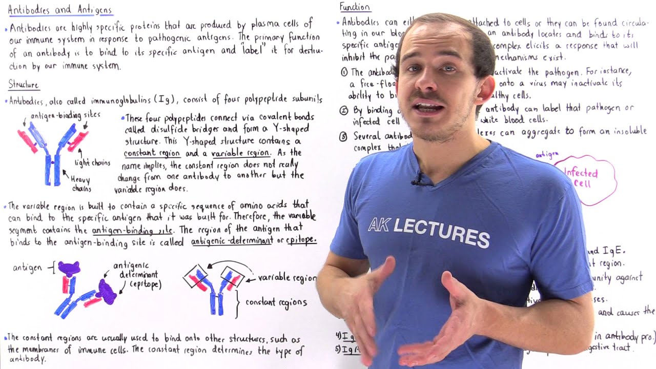
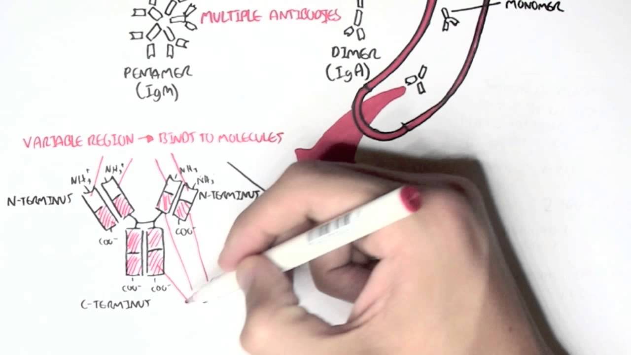
These Y-shaped molecules bind antigens at the tips of two identical arms. The rest of the antibody is made up of constant domains. The repeated patterns of protein structure in the constant domains are conserved—that is, they’re the same—in antibodies of the same class, and there are five of these antibody classes, or /isotypes/.
You might be wondering how the body can recognize a foreign substance that it’s /never/ encountered before. Well, the amino acid sequence where the antibody binds the antigen, at the tips here, is variable. While the B-cell is developing, the genes that code for the antigen binding region, which I’ve coloring slightly lighter here, are shuffled up like a deck of cards.
This produces many different amino acid sequences in this region here, called the variable domain. Each B-cell therefore produces a unique antibody, but all antibodies produced by that specific B-cell are the same. The first antibodies the B-cell produces are attached to the cell surface, and if that B-cell happens to bind an antigen, it can be stimulated to produce more antibodies, some of which can float freely in our bloodstream and lymphatic systems.
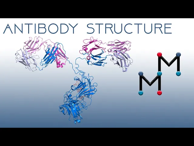
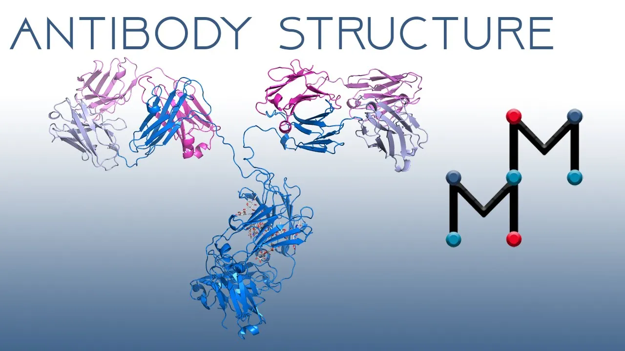
With that in mind, let’s take a deep dive into antibody structure. This is an immunoglobulin G, or IgG antibody that we’re looking at here. IgG is the main antibody in the blood, and the only one that can cross the placenta, transferring immunity from a mother to a fetus.
The antibody is made up of four different protein chains, two identical /light/ chains, shown here in magenta. The two larger chains, shown in blue, contain more amino acid residues and are therefore heavier than the light chains. These /heavy/ chains meet up to form a region called Fc.
Within the Fc, there are two identical carbohydrate chains, bound to the protein through a covalent bond with the side chain of an asparagine amino acid residue. In addition to stabilizing the immunoglobulin structure, these carbohydrates may play a role in antibody secretion and antigen elimination. Focusing on the antigen binding region for a moment, let’s recolor the antibody to show the domain that binds the antigen in a softer color scheme, but still show the coloring of the heavy and light chains.
Notice both the heavy and light chain contribute to the variable domain that binds the antigen. Now, there’s a repeating structural motif throughout the entire protein. This is the immunoglobulin fold.
I’ll apply a new color scheme to highlight each of the 12 immunoglobulin folds in this structure. Let’s focus in on a single motif to examine the fold in detail. The beta sheets twist and tangle, which makes it challenging to really understand the fold when it’s a solid color.
I’ll apply a rainbow color scheme, and let’s reorient this structure so we can follow the motif from the N-terminus to C-terminus. Conveniently, with the cartoon representation, we just need to follow the arrows! The immunoglobulin fold is made up of about 110 amino acid residues that form two tightly packed antiparallel beta sheets.
Starting with the blue N-terminal beta strand, A, we can follow the secondary structure through a short helical segment into strand B, shown in cyan, a light blue. Now we twist around into strand C, which connects to the short strand D through a flexible loop. Twisting back around, strand E connects to strand F through a short 3,10-helix.
We find the seventh strand, strand G, at the C-terminus. Finally, there is a disulfide bond covalently linking two cysteine residues in the B and F β-strands. This is the form of the immunoglobulin fold in every one of the constant domains.
The immunoglobulin fold is slightly different in the variable domains. Let’s check out one of those! Applying the rainbow color scheme once again, and focusing on the N-terminal beta strand, Here’s strand /A/.
A flexible loop connects this to strand B, and again, we’ll need to rotate our structure to see the connection to strand C, shown in blue green. Now, there’s quite a difference. This strand connects to two additional strands, which are named, by convention, C’ and C’’.
These continue into strands D and E, which link around to strands F and G, shown in orange and red at the C-terminal end. There are nine strands in the variable domain immunoglobulin fold, compared to seven in the constant domains. And here’s the disulfide bond linking the B and F strands.
Antibody structure is cool and all, but seeing an antibody interact with an antigen is /really/ interesting. So, let’s take a peek at the antibody–antigen interaction that’s made the immune system a topic of casual conversation, and explore a human antibody that binds to the SARS-CoV-2 coronavirus spike protein. The spike protein is located on the surface of the SARS-CoV-2 virus.
It binds to the ACE-2 receptor on human cells to cause infection. I’ve got a video on this interaction if you want to check it out, but this video is focused on antibodies. Here, we’re looking at a side view of the spike protein, and let’s switch from surface representation to cartoon to view specific interactions more easily.
Here’s the spike protein with an antibody, but this isn’t the real structure. I’ve overlapped the whole antibody we were looking at before with the antibody that binds to spike to help you orient yourself to this structure. You see, when studying proteins, structural biologists rarely look at the entire antibody because it’s difficult to get a good resolution structure and see everything clearly.
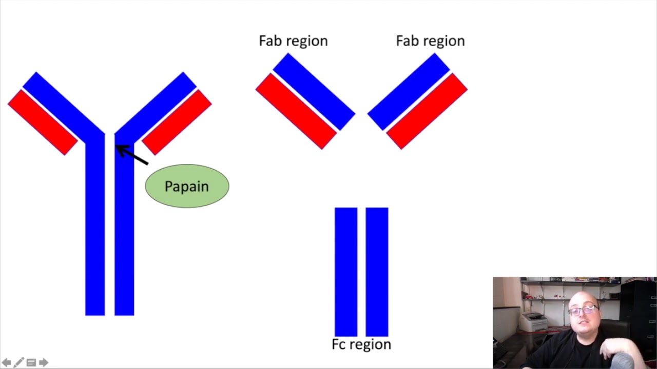
Instead, many antibodies are cleaved in specific locations using protease enzymes, and the structure is solved with an antibody fragment. A specific protease named papain will cleave the antibody right about here, lopping off the two antigen-binding fragments, called Fabs for short. The Fab consists of the variable domains from both the heavy and light chain, plus a constant domain from each, all conveniently held together by the disulfide bonds that make up the immunoglobulin fold.
After the protein is cleaved, the Fc domain can be separated from the Fab domains, and the Fabs alone are used in the binding experiment. Oh and by the way, Fc stands for “crystallizable fragment”—when researchers had just started to study the cleavage of antibodies with proteases, they found that this fragment was easy to crystallize. Coming back to the structure at hand, let’s switch over to the actual antibody Fab that’s bound to spike.
In this structure, there are actually three Fabs bound in three different regions of the spike protein. This particular antibody binds spike in an inactive conformation, preventing it from changing to the shape it needs to adopt to infect cells. But, let’s just focus on one Fab to explore the molecular interactions that allow this antibody to neutralize spike, and recolor spike white so the red oxygen atoms will be easy to see.
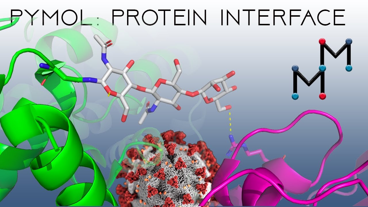
Zooming in on the antibody-spike interface, we’ll show the amino acids that can interact as sticks. Some of the interactions stabilizing the structure are hydrogen bonds, like this short, strong one that forms between the OH group on tyrosine and the negatively charged aspartate side chain. Remember, in these structures, the hydrogen atoms aren’t shown.
Another side chain/side chain interaction that promotes antibody binding is this hydrogen bond between this negatively charged glutamate and the hydrogen of the serine hydroxyl group. Side chain-/backbone/ interactions also promote binding, like the interaction of this positively charged lysine with this carbonyl group on the main protein chain. Though weaker, van der Waals interactions are also important in binding, and we see a network of these dispersion forces driving the interaction of this phenylalanine on spike with tyrosine residues on both the heavy and light chains of the antibody.
Here’s another one, this time between a phenylalanine from the heavy chain and a tryptophan on spike. And another, this time between a tryptophan and a valine. Antibody-antigen binding kicks off the immune response that allows the body to fight infection.
Part of this is the formation of more B-cells that produce the antibody. As the B-cells divide, some fine-tuning occurs. In a process called somatic hypermutation, the antigen-binding region undergoes additional mutations, which change some of the amino acids, particularly in loop regions called CDRs.
Some of these mutated antibodies won’t bind at all, but some will bind better, and the B-cells that produce these superior antibodies will become long-lived memory B-cells that can quickly respond to similar antigens in the future. Scientists can also facilitate the process of superior antibody production. Understanding molecular interactions can help us engineer antibodies that can serve as antivirals, antibiotics, and even cancer treatments.